
Abstract

“We are proud to recognize the work that is inspiring the next-generation of medical advances.”
During the past year, the JALA Editorial Board requested nominations for The JALA Ten in an effort to highlight the top 10 breakthroughs of the year that are making an impact on a diverse set of fields in biology and medicine. Following a selection process based on the expertise of internationally recognized authorities in automation, microfluidics, drug screening and delivery, and diagnostics, we are proud to announce The 2011 JALA Ten honorees. These discoveries represent the efforts of researchers from all over the world and cover disciplines that range from nanomaterials to drug delivery and pathogen detection. Covering the best in multidisciplinary research serves as a foundation for JALA, and we are proud to recognize the work that is inspiring the next generation of medical advances.
Dissolving Microneedles for Influenza Vaccination
By Sean P. Sullivan1, Dimitrios G. Koutsonanos2, Maria del Pilar Martin2, Jeong Woo Lee1, Vladimir Zarnitsyn1, Seong-O Choi1, Niren Murthy1, Richard W. Compans2, Ioanna Skountzou2, and Mark R. Prausnitz1 of 1Georgia Institute of Technology and 2Emory University School of Medicine, Atlanta, GA
Dissolving microneedles for transdermal drug delivery have been in development for many years. The work described here is the first demonstration of effective administration of a vaccination via this technology. Transdermal administration has been found to enhance the immune response to the vaccination, and the microneedle administration of described herein had a substantial increase in vaccination benefit as compared with a standard vaccination administration in animal models. The microneedle administration is significant for multiple reasons: (1) increased efficacy of vaccination—in this case, against the influenza virus, (2) a pain-free administration that will enhance patient compliance, (3) needle-less administration, thus eliminating a significant source of biomedical waste and potentially reducing the spread of disease through the secondary use of needles, and (4) the potential for the development of widespread immunization that does not require a medical professional for administration. This is particularly important in developing countries where access to medical professionals is often difficult. This can have widespread public health benefits and increase health and quality of life in these countries.

Image reprinted with permission from Macmillan Publishers Ltd: Nature Medicine. Sullivan, S. P., et al., Dissolving polymer microneedle patches for influenza vaccination. Nat. Med.
Photothermal Nanoblade for Large Cargo Delivery into Mammalian Cells
By Ting-Hsiang Wu, Tara Teslaa, Sheraz Kalim, Christopher T. French, Shahriar Moghadam, Randolph Wall, Jeffery F. Miller, Owen N. Witte, Michael A. Teitell, and Pei-Yu Chiou of the University of California, Los Angeles, CA
Currently there is no existing technology that allows rapid delivery of large objects into mammalian cells. This represents a major technological hurdle in various fundamental and translational problems. Using highly localized thermal bubbles induced by short laser pulse near a metal-coated microcapillary, researchers from University of California, Los Angeles have demonstrated a novel approach, which is called a photothermal nanoblade, for effective intracellular delivery large cargos, including DNA, RNA, polystyrene beads, and even live bacteria, with high cell viability. This novel photothermal nanoblade holds great promise for delivering large objects, such as large nanocomplexes, organelles, and pathogens that are currently untransferable into mammalian cells. The technology will solve a wide spectrum of biomedical problems and address new biological questions beyond current imagination.
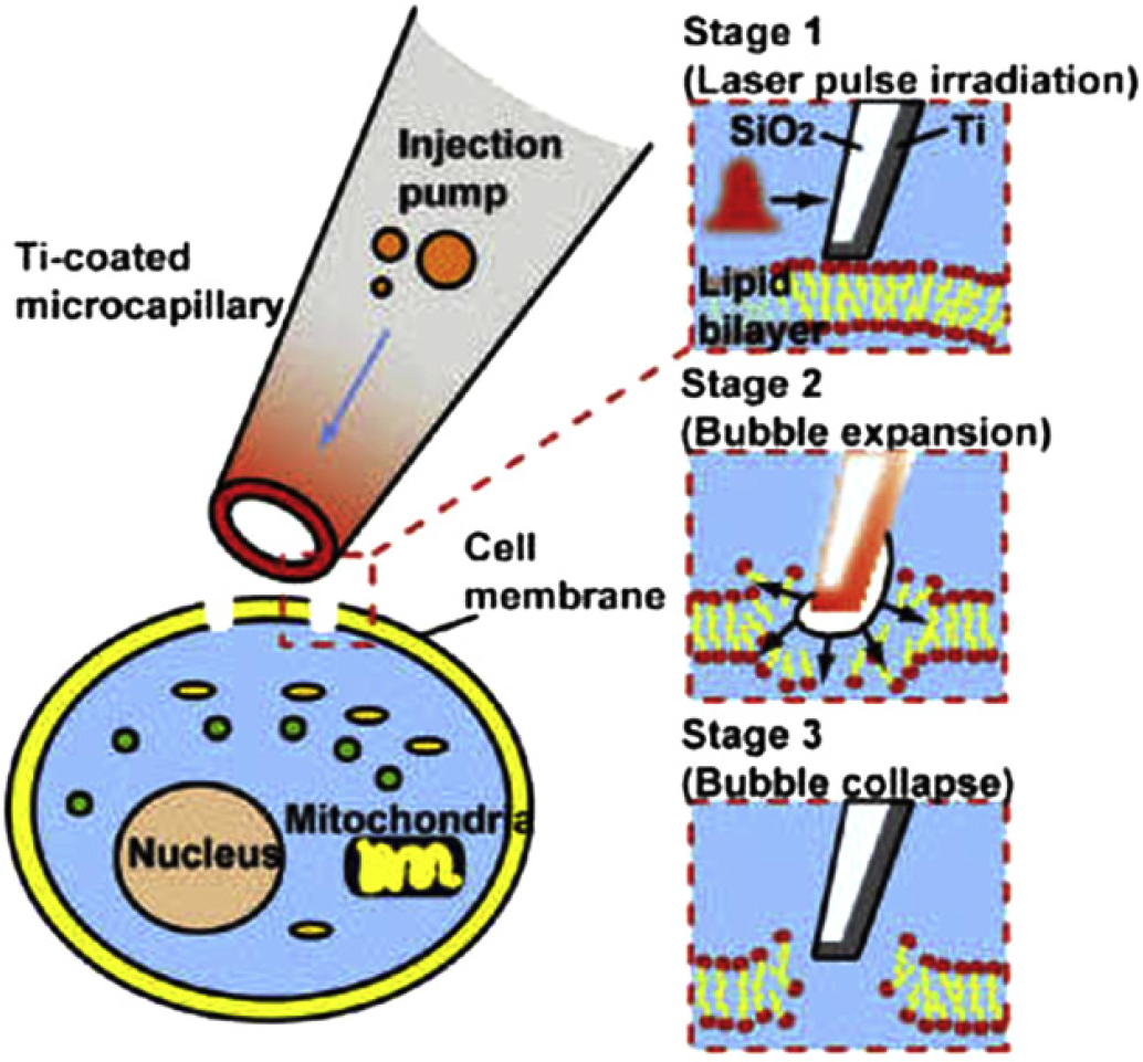
Image reprinted with permission from Wu, T. H.; Teslaa, T.; Kalim, S.; French, C. T.; Morghadam, S.; Wall, R.; Miller, J. F.; Witte, O. N.; Teitell, M. A.; Chiou, P. Y. Photothermal nanoblade for large cargo delivery into mammalian cells. Anal. Chem.
Wettability Patterning by UV-Initiated Graft Polymerization of Poly(acrylic acid) in Closed Microfluidic Systems of Complex Geometry
By Marc H. Schneider, Hervé Willaime, Yvette Tran, Fadhel Rezgui and Patrick Tabeling of ESPCI, Paris and Schlumberger, of Clamart, France
The ability to manipulate complex fluids in a microfluidic environment represents a key step toward the next generation laboratory automation systems for single molecules and single cells analyses. New perspectives in system biology and integrative biomedicine warrant major breakthroughs in microfluidic wettability control to truly realize the innovative technology in a laboratory automation setting. Using UV-initiated graft polymerization of poly(acrylic acid) in closed microfluidics, spatial modification of wettability with high resolution within enclosed microfluidic systems is now feasible and allows the creation of high-density microfluidic networks with complex geometries while yielding excellent wettability contrast. The versatile properties of this technology open new doors for next generation microfluidic automation.
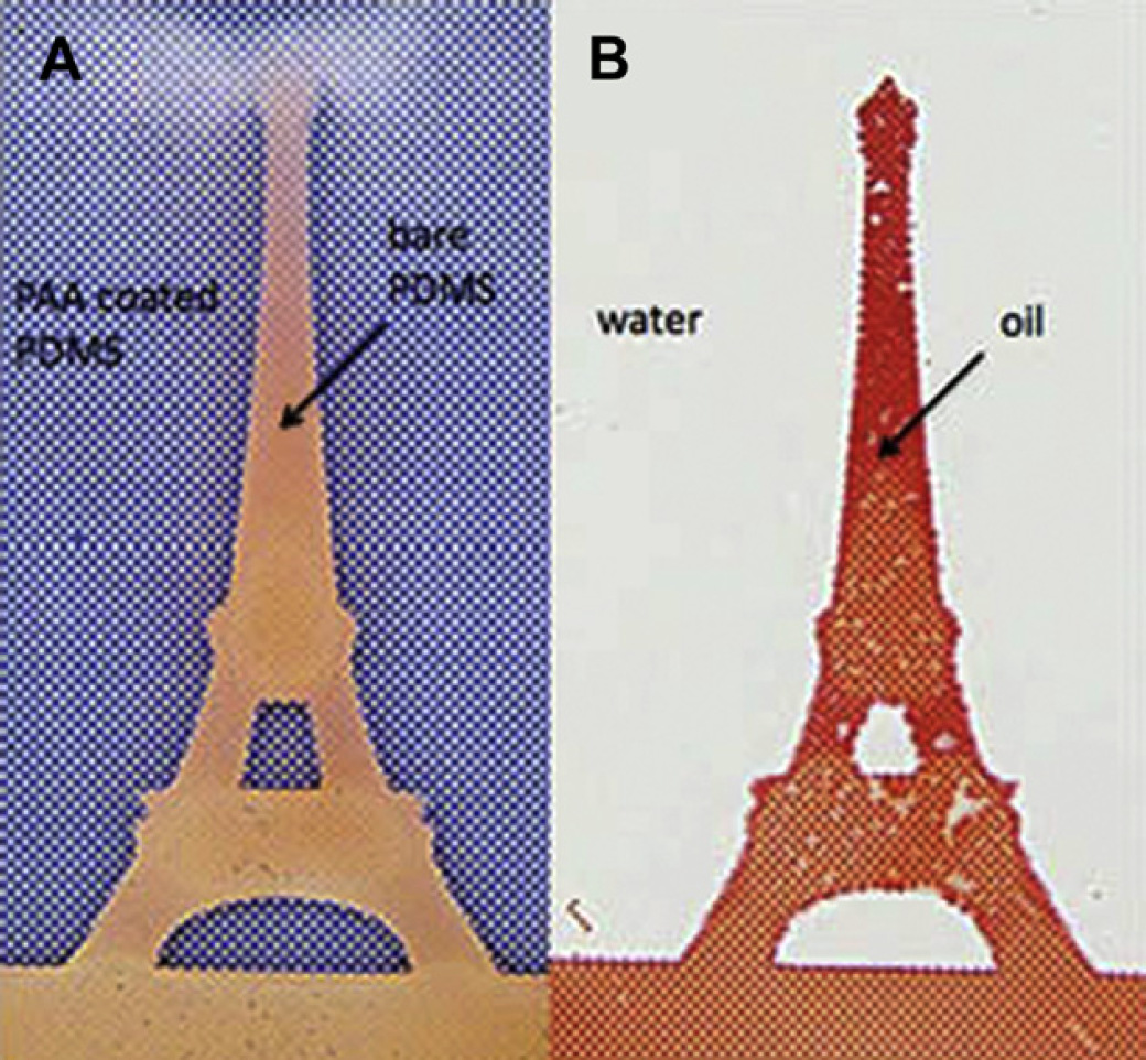
Image reprinted with permission from Schneider, M. H.; Willaime, H.; Tran, Y.; Rezgui, F.; Tabeling, P. Wettability patterning by UV-initiated graft polymerization of poly(acrylic acid) in closed microfluidic systems of complex geometry. Anal. Chem.
A Surface Topography Assisted Droplet Manipulation Platform for Biomarker Detection and Pathogen Identification
By Yi Zhang, Seungkyung Park, Kelvin Liu, Jennifer Tsuan, Samuel Yang and Tza-Huei “Jeff” Wang of Johns Hopkins University, Baltimore, MD
This work describes a droplet microfluidic, sample-to-answer platform for genetic detection of diseases, such as cancer and infectious diseases, using crude biological samples including blood. The platform exploited the dual functionality of silica superparamagnetic particles for solid-phase extraction of DNA and magnetic manipulation of droplets. This enabled the full integration of the entire sample preparation and genetic analysis process in droplets, including the steps of cell lysis, DNA binding, washing, elution, amplification, and detection. The microfluidic device was self-contained, with all reagents stored in droplets, thereby eliminating the need for fluidic coupling to external reagent reservoirs.

Zhang, Y.; Park, S.; Liu, K.; Tsuan, J.; Yang, S.; Wang, T. H. A surface topography assisted droplet manipulation platform for biomarker detection and pathogen identification. Lab Chip.
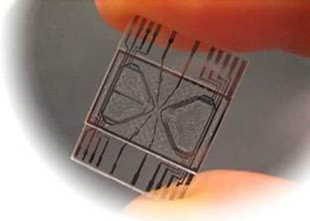
Fully Integrated Point-of-Care Blood Counting
By Cees van Berkel1, James D. Gwyer1, Steve Deane1, Nicolas Green2, Judith Holloway1, Veronica Hollis2 and Hywel Morgan2 of 1Philips Research Laboratories, Cambridge and 2University of Southampton, Southampton, UK
Point-of-care blood cell counts have the possibility to enable new types of diagnosis and treatment decisions in the emergency room, clinic, or during emergency transit. However, current blood cell counting systems have not been miniaturized to the point where they are practical in these cases. Hywel Morgan and others have recently demonstrated an electrical impedance-based microfluidic platform that combines sample processing and characterization and enumeration of a complete blood count in a single, integrated, and miniaturized system. This work has been validated in comparison with current systems using real samples. This is not a system that will work only in a laboratory but deals with all of the issues of automation required in preparing samples before analysis—all on a simple platform. Development of simple label-free approaches to perform a three-part differential measurement is poised to increase the availability and distribution of hematology analyzers for applications in the developed and developing world.
Van Berkel, C.; Gwyer, J. D.; Deane, S.; Green, N.; Holloway, J.; Hollis, V.; Morgan, H. Integrated systems for rapid point of care (PoC) blood cell analysis. Lab Chip.
Liquid Gradient Refractive Index Microlens for Biophotonic Applications
By Hua Huang, Xiaole Mao, Sz-Chin Steven Lin, Brian Kiraly, Yiping Huang, and Tony Jun Huang of The Pennsylvania State University, University Park, PA
Professor Tony Jun Huang's research group at The Pennsylvania State University pioneered a novel optofluidic microlens named the liquid gradient refractive index lens for variable focusing of light within a microfluidic device. The focusing of light was achieved through the gradient refractive index within the liquid medium, rather than via curved refractive lens surfaces. This novel on-chip light manipulation mechanism enables not only tuning of the focal distance (translation mode) but also shifting of the output light direction (swing mode), a second degree of freedom that has yet to be accomplished for in-plane tunable microlenses. This work could have revolutionary impact on a variety of lab-on-a-chip and biophotonic applications.

Huang, H.; Mao, X.; Lin, S. C.; Kiraly, B.; Huang, Y.; Huang, T. J. Tunable two-dimensional liquid gradient refractive index (L-GRIN) lens fro variable light focusing. Lab Chip.
Novel Strategies to Deagglomerate and Functionalize Nanodiamond Materials
By Yuejiang Liang1, Thomas Meinhardt1, Gerald Jarre1, Masaki Ozawa1, Pavo Vrdoljak1, Achim Schöll1, Friedrich Reinert1,2 and Anke Krueger1 of 1Julius-Maximilians-Universität Würzburg and 2Gemeinschaftslabor für Nanoanalytik, Karlsruhe, Germany
Nanodiamonds are emerging as promising platforms for applications ranging from drug delivery to imaging. However, because nanodiamonds are potentially prone to clustering, which can impact subsequent surface processing procedures to result in functionalized aggregates. Therefore, one of the key challenges toward their translation is based on developing new strategies to disperse nanodiamonds to yield comprehensively functionalized particles. Current methods based on ball milling can mechanically disperse particles but may potentially result in contamination. Recent work by Krueger and colleagues has shown that nanodiamond processing with aryl diazonium salts can be used to disperse particles down to sizes of ∼ 20–50 nm in diameter without the need for mechanical techniques. Furthermore, a diverse range of chemical groups can be carried by the aryl diazonium salts (e.g., COOH, SO3H), producing nanodiamond-COOH and nanodiamond-SO3H complexes that can be dispersed in water or physiological media. This work thus provides a broadly applicable approach toward further capabilities in grafting on the surfaces of dispersed nanodiamonds, further expanding their applications in biology, medicine, and beyond.

Liang, Y.; Meinhardt, T.; Jarre, G.; Ozawa, M.; Vrdoljak, P.; Schöll, A.; Reinert, F.; Krueger, A. Deagglomeration and surface modification of thermally annealed nanoscale diamond. J. Colloid Interface Sci.
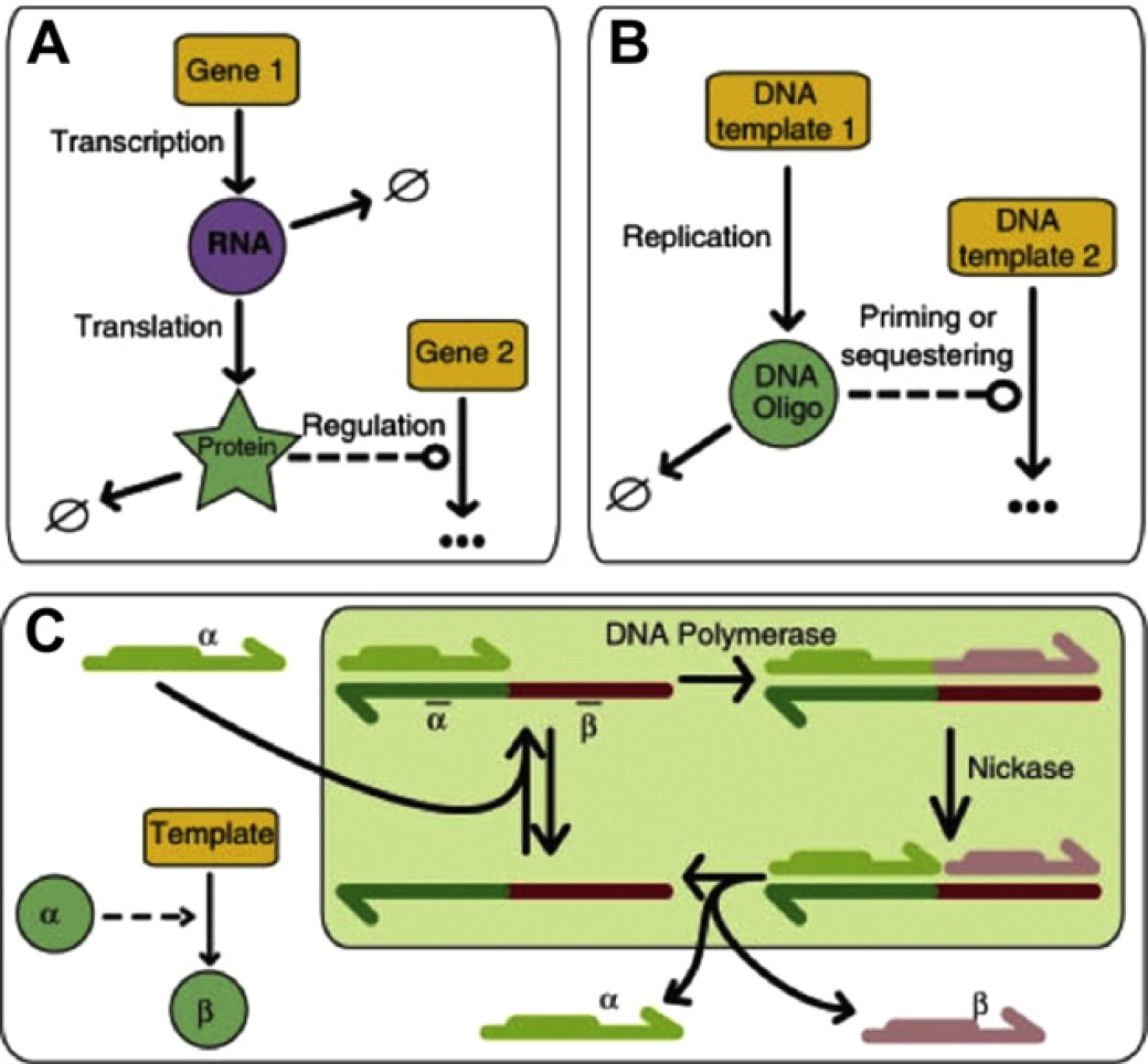
Programming an in Vitro DNA Oscillator Using a Molecular Networking Strategy
By Kevin Montagne1, Raphael Plasson2, Yasuyuki Sakai1, Teruo Fujii1 and Yannick Rondelez1 of 1LIMMS/CNRS-IIS, Institute of Industrial Science, University of Tokyo and 2Department of Applied Chemistry, Faculty of Science and Technology, Keio University, Yokohama, Japan
Living organisms perform and control complex behaviours by using webs of chemical reactions organized in precise networks. This powerful system concept, which is at the very core of biology, has recently become a new foundation for bioengineering. Remarkably, however, it is still extremely difficult to rationally create such network architectures in artificial, nonliving, and well-controlled settings. Researchers from Japan have recently introduced a method for such a purpose, on the basis of standard DNA biochemistry. This approach is demonstrated by assembling de novo an efficient chemical oscillator: they encode the wiring of the corresponding network in the sequence of small DNA templates and obtain the predicted dynamics. Their results show that the rational cascading of standard elements opens the possibility to implement complex behaviors in vitro. Because of the simple and well-controlled environment, the corresponding chemical network is easily amenable to quantitative mathematical analysis. These synthetic systems may thus accelerate the understanding of the underlying principles of biological dynamic modules.
Montagne, K.;Plasson, R.; Sakai, Y.; Fujii, T.; Rondelez, Y. Programming an in vitro DNA oscillator using a molecular networking strategy. Mol. Syst. Biol.
Novel Nanoresolution Label-Free Detection Probes using Fluorescent Nanodiamond
By V. Petrakova1,2, I. Kratochvilova2, F. Fendrych2, J. Kucka3, J. Stursa3, P. Cigler4, M. Ledvina4, A. Fišerová5, P. Kneppo1 and M. Nesládek6 of 1Czech Technical University in Prague, Biomedical Engineering, Kladno, Czech Republic and 2Institute of Physics, Academy of Sciences of the Czech Republic, Prague, Czech Republic and 3Institute of Nuclear Physics, Academy of Sciences of the Czech Republic, Prague, Czech Republic and 4Institute of Organic Chemistry and Biochemistry, Academy of Sciences of the Czech Republic, Prague, Czech Republic and 5Institute of Microbiology, Academy of Sciences of the Czech Republic, Prague, Czech Republic and 6IMOMEC division, IMEC, University Hasselt, B-3590 Diepenbeek, Belgium
A unique characteristic of nanodiamond (ND) particles is its stable fluorescence from engineered color centers (i.e., neutral nitrogen-vacancy [NV0] or negatively charged NV−) that makes (fluorescent nanodiamonds) FNDs interesting nanotools for stimulation tracking and detection of delivery events. Recently, Nesladek and coworkers have demonstrated principles of this novel method for remote optical monitoring of chemical processes in biological environments based on color changes from photoluminescent NV centers in FND. This method can thus open up a novel domain for contact-less high-resolution intracellular monitoring using standard confocal microscope working in the far field. This method is based on driving the NV luminescence chemically, by alternating the surface chemical potential, by interacting atoms and molecules with the FND surface. Because of the small FND size, the close proximity of NV centre from the surface is influenced by chemical potential of the environment and leads to changes in the ND energetics and intermingling with the electronic NV states. This allows construction of optical chemobiosensors operating in cells, visible in classical confocal microscopes. This phenomenon was demonstrated in oxidized and hydrogenated and fluorinated ND. Hydrogenation of NDs leads to quenching of fluorescence related to negatively charged (NV−) centers and by this way produces color shifts from NV− (636 nm) to neutral NV0 (575 nm) fluorescence. Although this effect, unlike the Förster resonance energy transfer, is detectable even in larger diamond particles such as 50–80 nm size nm, based on modeling, the reduction of diamond size to 5–10nm increases the magnitude of NV color shift phenomena by size effects. By using the charge interaction arising from molecular environment in the cells, FND fluorescence can be changed and make thus particular molecular interaction/delivery event optically visible.

Petráková, V.; Kratochvilova, I.; Fendrych, F.; Kučka, J.; Štursa, J.; Cígler, P.; Ledvina, M.; Fišerová, A.; Kneppo, P.; Nesládek, M. Novel nano-resolution label-free detection probes using fluorescent nanodiamond (FND). In press. Image reprinted with permission.
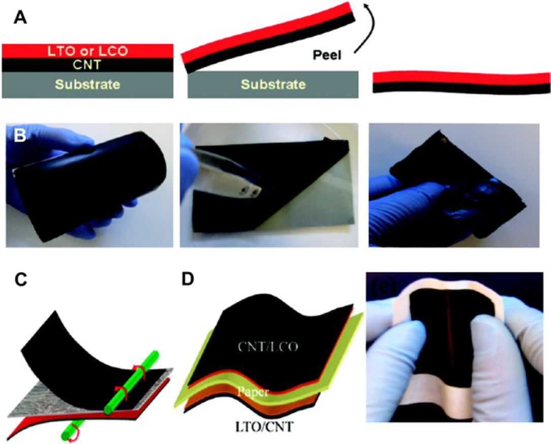
Robust Lithium Ion Paper Batteries
By Liangbing Hu, Hui Wu, Fabio La Mantia, Yuan Yang, and Yi Cui of Department of Materials Science and Engineering, Stanford University, Stanford, CA
The development of mechanically robust, high energy density, and ultra-portable batteries represents a high impact area of interest with broad applications. Recent work by Yi Cui and colleagues demonstrated the integration of lithium ion battery components (e.g., LTO or LCO) and freestanding carbon nanotube (CNT)-based current collectors for the anode and cathode. LCO/CNT or LTO/CNT double-layer films were synthesized and peeled off the substrate, and the double films were subsequently bound to the paper through a straightforward lamination process. In this study, the paper served as both a mechanical support for the device and a separator membrane that had a lower impedance than commercial standards. The lithium ion battery was capable of 300 recharge cycles while the devices were able to withstand significant bending. Furthermore, the devices were lightweight at 0.2 mg/cm2, only 300-μm thick, and exhibited a high energy density of 108 mWh/g, demonstrating their immense potential for a broad spectrum of portable energy applications.
Reprinted with permission from Hu, L.; Wu, H.; La Mantia, F.; Yang, Y.; Cui, Y. Thin, flexible secondary Li-ion paper batteries. ACS Nano.