
Abstract
Clinical report
A 3-year-old, neutered male domestic shorthair cat was presented for veterinary investigation of a painful right eye, which manifested as severe blepharospasm, serohaemorrhagic discharge and chemosis with third eyelid protrusion. The eye was examined under general anaesthesia. No corneal ulceration was evident but a 3 to 4 mm diameter conjunctival wound was visible at the temporal aspect of the globe, close to the limbus. Skull radiographs were taken and no foreign body could be detected.
Medical treatment was initiated with atropine sulfate SID (Atropine; Viatris), topical gentamicin QID (Clinagel-Vet; Ecuphar) and a systemic antibiotic (orbifloxacin 25 mg SID, Orbax; Schering-Plough). The cat was examined on three occasions by the veterinary surgeon over a 2-month period. The eye was no more painful and the chemosis reduced, but the third eyelid profusion remained. The local treatment regimen was switched to dexamethasone BID (Maxitrol; Alcon-Couvreur) and methylprednisolone 10 mg IM (Moderin Long Acting; Pfizer). The cat was then referred for further ophthalmic examination.
Ophthalmic examination
Ophthalmic examination revealed blepharospasm and lacrimation from the right eye. The pupillary light reflexes and the response to the menace test were normal. The eye movements had a normal amplitude but retropulsion of the globe was limited. The third eyelid was congested and protruding. No corneal lesion could be detected. The ophthalmic examination was otherwise normal.
Imaging
Orbital ultrasonography undertaken 3 months after initial presentation revealed increased echogenicity of the orbital fat with an additional linear hyperechoic structure (about 5 mm width) ventromedial to the globe. This structure, which was horizontal in orientation and approximately 7 mm long with acoustic shadowing, was situated beside the optic cone (which was not otherwise affected). Thus, the ultrasound examination confirmed the presence of an orbital cellulitis surrounding a possible foreign body.
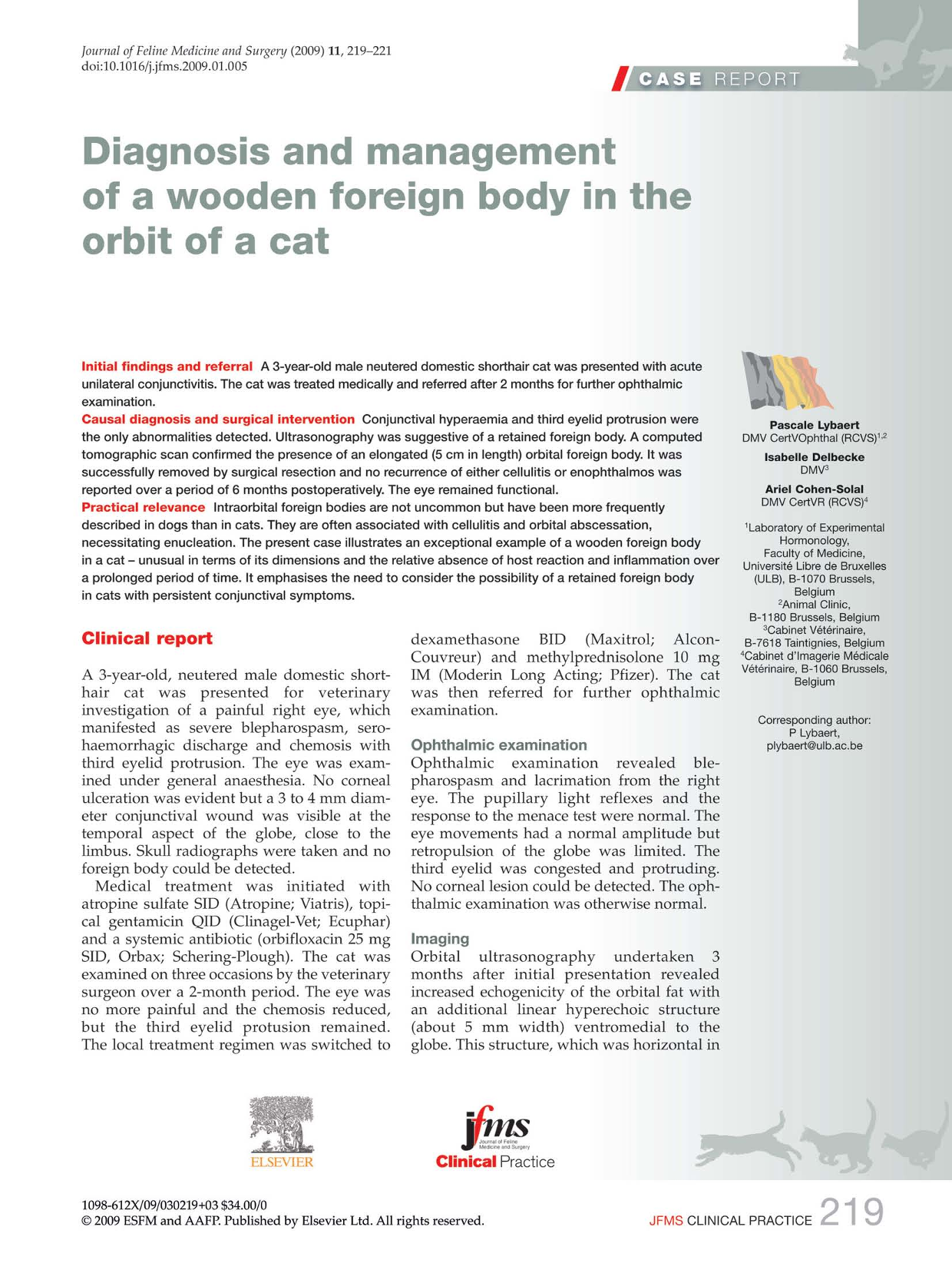
A computed tomographic (CT) scan was performed to evaluate the nature and extent of the putative foreign body. A well-defined thick linear foreign body was visible on plain views on the medial aspect of the orbital floor (Fig la). The rostral limit of the foreign body was situated close to the ventral orbital rim and its caudal limit extended alongside the right sphenoidal wing (Fig lb). The globe was slightly deviated dor-sally but otherwise was normal in appearance. No periosteal or lytic reaction was evident. The foreign body, as evaluated by the CT scan, measured 5 cm in length, and approximately 7 mm in diameter at its cranial aspect (Fig 1c) and 3 mm at its caudal aspect.

Computed tomographic scans: transverse (a), sagittal (b) and dorsal (c) plans. A well-defined linear foreign body (arrowheads] is visible, lying in the ventral portion of the orbit
A further scan, performed after iodine contrast medium injection, disclosed marked enhancement of the soft tissue surrounding the foreign body. The other skull structures were normal. The right submaxillary lymph node was slightly enlarged.
Surgery
Surgical resection under general anaesthesia was performed 2 days later. A lateral canthotomy allowed close examination of the inner aspect of the third eyelid. The anterior aspect of what appeared to be a wooden foreign body, deep in the conjunctival fornix close to the ventral aspect of the globe, was visible. The extremity of the foreign body was grasped with forceps and pulled gently outwards (Fig 2a, b). Examination of the foreign body confirmed its wooden nature and the dimensions as evaluated by the CT scan (Fig 2b). No purulent material was noted. The penetration site was left unsutured and the lateral canthotomy was closed with simple interrupted sutures.

(a,b) Surgical removal of the foreign body. (c) The foreign body was wooden in nature and its dimensions correlated well with the estimation based on the CT scan. Scale bar — 1 cm
Post-surgical treatment included topical antibiosis with ciprofloxacin QID (Ciloxan; Alcon-Couvreur), systemic antibiosis with amoxicillin 40 mg/clavulanic acid 10 mg BID (Synulox; Pfizer) for 7 days and systemic anti-inflammatory treatment with ketoprofen 5 mg SID (Ketofen; Merial) for 3 days.
Recovery was uneventful and no recurrence of orbital cellulitis or enophthalmos was reported 6 months postoperatively. The cat's vision was not altered and he was otherwise well (Fig 3).

The cat 3 months postoperatively
Discussion
Intraorbital foreign bodies are not uncommon but have been reported more frequently in dogs than in cats. 1 Organic foreign bodies mainly consist of migrating grass awns,1–3 although wooden materials and porcupine quills have also been encountered. 4 In the present case, the foreign body appeared to be a splinter of dead wood (Fig 2), possibly from a wooden box, plank, railing or item of old furniture.
Orbital (including intraocular) foreign bodies, have been reported to have a poor prognosis. 2 They are commonly associated with cellulitis and orbital abscessation, culminating in the need for enucleation.2,5 Moreover, plant and wooden materials may carry soil-borne bacteria or fungi that can potentially cause panophthalmitis. 5 Migration of an organic foreign body may also lead to ocular penetration and severe lesions. 2 There is a report of non-specific injury to the central nervous system of a man associated with a retained wooden golf tee. 5
The present report details an exceptional case of a wooden foreign body in the orbit of a cat — unusual in terms of its dimensions and the relative absence of inflammatory reaction and infection in the host over a prolonged period of time. A similar case has been reported in a 46-year-old man with a retained orbital wooden twig, which remained undetected for 14 weeks. 6 Such a presentation emphasises the need to include a possible retained foreign body in the differential diagnosis of cats with conjunctivitis, especially when a conjunctival laceration is detected. 6
The difficulty of diagnosing a wooden foreign body retained inside the orbit or cranium has been much discussed.5–8 In the present clinical case, orbital ultrasonography was clearly suggestive of a retained foreign body. Ultrasonography has indeed proven to be effective in detecting a range of organic foreign bodies, 9 but has not been successful in all cases. 2 An additional CT scan was decisive in this case, both confirming the presence of a foreign body and determining its dimensions and exact location prior to surgery.