75. Suppression of atherosclerotic changes in cca of SHR-SP with a HMG-coa reductase inhibitor
S. Nagotani, K. Deguchi, T. Yamashita, V. Lukic-Panin, M. Takamiya, T. Kamiya and K. Abe
Department of Neurology, Okayama University, Okayama, Japan
Objectives: Statin reduces cerebrovascular events independent of its cholesterol lowering effect. We hypothesized statin inhibits early atherosclerotic change in common carotid artery (CCA), and investigated its effect on lectin-like oxidized-LDL receptor-1 (LOX-1) and monocyte chemoattractant protein-1 (MCP-1) expression, both of which are early atherosclerotic markers.
Methods: Stroke-prone spontaneous hypertensive rats (SHR-SP) were divided into 3 groups. Each group was treated with vehicle or simvastatin for 2 or 4 weeks and fed high fatty food and 1% NaCl water for 2 weeks, and CCA was removed. LOX-1 and MCP-1 expression as well as macrophage infiltration were histologically investigated. Lipid deposition was also investigated by Sudan III staining.
Results: Simvastatin groups showed significantly smaller amount of lipid deposition and LOX-1 and MCP-1 expressions, independent of serum lipid levels. Macrophage infiltration was also inhibited.
Conclusions: Simvastatin suppresed atherosclerotic change in CCA of hypertensive rats with high fatty diet. Reduction of cerebrovascular event by statins may be brought by the direct inhibition against atherosclerotic change.
Representative photomicrographs of immunohistochemistry for LOX-1 and MCP-1 and Sudan III staining. Note strong expression of LOX-1 and MCP-1 and lipid deposition in the vessel wall of the vehicle group. Simvastatin decreased LOX-1 and MCP-1 expression and lipid deposition in a treatment-period dependent manner.
139. MRNA granules form in penumbral neurons following focal ischemia
D. DeGracia, M.K. Lewis, N.N. Rizk and J.C. Dunbar
Department of Physiology, Wayne State University, Detroit, Michigan, USA
Objectives: Irreversible Translation arrest (TA) following global ischemia TA correlates with the delayed death of vulnerable neurons. Stroke, or focal ischemia, results in a pathophysiology that shares some features with, but has significant differences from, global ischemia. Stroke results in immediate necrosis of brain tissue, while a similar type of delayed neuronal death occurs in adjacent penumbral tissue. Others have shown that penumbral neurons undergo post-ischemic TA. We therefore sought to investigate if similar alterations to translation regulatory systems occur following focal ischemia as have been observed following global ischemia.
Methods: We evaluated markers of stress granules (SGs) and mRNA sequestration using double labeling immunofluorescence histochemistry (IC) and in situ fluorescent immunohistochemistry (FISH). Focal ischemia was produced using normothermic middle cerebral artery occlusion (MCAO) by suture occlusion of the distal MCA. Experimental groups (n = 3 to 5/group) included 2 h MCAO+24 h reperfusion, 3 h ischemia only, and 4 h ischemia only. Stroke volume was characterized by TTC staining. Slices from perfusion-fixed animals were double-labeled for poly-adenylated mRNAs (via FISH) and the following protein markers (via IC): PABP, S6, TIA-1, and TTP. In vitro and in vivo translation rates were measured by standard methods.
Results: 2 h MCAO resulted in ipsilateral necrosis of striatal and cortical tissue at 24 h reperfusion. At 3 h and 4 h ischemia-only, pre-necrotic cells were shrunken and pycnotic but did not display evidence of changes in SG number or composition, or evidence of mRNA granulation. Neurons outside the core showed graded effects. Those adjacent to the core showed mRNAs granules that colocalized with PABP but not S6, TIA-1 or TTP. mRNA granules were most prominent in layer II cortical pyramidal neurons. The presence of mRNA granules correlated with decreased translation rates. Ipsilateral cortical neurons distal to the core and contralateral neurons did not show evidence of mRNA granules. The number of SGs compared to controls in penumbral neurons was a function of distance from the lesioned area on the ipsilateral side.
Conclusions: Neuronal necrosis occurred independent of morphological changes in translational machinery. Penumbral neurons showed graded effects with respect to mRNA granule formation. Our data suggests that mRNA granules contribute to the prolonged TA in penumbral neurons following focal cerebral ischemia as we have shown occurs after global cerebral ischemia.
170. Blood cell microRNA expression profile of neurologic diseases: a pilot microrna array study
D. Liu, Y. Tian, B. Ander, H. Xu, B. Stamova and F. Sharp
Department of Neurology and the M.I.N.D. Institute, University of California at Davis, Sacramento, California, USA
MicroRNAs (miRNAs) are a recently discovered class of endogenous, small non-coding RNAs that regulate gene expression and have a critical role in many biological and pathological processes. Here we investigated the miRNA expression profile in blood cells after brain injuries. Adult rats were subjected to ischemic strokes, hemorrhagic strokes, sham surgeries, kainate-induced seizures, compared with controls. The blood cell miRNA expression patterns were assessed 24 h later, using Taqman rodent miRNA arrays. Results showed that many miRNAs were upregulated and a few ones were downregulated at least twofold in blood cells after each experimental condition. miRNA response patterns were different for each condition. These results demonstrate the possible use of blood cell miRNAs as biomarkers and may be helpful to explain the results of our previous blood cell mRNA profile studies that many genes were downregulated of in these above conditions.
184. Intracellular calcium levels and vasoconstriction induced by 5-CT and s6c in rat after cerebral ischemia
H. Ahnstedt, R. Waldsee and L. Edvinsson
Clinical Sciences, Experimental Vascular Research, Lund University, Lund, Sweden
Objective: Middle cerebral artery occlusion (MCAO) in rat increases the contractile responses of the middle cerebral arteries (MCAs) induced by different vasoconstrictor mediators and decreases the blood flow that follows the brain ischemia. Previous studies have shown that there is up-regulation of endothelin type B (ETB) receptors1 after MCAO and up-regulation of 5-HT1B receptors in MCAs after subarachnoid hemorrhage.2 The objective of this study was to evaluate the intracellular calcium changes that occur during contraction induced by the 5-HT1 receptor agonist 5-carboxamidotryptamine (5-CT) and the ETB receptor agonist sarafotoxin 6c (S6c) in rat MCAs after MCAO.
Methods: MCAO for 90 mins and reperfusion for 48 h in rat were followed by evaluation of ischemic brain damage by TTC staining and neurological examination. The MCAs were removed and mounted in sensitive myographs and the contractile responses to S6c or 5-CT were studied in the right MCA (stroke) and the left MCA (control). Intracellular calcium measurements using FURA-2 were applied on segments of MCAs and calcium changes and contraction were evaluated simultaneously.
Results: Brain damage calculations after MCAO confirmed a mean brain damage of 15.6%±1.9%. The neurological deficit score, using an established scoring system of 0–5 to assess the neurological behavior of the animals,3 was about 4 at the time of reperfusion and 2 after 48 h. Pharmacological results showed that S6c induced contraction in MCAO arteries (25.3%±11.3%) but not in control arteries (4.1%±1.3%) relative to 63.5 mol/L potassium contraction. The response to 5-CT decreased after MCAO; 53.2%±4.8% in control arteries compared to 16.8%±8.4% in MCAO arteries. The intracellular calcium concentration at resting level was 84.8±17.2 nmol/L; contraction induced by 5-CT and S6c increased the plateau calcium level (in nmol/L) to 111.9±22.4 and 130.3±24.5, respectively.
Conclusion: Our data shows that MCAO results in increased contractile responses mediated by ETB receptors. Contradictory to what have been seen after subarachnoid hemorrhage, cerebral ischemia results in decreased contractile responses induced by 5-CT. There was no significant change in the intracellular calcium handling between control arteries and MCAO arteries suggesting that altered contractile responses mediated by ETB and 5-HT1 receptors are not due to calcium itself.
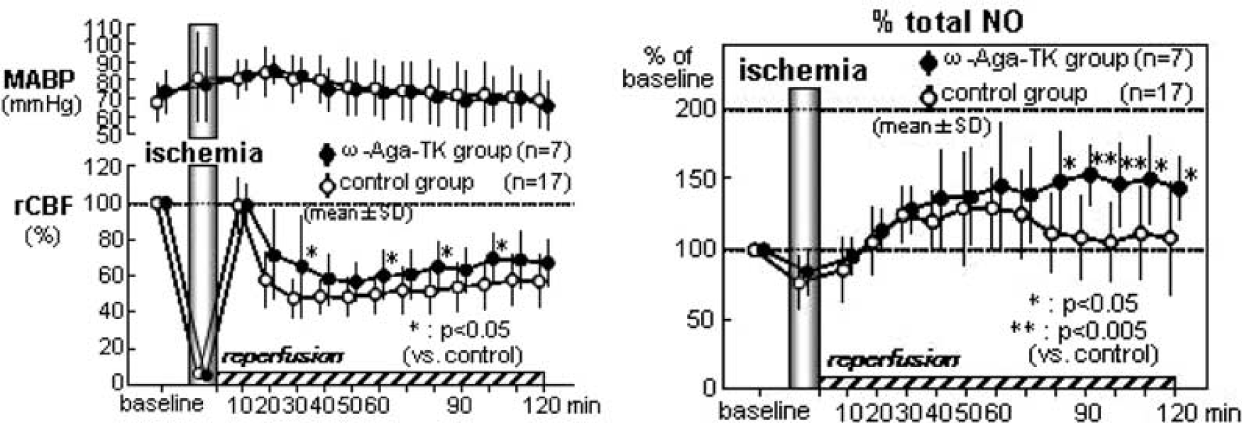
198. Effects of P-type calcium channel block on nitric oxide production during cerebral ischemia and reperfusion in mice
T. Ohkubo, Y. Asano, K. Hattori, D. Furuya, T. Shimazu, H. Nagoya, M. Yamazato, Y. Ito, Y. Kato and N. Araki
Department of Neurology, Saitama Medical University, Saitama, Japan
Objectives: Nitric oxide (NO) plays an important role in the pathogenesis of neuronal injury during cerebral ischemia. P-type calcium channels are present in the brain. We investigated the effects of P-type calcium channel blocker on NO production during cerebral ischemia and reperfusion.
Methods: Twenty four C57BL/6 mice were used in the study. ω-Agatoxin-TK (0.5 μg/kg), P-type calcium channel blocker, was administered by intraperitoneal injection at 30 mins before cerebral ischemia in 7 mice (ω-Aga group), and the drug was not administered in the remaining 17 mice (control group). The animals were anesthetized with 2% halothane and maintained with 0.5% to 1% halothane. NO production was continuously monitored by in vivo microdialysis. A microdialysis probe was inserted into the left striatum and perfused with Ringer's solution at a constant rate of 2 μl/mins. After 2 h equilibrium period, fractions were collected every 10 mins. A laser Doppler probe was placed on the right skull surface. Global ischemia was produced by clipping both common carotid arteries using Zen clip for 10 mins. The levels of nitrite (NO2−) and nitrate (NO3−) in the dialysate samples were measured by the Griess reaction.
Results:
Blood Pressure: No significant differences were observed between ω-Aga group and control group.
Cerebral Blood Flow (CBF) (Figure 1): CBF decreased to 3.7%±2.0% (mean±s.d.) in ω-Aga group and 4.2±1.5 in control group during ischemia. After reperfusion, CBF transiently returned to baseline values and then gradually decreased significantly in both groups. In ω-Aga group, CBF at 30 mins (65.4%±27.7*%), 60 mins (60.4±12.9*), 80 mins (65.3±13.3*) and 120 mins (67.3±12.1*) after reperfusion were significantly higher than those in control group (47.7±10.2, 50.2±10.1, 51.6±12.2, 55.5±13.1, respectively).
NO Metabolism (Figure 2): In ω-Aga group, the change rate of NO2− levels during ischemia (106.3±31.9*%), at 10 mins (119.8±21.8*) and at 80 mins (105.5±35.2**), and at 120 mins (102.7±23.6*) after reperfusion were significantly higher than those in control group (80.4±23.5, 88.8±31.3, 60.5±21.6, 64.2±37.7, respectively). As for the change rate of NO3− levers, no significant differences were observed between the two groups. The change rate of total NO (NO2−+NO3−) levels at 80 mins (147.9±34.6*), and at 120 mins (143.2±22.1*) in ω-Aga group were significantly higher than those in control group (111.8±27.3, 108.5±39.0, respectively) [*P<0.05, **P<0.005].
Effects of ω-agatoxin.
Conclusions: The above data indicate that ω-Agatoxin-TK attenuates the decrease of cerebral blood flow following reperfusion and affects the NO production after reperfusion. Although it has been reported that N-type calcium channel blocker cilnicipine increased eNOS expression, the relationship between NOS and P-/Q-type calcium channel is not clear. These data suggest P-type calcium channel inhibition may exert the brain protective effect through the influence on cerebral blood flow and NO metabolism.
240. Brief focal cerebral ischemia induces IL-1 β protein expression in rats
X. Zhan, B. Ander and F. Sharp
Neurology, MIND/UC Davis, Sacramento, California, USA
Objective: Transient ischemic attack (TIA) is a common cerebrovascular disorder. TIAs are under recognized, under reported and under treated though as many as a third of classical TIA patients eventually go on to have ischemic strokes.1 Our previous study demonstrated that 5 or 10 mins of brief focal ischemia caused microinfarctions and an inflammatory response in rat brains.2 Since cytokines can contribute to the cellular damage and inflammatory response seen following a stroke, we hypothesized that the expression of cytokines might be increased following very brief focal ischemia that simulates TIAs occurring in humans.
Methods: Male Sprague-Dawley rats weighing 280 to 320 g (Charles River Labs, USA) were used in this study (n = 12). Focal cerebral ischemia was produced by occluding the middle cerebral artery (MCA) using the intraluminal suture technique. Rats subjected to 5 mins or 10 mins of brief focal ischemia were allowed to survive 24 h. A stroke group receiving 2 h focal ischemia was used to compare to the brief focal ischemia treatment. Sham-operated rats were subjected to the identical surgical protocol except that no suture was inserted. After a 24 h period of reperfusion, cortex and basal ganglia in the ischemic hemisphere were dissected and frozen at −70 °C. Frozen tissues were homogenized in ice-cold buffer containing a complete protease inhibitor mixture. The homogenates were centrifuged at 14,000 × g for 30 mins at 4°C, and the pellet was discarded. The protein (20 m g) in the supernatant was used for ELISA analysis for IL-1β, IL-6, TNF-α, and IFN-g. Data were calculated as fold changes and expressed as mean±SE. One-way ANOVA was performed with a Student-Neuman-Keuls post hoc test. A P value of less than 0.05 was considered statistically significant.
Results: IL-1 β levels were increased significantly by 77.48±0.94 (P<0.05) fold, and 166.21±0.47 (P<0.05) fold at 24 h following 10 mins and 2 h of ischemia respectively. The increase (by 2.34±0.22 fold) at 24 h following 5 mins of ischemia was not significant. IL-6 levels were increased by 3.22±0.42, 5.12±0.46, and 6.81±0.30 fold; IFN-g levels were increased by 1.62±1.24, 1.88±0.49, and 1.68±1.70 fold; TNF-α levels were increased by 0.73±0.60, 2.56±0.64, and 3.11±0.34 fold at 24 h following 5 mins, 10 mins, and 2 h ischemia respectively. The changes in IL-6, IFN-g, and TNF-a at 24 h following 5 mins and 10 mins of focal ischemia were not significant.
Conclusion Focal ischemia of 10 mins duration, that simulates a TIA that occurs in humans, increases IL-1 β expression in brain 24 h later. Thus, ischemia without infarction can increase this cytokine in brain.
Acknowledgments: Department of Neurology and MIND Institute, University of California at Davis; and supported in part by NS054652.
249. Treatment with angiotensin II receptor blocker, olmesartan, recovers protective effect of chronic mild hypoperfusion aftery mca occluion in hypertensive rats
E. Omura-Matsuoka1, Y. Yagita2, T. Sasaki1, Y. Terasaki1, N. Oyama1, Y. Sugiyama2, S. Okazaki2 and K. Kitagawa2
1Cardiovascular Medicine; 2Neurology, Osaka University Graduate School of Medicine, Suita, Japan
Objectives: We showed occlusion of unilateral common carotid artery (CCAO) for two weeks reduced infarct size after occlusion of middle cerebral artery (MCAO) in Wistar rats. The purpose of this study is to show whether CCAO for two weeks reduces infarct size after MCAO also in spontaneously hypertensive rats (SHR), and to clarify the effect of administration of angiotensin II AT1 receptor blocker (ARB), olmesartan, in SHR.
Methods: < Wistar vs. SHR>In adult male Wistar rats (n = 28) or SHR (n = 18), each animal was anesthetized with halothane. Pre-CCAO group rats received the left CCAO and the left MCA was subsequently occluded permanently 2 weeks later (n = 22). Sham group rats received only exposure of the left CCA and MCAO 2 weeks later (n = 24). The CBF change during MCAO was recorded by laser-Doppler flowmetry. Infarct size and neurological deficit were determined 2 days after MCAO.
<ARB on SHR> In adult male SHR, CCAO group rats received the left CCAO and were administered orally vehicle (VE) or olmesartan (OL) 5 mg/kg per day for 13 days before the left MCAO. Sham group rats received only exposure of the left CCA and oral administration of VE or OL for 13 days before subsequent MCAO. They were devided into 4 groups; Sham-VE group (n = 8), Sham-OL group (n = 9), CCAO-VE group (n = 10) and CCAO-OL group (n = 10). The CBF change during MCAO, infarct size and neurological deficit were also evaluated.
Results: In Wistar rats, infarct size was significantly attenuated in pre-CCAO group (126.5±111.0 mm3) compared with sham group (328.8±81.9 mm3). Pre-CCAO group also showed better recovery in neurological findings than sham group. Cortical perfusion after MCAO was significantly preserved in pre-CCAO group (51.8%±10.8%) compared with sham group (37.8%±8.5%). However, in SHR, infarct size was almost identical between pre-CCAO and sham groups. Administration of olmesartan 5 mg/kg for 13 days reduced systolic blood pressure about 30 mm Hg (Sham-VE; 197.0±6.9 mm Hg, CCAO-VE; 207.3±13.0 mm Hg, Sham-OL; 169.9±12.3 mmHg, CCAO-OL; 175.5±16.9 mmHg). CCAO-OL group showed significant reduction of infarction volume (192.3±70.6 mm3) compared with Sham-VE (321.1±54.8 mm3), Sham-OL group (305.9±54.4 mm3) or CCAO-VE group (283.9±65.2 mm3). CCAO-OL group also showed best neurological score among 4 groups. Cortical perfusion after MCAO was significantly preserved in CCAO-OL group (33.9%±6.5%) compared with Sham-VE group (22.7%±8.6%) or CCAO-VE group (24.4%±9.1%).
Conclusions: Chronic CCAO preserved cortical perfusion and attenuated infarct size after MCAO in Wistar rats, but the protective effect of CCAO was not found in SHR. In SHR, the administration of ARB after CCAO induced mild reduction of cerebral blood flow, reduction of ischemic insult and better neurological recovery. Administration of ARB for 2 weeks after CCAO induced ischemic tolerance also in SHR.
268. Cortical microhemorrhages reduce stimulus-evoked calcium responses in nearby neurons
F.A. Cianchetti, N. Nishimura and C.B. Schaffer
Biomedical Engineering, Cornell University, Ithaca, New York, USA
Objectives: Microhemorrhages are a common feature of the aging cerebral cortex and increased incidence of such lesions appears to be correlated to cognitive dysfunction.1 Here, we investigate the changes in the response of somatosensory neurons to a peripheral stimulus after hemorrhage of a nearby microvessel.
Methods: We use in vivo two-photon excited fluorescence microscopy to image vasculature as well as cell-resolved neural activity in the brain of urethane-anesthetized rats. Blood vessels are labeled with an intravenous injection of tetramethylrhodamine-dextran and neural activity is imaged using the calcium-sensitive fluorescent dye Oregon Green BAPTA, which is bulk loaded into cells in somatosensory cortex.2 Astrocytes are labeled with surlforhodamine 1013 so that neurons and glia can be distinguished. We use 100-fs duration, ∼1-μJ, 800-nm laser pulses to selectively injure a specifically targeted arteriole, leading to a 50 to 200-μm diameter hemorrhage.4 We monitor intracellular calcium transients in response to an electric stimulus to the hind paw at baseline (Figure 1A) and at 5 to 30 mins after inducing a microhemorrhage for neurons and neuropil within 200 μm of the hemorrhage site (Figure 1B).
Results: To characterize the neural response, we calculate the normalized fluorescence change, dF/F0, over a 4-s window around the stimulus. An average response to 10 stimuli is determined for each neuron or region of neuropil. We then compare the amplitude of the average response at baseline (Figure 1C) to that after a microhemorrhage (Figure 1D). We find that the average amplitude of the calcium response drops to 50% of baseline after the microhemorrhage, while no change is observed in controls where no hemorrhage is induced (P<0.0001, 30 (15) neurons and neuropil regions across 4 hemorrhaged (control) rats, t-test).
Two-photon imaging of stimulus evoked calcium dynamics after microhemorrhage. Blood vessels (red) and cell bodies (green) (A) before and (B) after triggering a hemorrhage in the target arteriole using femtosecond laser ablation (circle in (A)). Stimulus evoked calcium transients, before (C) and after (D) the microhemorrhage.
Conclusions: We have shown that a small hemorrhage in the brain adversely impacts the functionality of nearby neurons. In most of the cases we examined, the amplitude of the evoked neural response is significantly reduced, and in a few instances the response is eliminated completely. This decrease in neural response could play a role in the loss of cognitive function following brain microhemorrhages.
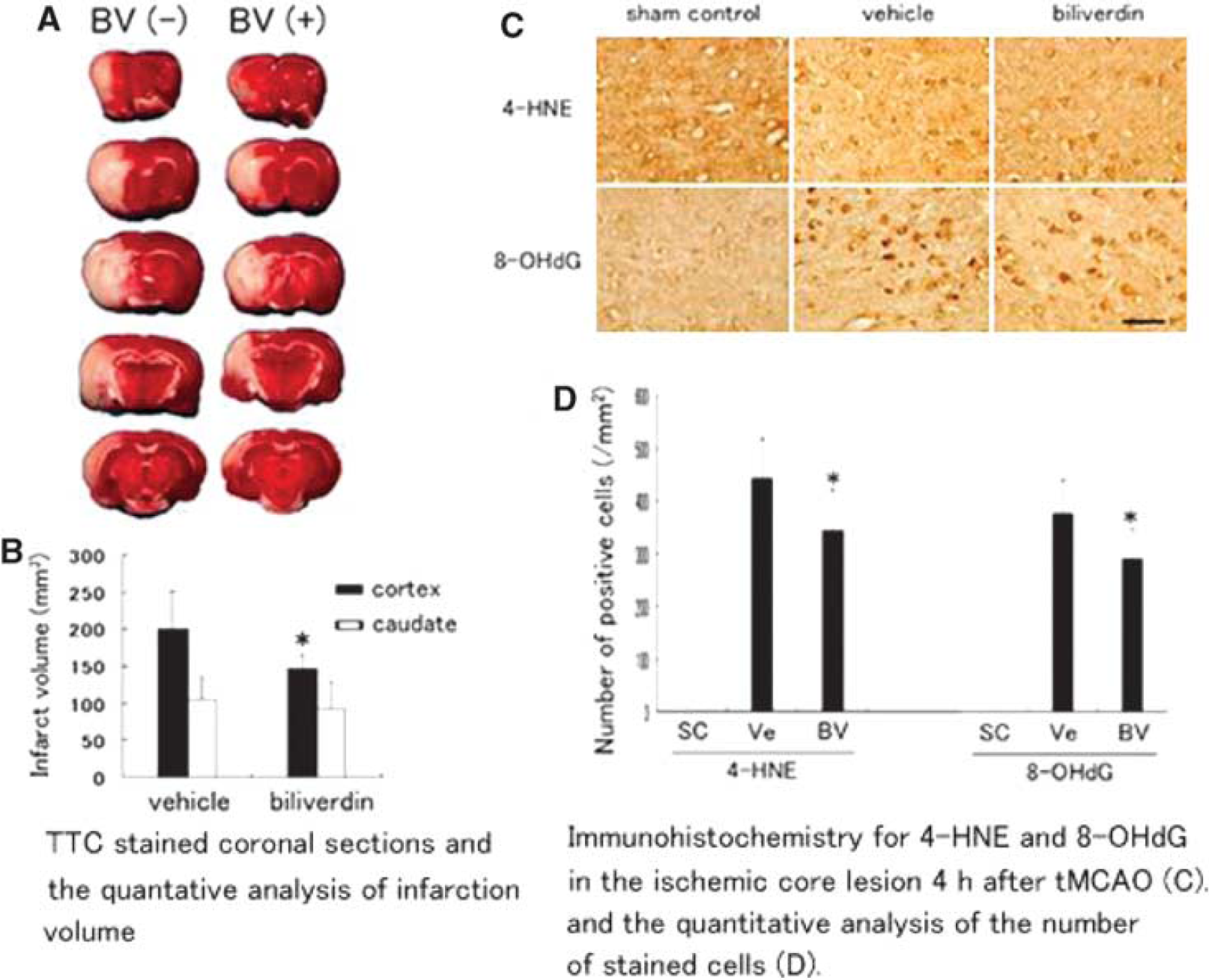
291. Reduction of cerebral infarction in rats by biliverdin associated with amelioration of oxidative stress
K. Deguchi1, T. Yamashita1, S. Nagotani1, M. Takamiya1, K. Tomiyama2, N. Morimoto1, M. Miyazaki3, N.-H. Huh3, A. Nakao2, T. Kamiya1 and K. Abe1
1Department of Neurology, Okayama University, Okayama, Japan; 2Department of Surgery, University of Pittsburgh, Pittsburgh, Pennsylvania, USA; 3Department of Cell Biology, Okayama University, Okayama, Japan
Background and aims: Biliverdin (BV), one of the byproducts of heme catalysis through heme oxygenase (HO) system, is a scavenger of reactive oxygen species. We hypothesized that BV treatment could protect rat brain cells from oxidative injuries via its anti-oxidant efficacies.
Methods: Cerebral infarction was induced by transient middle cerebral artery occlusion (tMCAO) for 90 mins, followed by reperfusion. BV or vehicle was administered intraperitoneally immediately after reperfusion. The size of the cerebral infarction 2 days after tMCAO was evaluated by 2,3,5-triphenyltetrazolium chloride (TTC) stain. Superoxide generation 4 h after tMCAO was determined by detection of oxidized hydroethidine. In addition, the oxidative impairment of neurons were immunohistochemically assessed by stain for lipid peroxidation with 4-hydroxy-2-nonenal (4-HNE) and damaged DNA with 8-hydroxy-2-deoxyguanosine (8-OHdG).
Results: BV treatment significantly reduced infarct volume of the cerebral cortices associated with less superoxide production and decreased oxidative injuries of brain cells.
Conclusions: The present study demonstrated that treatment with BV ameliorated the oxidative injuries on neurons and decreased brain infarct size in rat tMCAO model.
TTC-stained coronal sections 2 days after tMCAO with or without BV treatment (A) and the quantitative analysis of infarct volume (B). Ve: vehicle controls, BV: biliverdin. (C) Immunohistochemistry for 4-HNE and 8-OHdG in the ischemic core lesion 4 h after tMCAO. (D) Note reduction of the number of stained cells in the BV-treated group compared with the vehicle-treated group (*P<0.05, scale bars: 50 μm). SC: sham controls, Ve: vehicle controls, BV: biliverdin.
313. Affective and cognitive consequences of small cerebral infarcts
G. Neigh and M. Shurte
1Psychiatry and Behavioral Sciences, Emory University, Atlanta, Georgia, USA
Objective: Together, late-life major depression and subsyndromal depression rob more than 15% of the elderly population of personal happiness and exacerbate comorbid conditions.1 The most serious consequence of late-life depression is premature death due to increased mortality following myocardial infarction and stroke as well as an increased rate of suicide.2 Although the etiology of late-life depression is not fully understood, it is associated with vascular pathology. Silent cerebral infarcts (SCI) have been identified in over 50% of patients with late onset depression,3 and these lesions are associated with more severe symptoms,4 more hospital admissions for depression, and longer hospitalizations for depression.5 Due to the inherent limitations of human research, it is unknown whether vascular changes in the elderly are causative of depressive behaviors or an unrelated but co-occurring event. Because small cerebral infarcts have been repeatedly associated with increased occurrence and severity of symptoms of depression, the current work tests the hypothesis that small cerebral infarcts induce anxiety-like and depressive-like behaviors in a rat model.
Methods: Male rats (3 or 18 mos old) were anesthetized and microembolism or SHAM procedures were performed. Briefly, the left or right common carotid was exposed and microbeads (50 μm in diameter) were injected via a 30-G needle. After 14d of recovery from the procedure, rats were tested for both depressive-like and anxiety-like behaviors. Tests included: social interaction, elevated plus maze, open field, sucrose preference, and forced swim.
Results: Data indicate that rats that received microembolism infarcts demonstrate an increase in depressive-like behavior, as measured by deficits in sucrose preference and forced swim activity, as compared to SHAM operated rats. Results from the full panel of behavioral tests in both age groups will be presented. Analysis of the brain tissue from these rats will determine the role of lesion laterality and location in generation of behavioral changes.
Conclusions: The data collected to date indicate that experimental induction of small cerebral infarcts is sufficient to induce depressive-like behavior. This suggests that small cerebral infarcts may be one underlying cause of late life depression in the elderly population. A better understanding of the behavioral consequences of small cerebral infarcts will provide insight into the neurobiology of vascularly-induced behavioral changes and guide the development of novel treatment strategies.
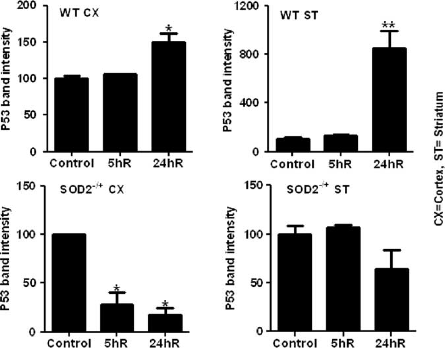
345. Differential correlation of P53 and ros determines hyperglycemic ischemic brain damage in wild-type and SOD2 heterozygous knockout mice
S.L. Mehta1, W. Chen2, Y. Lin3, P. Lolla1, L. Cao4, P.H. Chan5 and P.A. Li1
1Pharmaceutical Sciences/BRITE, North Carolina Central University, Durham, North Carolina, USA; 2Department of Laser Therapy, Ningxia Medical University, Yinchuan, China; 3University of Hawaii, Honolulu, Honolulu, USA; 4McMaster University, Ontario, Ontario, Canada; 5Stanford University School of Medicine, California, USA
Objectives: Hyperglycemia negatively determines cerebral stroke outcome and indeed suggested to accelerate the infarction,1,2 although the underlying mechanism is not clear. The reactive oxygen species (ROS) production is one of the critical factors implicated in the stroke pathogenesis and tumor suppressor protein, p53, has been linked to neuronal cell death and ROS levels. The objective of this study is to examine the expression of p53 and its association with ROS and ischemic brain damage under hyperglycemic condition in wild-type (WT) and manganese superoxide dismutase (SOD2) heterozygous knockout (−/+) mice.
Methods: Male SOD2−/+ and CD1 WT mice (24 to 26 g) were made hyperglycemic 30 mins prior to middle cerebral artery occlusion (MCAO) with a single injection of glucose (25%). Animals were killed at various times of reperfusion (0.5 to 24 h) for assessment of brain damage, ROS production, p53 and cyclin G1 (CG1) protein levels.
Results: The results showed that hyperglycemia enhanced the ischemic brain damage in SOD2−/+ mice. The damage was significantly greater in SOD2−/+ as compared to WT and well correlated with the ROS production. The ROS production gradually and significantly increased in cortical and striatal area of WT mice, which was correlated with the elevation of p53 and survival factor, CG1. In contrast, ROS production showed abrupt elevation early during recirculation and decrease thereafter in SOD2−/+ animals. Similarly, p53 and CG1 expression decreased in cortical and striatal area of SOD2−/+ animals suggesting the down regulation of these factors in SOD compromised brain.
Conclusions: These results suggests that hyperglycemic ischemic brain damage is linked to ROS and p53 in WT, whereas, p53 plays no role in progression of damage in SOD2−/+ animals. Thus, association of p53 and ROS appears to depend upon the severity of ischemic stress.
p53 protein expression in cortex and striatum.
352. A model of recurrent stroke: mri and histological changes in rats subjected to an initial minor and subsequent moderate insult
M. Qiao1, Z. Zhao2, P.A. Barber2, D. Barua2, T. Foniok1, D. Kirk3, S. Sun2 and U.I. Tuor1,3
1MR Technology, Institute for Biodiagnostics (West), National Research Council Canada; 2Department of Neuroscience; 3Experimental Imaging Centre, University of Calgary, Calgary, Alberta, Canada
Objectives: There is a high risk of recurrent stroke following a transient ischemic attack (TIA) or minor stroke.1,2 Such patients would be ideal for treatment with neuroprotective agents after their initial insult. However, the cellular responses to a second stroke following an initial minor stroke are poorly understood. Indeed, animal models of recurrent stroke are lacking. The present study hypothesized that following a moderate stroke preceded by a minor stroke, the subsequent brain injury, examined with magnetic resonance imaging (MRI) and histological techniques, would either be exacerbated (if insults were additive) or reduced (if there was a pre-conditioning neuroprotective effect).
Methods: An initial mild transient focal cerebral ischemia was induced in rats by occluding the middle cerebral artery (MCAO) with a microclip for 40 mins along with transient concurrent occlusion of both carotid arteries. Stroke severity was confirmed using MRI 2 days later and at three days a second moderate stroke was produced (60 mins). Seven days later MRI and then histology was performed. Subgroups of animals (n = 23) with either a single mild stroke, a single moderate stroke or a combination of mild and moderate stroke were compared. Brain injury was graded from 0 to 4 in 4 regions of an anterior section stained with hematoxylin and eosin (HE) providing a cumulative injury score. Sections were also stained for astrocytosis (GFAP) and microgliosis (ED1) immunohistologically.
Results: In rats 48 h following a mild stroke, T2 maps appeared normal despite evidence of scattered cell death, modest increased GFAP labeling and no changes in ED1 or MAP2 (Figures 1 and 2). In contrast, MRI scans in rats with 60 min-MCAO had hyperintense areas in cortex corresponding to extensive cell death or infarction. Loss of MAP2 and GFAP staining were observed in the core of infarct with extensive labeling for ED1 and GFAP in the peri-infarct area. The recurrent stroke with a 40 mins MCAO and a subsequent 60 mins MCAO 3 days later resulted in a higher cumulative injury score than what was observed following a single mild MCAO (Figure 2). Changes in MRI, MAP2, GFAP and ED1 tended to be greater than that observed in the group with a single moderate MCAO.
Changes in MRI and histology in the brains exposed to either a single middle cerebral artery occlusion (MCAO), a single moderate MCAO or a combination (recurrent) MCAO. T2 changes were shown as hyperintensity and solid black colour or dots showing tissue as infarct or scattered cell death, respectively.
Cumulative histological score from the brains with either a single MCAO, a single moderate MCAO or a combination (recurrent) stroke. **P<0.01 vs the value of 0; #P<0.05 vs the single mild using a rank test.
Conclusions: Unlike preconditioning studies, where periods of ischemia without permanent injury result in neuroprotection against a subsequent ischemic insult, we demonstrate that a minor stroke resulting in mild injury prior to a more substantial stroke could enhance the subsequent ischemic injury. This model of recurrent stroke should provide a valuable platform to test neuroprotective agents suitable for reducing stroke severity following a mild stroke or transient ischemic attack (Funded by CIRH).
373. Combination treatment with ethyl pyruvate and aspirin enhances neuroptotection in the postischemic brain
S.-W. Kim1, J.-H. Shin1, J.-Y. Jeong1, Y. Jin1, I.-D. Kim1, P.-L. Han2 and J.-K. Lee1
1Anatomy, Inha University School of Medicine, Inchon; 2Nano Sciences and Brain Disease Research Institute, Ewha Womans University, Seoul, South Korea
Background and purpose: Ethyl pyruvate, acts as an anti-inflammatory molecule in various pathological conditions, including cerebral ischemia. Aspirin has been reported to confer neuroprotection to the ischemic brain, whose protective effect has been attributed to the anti-platelet action, NMDA- or Zn2+-induced neurotoxicity aspirin also has direct neuroprotective effects, including NF-kB inhibition. In this study, we examined enhanced neuroprotective effects of combination treatment with ethyl pyruvate and aspirin in a rat cerebral ischemia model with middle cerebral artery occlusion (MCAO).
Methods: Male Sprague-Dawley rats were subjected to 1 h of MCAO. Ethyl pyruvate alone or in combination with aspirin was administered at various time points before or after MCAO. The changes in brain infarction, neurological deficits, microglia activation and pro-inflammatory cytokine expression were evaluated. Primary microglial cultures and primary cortical cultures were used to elucidate the underlying molecular mechanism of enhanced neroprotective effect of combination treatment.
Results: Ethyl pyruvate dose-dependently suppressed infarct volume in the post-ischemic brain, wherein intravenous administration of 5 mg/kg ethyl pyruvate 30 mins after the occlusion reduced infarct volume to 34.5%±15.5% (n = 6, P<0.01) of that of the untreated control. In combination with aspirin (5 mg/kg. i.v.), the neuroprotective effect was enhanced, resulting in 16.0%±5.9% (n = 6, P<0.01) infarct volume. The time window for synergistic neuroprotection by ethyl pyruvate and aspirin extended to 9 h post-MCAO. The synergistic neuroprotection was accompanied by suppression of motor and other neurological deficits. Inflammatory processes were notably suppressed by the combination treatment in the postischemic brain and primary microglia cultures. wherein ethyl pyruvate and aspirin modulate NF-kB signaling differentially. Similar enhancement in neuroprotective effect and differential modulation of NF-kB signaling pathway was also observed in oxygen-glucose deprivation-treated primary cortical cultures.
Conclusions: Combination treatment of ethyl pyruvate and aspirin affords synergistic neuroprotection in the postischemic brain with a wide therapeutic window, in part via differential modulation of the NF-kB signaling pathway.
396. Brain damage and functional deficits evolution following permanent or 3 h-transient ischemia in marmosets: studies with behavioral tests, mri and immunohistochemistry
E. Bihel1, J. Toutain1, P. Pro-Sistiaga2, M. Bernaudin1, S. Roussel1 and O. Touzani1
1UMR-CINAPS 6232, CERVOxy Team ‘Hypoxia and Cerebrovascular Pathophysiology’, CNRS, CEA, Université de Caen Basse Normandie and Université Paris Descartes, Caen, France; 2Human Neuroanatomy Laboratory, Department of Health Sciences and C.R.I.B., School of Medicine, University of Castilla-La Mancha, Albacete, Spain
Objectives: Based on behavioral tests, magnetic resonance imaging (MRI) and immunohistochemistry, the aim of our study was to characterize, at both the acute and the chronic stages, the evolution of functional deficits and brain damage in the marmoset subjected to permanent or 3 h transient ischemia induced by intraluminal occlusion of middle cerebral artery (MCAO).1
Methods: Six and five marmosets were subjected to transient (tMCAO) and permanent (pMCAO) intraluminal MCAO,1 respectively. During 45 days after the occlusion, a battery of behavioral tests has been performed weekly to quantify the sensorimotor deficits. These tests included neurological score, tactile simulation, hill and valley staircase, adhesive removal task, six tubes choice, and reaching up tube2. Each animal underwent 3 sessions of MRI (7T, Pharmascan; Bruker) at 60 min, 8 days and 45 days following MCAO. In each session, diffusion weighted imaging (DWI), T2 and T2*-MRI sequences were acquired. NeuN, GFAP and NeuN/BrdU labeling were undertaken at 45 days to assess neuronal loss, astrogliosis and neurogenesis, respectively.
Results: Behavior: Unilateral motor impairment of the contralateral forelimb and neglect of contralateral body side were observed in all subjects, with more severe deficit in pMCAO group. These deficits were long-lasting despite a partial recovery in tMCAO group. In pMCAO group, this partial recovery was significantly more delayed in most tests analyzed.
Brain lesion: MRI data showed that the lesion affected cortical and subcortical structures at the acute and sub-acute stages. At 60 mins, the volume of the DWI lesion was not significantly different between tMCAO and pMCAO groups (298±165 mmł and 208±116 mmł respectively). At 8 days after the occlusion, the T2-MRI-defined lesion was larger in pMCAO group (462±348 mm3) compared to tMCAO group (123±86 mmł Mann Whitney test, P = 0.04). At 45 days, a hyperintense signal in T2-MRI was visible in pMCAO group (28±22 mm3) but not in tMCAO one. However, a hemispheric atrophy was observed in the two groups without statistically significant difference (4.8%±1.7% and 3.1%±1.5%, respectively for pMCAO and tMCAO). In the 3 sessions of MRI, T2* sequence did not revealed any cerebral hemorrhage.
Cellular reaction: NeuN and GFAP labeling revealed, at 45 days post-MCAO, a widespread neuronal loss and associated astrogliosis in the ipsilateral hemisphere in greater extend in pMCAO group. Compared to SHAM animals, those subjected to ischemia showed newly generated neurons, as attested by NeuN/BrdU double labeling, in the vicinity of the initial lesion, in particular in the caudate nucleus (Mann Whitney test P = 0.06). However, no difference between pMCAO and tMCAO groups was noted.
Conclusions: The data show that intraluminal MCAO in the marmoset results in widespread brain damage and long-lasting functional deficits that can be reduced by reperfusion. Moreover, this work revealed the existence of a post-ischemic neurogenesis in this model. Altogether, the results show that this model of brain ischemia in the marmoset could be considered as suitable to test new therapies against stroke.
418. Effects of betulinic acid in a rat model of cerebral venous ischemia
A. Heimann1, K. Horiuchi1,2, B. Alessandri1, H. Li3 and O. Kempski1
1Institute for Neurosurgical Pathophysiology, Universitätsmedizin, Johannes Gutenberg-University, Mainz, Germany; 2Department of Neurosurgery, Nara Medical University, Nara, Japan; 3Department of Pharmacology, Johannes Gutenberg-University, Mainz, Germany
Background and aims: Betulinic acid, one of three triterpenoids which are isolated from traditional Chinese medical herbs, has been shown to up-regulate eNOS and to reduce the NADPH oxidase expression (J Pharmacol Exp Ther 2007;322(2):836–42). Furthermore betulinic acid sulfate is a potent inhibitor of the classical pathway of the complement system and a weak inhibitor of the alternative pathway (Bioorg Med Chem 2007;15(10):3489–98). Thus, betulinic acid may be useful in the therapy of focal cerebral ischemia. Therefore this study was designed to investigate whether betulinic acid has neuroprotective potential in a rat model of focal venous ischemia.
Methods: 18 male Wistar rats were randomized into two groups, anaesthetized, intubated and ventilated. The tail artery was canulated for blood pressure monitoring and blood gas control. Regional cerebral blood flow (rCBF) was assessed by laser Doppler scanning and tissue impedance was measured to monitor cell swelling and cortical spreading depression in the ipsilateral hemisphere. After stable baseline conditions two adjacent bridging veins were occluded photochemically (2-VO). During the initial 75 mins after 2-VO ten cortical spreading depressions were elicited by intracortical injection of 150 mmol/L KCl as metabolic challenge. Vehicle (n = 9; DMSO) or betulinic acid (n = 9; 30 mg/kg b.w.) was administered for seven days by daily gavage, before animals were euthanatized for histological infarct evaluation (HE-staining).
Results: Body weight, arterial blood pressure and blood gases did not differ between the experimental groups. At baseline rCBF ranged between 50.01±6.3 LD-units (vehicle) and 42.32±6.8 LD-units (betulinic acid) and was significantly decreased after 2-VO to 44% and 50% of baseline, respectively (Figure 1A). During the initial 75 mins after 2-VO cortical spreading depressions occurred 12.7±0.7 times in the vehicle group and 12.4±1.3 times in the betulinc acid group (ns.). Quantitative histological evaluation showed a significant infarct reduction from 3.81±2.21 mm3 in the vehicle to 1.90±1.1 mm3 in the betulinc acid group (Figure 1B).
Conclusion: Betulinic acid has a neuroprotective effect in a rat model of venous focal cerebral ischemia. The inhibition of the complement system is a possible mechanism of action, since in the same model a C-1 inhibitor also reduced infarct size (BrainRes 1999;838:210–213). Further studies have to determine the degree of contribution of complement inhibition and NO production to the observed infarct reduction.
421. Change of expression of orexin in the rat brain after foal cerebral ischemia
E. Kitamura, N. Kanazawa, S. Maruyama, J. Yonekura, R. Masuda, K. Koizumui, J. Hmada and F. Sakai
Kitasato University School of Medicine, Sagamihara, Japan
Objective: Orexin is known as a neuropeptide which controls feeding behavior, arousal or sleep behavior and has a function on maintenance of homeostasis. There are only few dozens of orexin containing neurons in hypothalamus. The orexinergic fibers are widely distributed in spinal cord and brain.1 Also orexin has some function on the brainstem neuronal nuclei related with the pathophysiological mechanism of migraine and brain ischemia.2,3 But the role of orexin in brain ischemia remains incompletely understood. In the present study, we investigated the expression of orexin in the rat brain after middle cerebral artery occlusion.
Methods: Twenty-eight male Sprague Dawley rats (350 to 500 g) were anesthetized with isoflurane. An intraluminal occluder was made of 4–0 surgical nylon monofilament coated with silicone. An occluder was inserted through the left common carotid artery to occlude middle cerebral artery. At 120 mins after the occlusion, the brain was recirculated. At 3(n = 7), 6(n = 7), 12(n = 7) and 24 h (n = 7) after recirculation, rats were perfused with heparinized 0.1M PBS and Zamboni's fixative. After enucleating the brains, we soaked them in 30% sucrose for 3 days. 10-μm-thick frozen section stained with hematoxylin and eosin. And we used immunohistochemical staing. Double immunofluorescence labeling for orexin-A and MAP2 (Microtubule Associated Protein) was performed to investigate the expression of orexin in rat cortex and hypothalamus (primary antibody: anti orexin-A polyclonal rabbit antibody, anti MAP2 monoclonal mouse antibody). For statistical analysis, we used paired t-test.
Results: In all rats subjected to middle cerebral artery occlusion, an ischemic lesion was detected in the ipsilateral hemisphere on coronal sections stained with hematoxylin and eosin. In the cortex, there was no difference in the expression of orexin-A between ischemic hemisphere and contralateraral non ischemic hemisphere. In the hypothalamus, there was no significantly different in the number of neurons which expressed orexin-A at 3, 6 and 12 h after recirculation. But at 24 h recirculation, there was a significant difference in the number of neurons which expressed orexin-A in the hypothalamus. The number of neurons (36.7±19.6) which expressed orexin-A in the ischemic side was significantly larger comparing with the non-ischemic side (25.7±15.3) (P<0.01).
Conclusions: Our study suggested that at 24 h after recirculation, in the hypothalamus, the number of the neurons which expressed orexin-A of the ischemic side was larger than that of the non ischemic side. Cortical spreading depression (CSD) was induced following focal brain ischemia. We have already shown that the intracerebroventricular administration of orexin inhibited CSD in the rat(in preparation). Given these reports, in the present study, it was suggested that orexin-A may give some influence on the pathophysiologial mechanism of focal cerebral ischemia.
425. Low-frequency ultrasound (60 KHZ) with varied duty cycle: effects on ischemic brain tissue and the inner ear
P. Reuter1, J. Masomi1, H. Kuntze1, I. Fischer2, A.-O. Viertmann1, J. Marx1, K. Helling2, C. Sommer3, B. Alessandri4, O. Kempski4 and M. Nedelmann5
1Neurology; 2Department of Otolaryngology; 3Neuropathology; 4Insitute of Neurosurgical Pathophysiology, Johannes Gutenberg-University, Mainz; 5Neurology, Justus-Liebig University, Giessen, Germany
Background and aims: The thrombolytic activity of ultrasound, but also the safety of ultrasound thrombolysis depends on variation of technical ultrasound parameters. In a recent in vitro study, a longer duration of the duty cycle [relationship of the on-phase to the total duration of an ultrasound pulse] was shown to improve thrombolytic efficacy of ultrasound.1 The aim of our study was to examine the effect of different duty cycle settings in vivo on brain tissue after occlusion of the middle cerebral artery.
Methods: To investigate the effects of 60 kHz pulsed ultrasound, rats were transcranially treated following reperfusion of the middle cerebral artery (90 mins after induction of ischemia by use of the suture model). The duty cycle was set at 20% and 80%, while keeping time average intensity constant. Analysis included measurement of infarct volume (TTC-staining) and functional-neurological evaluation. An additional group of healthy animals was insonated with the same ultrasound setup, and acoustically evoked potentials were measured to examine side effects on the auditory system.
Results: Ultrasound at short duty cycle setting resulted in a significant increase of the ischemic lesion volume compared to control animals. This negative side effect could not be detected in prolonged on-phase pulsation (resulting in reduction of energy spikes within the pulse). Furthermore, insonation resulted in a reduction of auditory function (reduction of the auditory threshold level up to 40 dB).
Conclusions: The results demonstrate that longer duty cycles not only positively influence the therapeutic effects but are also beneficial with regard to safety issues. The findings may serve as a basis for future developments of prototype therapeutic ultrasound devices. The study is first to describe side effects on the auditory system, which may further limit clinical utility of low frequency ultrasound in therapeutic applications. Further histological investigation of the tissue (brain and auditory system) may give more insight into the nature of the observed side effects.
426. A novel modified method and its confirmation of injection into CSF via the cerebellomedullary cistern in mice
Y. Chen1,2, A. Ito1 and N. Saito1
1Department of Neurosurgery, Faculty of Medicine, University of Tokyo, Tokyo, Japan; 2Department of Neurosurgery, Sir Run Run Shaw Hospital, Medical College of Zhejiang University, Hangzhou, China
Background and aims: Central administrations of delivering drugs and chemotherapeutic agents are very difficult to be applied in mice related to the small size. Injection into the cerebellomedullary (CM) cistern is one of the options of central administrations. As its location is relatively fixed, it has potent advantantage than intraventricular administration under some pathological state, especially in supratentorial lesion models. We aimed to obtain a more accurate method for Injection into the CM cistern in mice.
Methods: We modified the method firstly introduced by Ueda et al. We choose the prone position with nape elevation and extension under inhalation anesthesia. The nape of the neck was incised at the midline. The hand-made curved tip of a 27 gauge dental needle was inserted into the cleft between the occiput and the atlas vertebra through the muscles and ligaments, being warranted on the line between sagittal suture and midline of nape. A volume of 6 μl methylene blue aqueous solution was injected slowly into the CM cistern to check accuracy. Mice were sacrificed 1 h(n = 8), 6 h (n = 6) or 24 h (n = 6) after injection.
Results: Twenty C57BL/6 mice were used to check our modified method and all succeeded without any serious vital or neurological deficits. The dye in the injection place and the intracranial distribution (CM cistern, ventral cisterns, trigerminal nerve and optic nerve roots) could be recognized within 6 h after injection. The dye was disappeared in all places while being checked 24 h after injection.
Conclusions: Our new method of injection into CM cistern is easy to be grasped and can become a common method to examine chemical substances' effects on central nerve system in mice. It is no use for dye injection along with the drug procedure for confirmation if mice were sacrificed beyond 6 h after injection.
434. Disturbed K+-channel activity of hippocampal CA1 neurons during cyanide-induced anoxia in a mouse model of Rett syndrome
M. Kron and M. Müller
Zentrum Physiologie und Pathophysiologie, Universität Göttingen, Göttingen, Germany
Background and aims: Rett syndrome is a neurodevelopmental disorder caused by mutations in the X-chromosomal MECP2-gene. Mecp2−/y knockout mice—a model of Rett syndrome—show an imbalance of inhibitory and excitatory synaptic transmission.1 Recently, we unveiled an enhanced hypoxia susceptibility of the hippocampus of Mecp2−/y mice which seems to arise from disturbed potassium fluxes.2 The present study aims to identify dysfunctional K+-channels.
Methods: Experiments were conducted in acute hippocampal slices of Mecp2−/y and wildtype (WT) males (postnatal day 40). Sharp-electrode recordings were performed in CA1 pyramidal neurons to quantify changes in membrane potential and input resistance in response to 1 mol/L cyanide (chemical anoxia) under control conditions and in the presence of glibenclamide or charybdotoxin. Single-channel recordings were performed in inside-out and cell-attached patches.
Purpose: Identification of disturbed neuronal K+-channels in Rett syndrome.
Results: Intracellular recordings did not reveal significant differences in pyramidal cell resting membrane potential (−56.7±3.8 mV versus −58.7±12.1 mV) and input resistance (86±40 M versus 74.8±27.3 M) between WT and Mecp2−/y pyramidal neurons. Cyanide elicited an initial hyperpolarization (−7.1±3.7 mV) and decreased the input resistance (−34%) in WT neurons. In Mecp2−/y neurons both the hyperpolarization and decrease in input resistance were dampened by ∼50%. These initial responses were followed by a progressive (terminal) depolarization. In the presence of the BK-channel blocker charybdotoxin (10 nmol/L), cyanide caused an initial depolarization in WT (5.1±3.8 mV) and Mecp2-deficient pyramidal neurons (4.5±4.8 mV). In WT neurons, the subsequent hyperpolarization became more pronounced (−10.2±5.5 mV) but remained unchanged in Mecp2−/y neurons. In the presence of the KATP-channel blocker glibenclamide (50 to 100 μmol/L), cyanide caused an initial depolarization in WT (2.9±5.7 mV) that also occurred in Mecp2−/y pyramidal neurons (4.4±4.2 mV). The subsequent hyperpolarization was virtually unchanged in WT neurons but was detectable in only 55% of the recorded Mecp−/y neurons. To assess the modified function of K+-channels during anoxia in more detail, single-channel analyses were performed. In cell-attached patches, cyanide caused a pronounced activation of a tolbutamide-insensitive intermediate-conductance (80 pS) K+-channel in WT neurons, but only a moderate activation in Mecp2−/y neurons. Basic BK-channel properties were unchanged, but BK-channels became massively activated in Mecp2−/y neurons during the terminal depolarization.
Conclusions: The enhanced hypoxia susceptibility of the Mecp2−/y hippocampus seems to arise from disturbed K+-channel function. As a result, the cyanide-induced hyperpolarization is weakened. Accordingly, during early hypoxia, the membrane potential is stabilized less efficiently. Also the ability to compensate pharmacological KATP-blockade during metabolic arrest seems impaired. The enhanced activation of the BK-channel in Mecp2−/y neurons may suggest more pronounced intracellular Ca2+-rises during anoxia.
Supported by the DFG (CMPB).
440. Effect of Imidapril on cerebral production of nitric oxide during cerebral ischemian and reperfusion in mice
H. Nagoya, T. Ohkubo, Y. Asano, K. Hattori, T. Shimazu, M. Yamazato, Y. Kato, Y. Ito and N. Araki
Neurology, Saitama Medical University, Saitama, Japan
Objectives: It is suggested that the angiotensin-converting enzyme (ACE) inhibitors shift the autoreguration curve to left, and has antiarteriosclerotic effects, and protectect against the cerebral ischemia. We investigated the effect of Imidapril on the cerebral nitric oxide production in the C57BL/6 mice in steady state and during ischemia and reperfusion.
Methods: Twenty male C57BL/6 mice were used: control group [n = 10], and Imidapril group (Imidapril was administered 1 mg/kg a day for two weeks) [n = 10]. The animals were anesthetized with 2% halothane and maintained with 0.5% to 1% halothane. NO production was continuously monitored by in vivo microdialysis. A microdialysis probe was inserted into the left striatum and perfused with Ringer's solution at a constant rate of 2 μl/mins. After 2 h equilibrium period, fractions were collected every 10 mins. A laser Doppler probe was placed on the right skull surface. Global ischemia was produced by clipping both common carotid arteries using Zen clip for 10 mins. The levels of nitrite (NO2−) and nitrate (NO3−) in the dialysate samples were measured by the Griess reaction.
Results:
Mean blood pressure; There were no significant differences between the two groups before cerebral ischemia and during ischemia. The Imidapril group showed significantly lower BP than that of the control group after reperfusion.
Cerebral blood flow; There was no significant difference between the two groups.
NO2−: The level of NO2− in the Imidapril group [2.34±0.71 μmol/ L(mean±s.d.)] before ischemia was significantly higher than that of control group [1.35±0.47μmol/ L] (P<0.05). There was no significant difference between the two groups during ischemia and after reperfusion (Figure 1).
NO3−; There was no significant difference between the two groups.
Total NO (NO2−+NO3−): The level of total NO in the Imidapril group [3.95±1.13 μmol/ L(mean±s.d.)] before ischemia was significantly higher than that of control group [2.84±0.85 μmol/L] (P<0.05) (Figure 2).
Production of nitric oxide.
Conclusion: This study suggests that Imidapril increases NO production in the baseline level (before cerebral ischaemia). As Imidapril increases the baseline level, it is difficult to analyze the effect of imidapril on the NO production during ischemia and reperfusion.
441. Effect of pterygopalatine arterial blood flow cannot be ignored in a mouse model of intraluminal suture middle cerebral artery occlusion
Y. Chen1,2, A. Ito1, K. Takai1 and N. Saito1
1Department of Neurosurgery, Faculty of Medicine, University of Tokyo, Tokyo, Japan; 2Department of Neurosurgery, Sir Run Run Shaw Hospital, Medical College of Zhejiang University, Hangzhou, China
Background and aims: The mouse model of intraluminal suture middle cerebral artery occlusion (MCAO) is still associated with several issues, such as variability of infarction volume and survival. Collateral blood flow and surgical methods contribute to these problems. To produce MCAO in mice, the pterygopalatine artery (PPA) is not normally cut or blocked because of technical demands related to the position of this artery and the size of the mouse. The effect of blood flow in the (PPA) in the mouse MCAO model was evaluated.
Methods: While producing mouse MCAO models using commercially available silicone-coated monofilaments, we temporarily occluded the common carotid artery (CCA) or PPA to determine whether cerebral blood flow (CBF) values, infarct size and the stability of the model would be affected. Forty male C57BL/6 mice were divided into 3 groups: MCAO with blocked CCA blood flow (MCAO-C; n = 12), MCAO with blocked PPA blood flow (MCAO-P; n = 16) and MCAO without either CCA or PPA blood flow blockage (MCAO-U; n = 12).
Results: The CBF values were significantly higher during occlusion in the MCAO-U than in the other two groups (P<0.001). We stained whole brains from each group at 24 h after reperfusion with 2% 2,3,5-triphenyltetrazolium chloride. Although mean infarct volume did not obviously differ between the MCAO-U and other two groups, infarct volumes varied significantly more within the MCAO-U, than in the other two groups (P<0.05).
Conclusions: Collateral circulation from the PPA to the brain significantly influences the mouse MCAO model, and cannot be ignored. An approximately consistent mouse MCAO model can be generated using commercially available silicone-coated sutures while blocking PPA blood flow during occlusion.
Staining with TTC. All sections from 3 groups.
444. Hemin protects against photothrombotic cortical ischemic injury in mice
J. You, H. Lee, S.J. Kim, C.D. Kim and W.S. Lee
Department of Pharmacology and MRCITR, Pusan National University School of Medicine, Yangsan, South Korea
This study aimed to investigate whether hemin can reduce mouse brain injury and facilitate the recovery following photothrombotic cortical ischemia in mice. Male C57BL/6 mice were anesthetized and systemically administered Rose Bengal. Permanent focal ischemia was induced in the medial frontal and somatosensory cortices by irradiating the skull with a cold light laser. Animals were treated with hemin and zinc protoporphyrin (ZnPP) 1 h after photothrombosis, and were sacrificed 24 h after ischemic insult. Hemin caused a significant reduction in the infarct size, the Evans blue extravasation index, and immunoreacitivities of PARP, but was without effect on HIF-1α expression, and furthermore induced a significant increase in the immunoreactivities of neuroglobin and heme oxygenase-1 (HO-1) in the ischemic region. These effects were reversed by co-treatment with ZnPP, a HO-1 inhibitor. It is suggested that hemin can facilitate the recovery following photothrombotic cortical ischemia via expression of neuroglobin and HO-1 proteins, and thereby indicating advantages in the therapeutic strategy for cerebral ischemia.
463. Endonuclease g does not play an obligatory role in PARP-activated cell death after transient focal cerebral ischemia
J. Zhang1, Z. Xu1, K. David2, X. Li1, K. Kibler1, T.M. Dawson2, V.L. Dawson2 and R.C. Koehler1
1Department of Anesthesiology/Critical Care Medicine; 2The Institute for Cell Engineering, Johns Hopkins University, Baltimore, Maryland, USA
Objectives: Activation of poly (ADP-ribose) polymerase (PARP) and subsequent translocation of apoptosis-inducing factor (AIF) contribute to neuronal injury from NMDA, oxygen-glucose deprivation, and stroke. In C. elegans, an analog of endonuclease G (Endo G) has been implicated in the DNA fragmentation induced by translocation of AIF to the nucleus. The present objective was to investigate if Endo G plays an obligatory role in the PARP dependent injury from middle cerebral artery occlusion (MCAO).
Methods: Endo G knockout and wild type (WT) mice were used to investigate the function of Endo G gene. Using isoflurane anesthesia, 90 mins of MCAO was performed with the intraluminal filament technique. After 72 h of reperfusion, infarct volume was analyzed. In some groups, saline vehicle or DR2313, a PARP inhibitor, was infused intravenously before MCAO (1 ml/kg; 10 mg/kg), at reperfusion (1 ml/kg; 10 mg/kg), and through the first 4.5 h of reperfusion (1 ml/kg/h; 10 mg/kg/h).
Results: Infarct volume was not different between male WT (47%±6% of hemisphere; ±s.d.) and Endo G knockout (49%±8%) mice or between female WT (37%±16%) and Endo G knockout (35%±9%) mice. Latex casts indicated a similar arterial distribution in WT and knockout mice, and a similar diameter of the posterior communicating artery. Furthermore, the reduction in laser-Doppler flux during MCAO was not different between WT and knockouts. When male mice were treated with DR2313, infarct volume was decreased to a similar extent in WT mice (saline = 44%±14%; DR2313 = 22%±16%) and in Endo G knockout mice (saline = 57%±15%; DR2313 = 22%±17%). The decrease in laser-Doppler flux was not influenced by DR2313 infusion.
Conclusion: These data demonstrate that Endo G is not required for the pathogenesis of transient focal ischemia in either male or female mice. Because the injury in Endo G null mice remains dependent on PARP, as demonstrated by tissue rescue with a PARP inhibitor, Endo G is not obligatory for executing PARP-dependent injury during stroke.
469. Potassium disturbances at the ‘ischemic edge’ in rat experimental focal ischemia
A. Kharlamov1, V.E. Yushmanov1 and S.C. Jones1,2,3
1Anesthesiology; 2Anesthesiology and Neurology, Allegheny-Singer Research Institute; 3Radiology, Univ of Pittsburgh, Pittsburgh, Pennsylvania, USA
Aims: Maximum changes of sodium,1 MAP2,2 water and K+3,4 and transient BBB disruption5 occur at the edge of the ischemic core after experimental focal ischemia. These phenomena may be related to higher levels of ‘trickle’ blood flow that result in greater degrees of swelling in the peripheral areas of the ischemic core. Here we hypothesize that the ‘ischemic edge’ (IE) differs depending on position (dorsal or ventral) and ischemic model.
Methods: Experimental focal ischemia was induced by MCA transection and bilateral common carotid artery or suture occlusion in 14 Sprague-Dawley rats (isoflurane/N2O) for 2.5 to 5 h. Brains were sectioned for quantitative K+ histochemistry and micro-punched using our previous procedures.4 2D images of brain K+ concentration, [K+]br, were used to extract [K+]br profiles along the cortical ribbon using MCID software and to provide in the one suture model animal a 3D image of [K+]br in the entire brain.
Results: There were prominent decreases in [K+]br in all animals at the edges of the ischemic core. The frequency distribution of [K+]br at these edges showed a bimodal distribution with peaks at 85 and 103% of ischemic core [K+]br with the minimum at 92.5%. An IE was defined by having a [K+]br less than this ‘trough’ value. In the MCAT animals, the distances from the edge of the normal cortex to the dorsal and ventral IE regions were 2.1±0.1 mm and 2.6±0.2 mm, respectively (P = 0.041, unpaired t-test).
From the suture model animal, a 3D reconstruction from K+-stained coronal slices (IE with [K+]br< 56 mol/L in yellow) in the Figure indicates that the IE completely surrounds the ischemic core but that the rostral IE (right) is narrower than the caudal IE (left).
Conclusions: We conclude from 2D evidence that the low level of [K+]br which defines the dorsal IE is closer to normal cortex than the ventral IE, suggesting lower dorsal collateral potential. The 3D study suggests that there are differences between IE characteristics due to the model of ischemia. The low K+ in the IE is not normally recognized as a feature of ischemic pathology, and its characteristics and their relation to the associated exaggerated progression of ischemic pathology are important unexplored concepts that need further study. These characteristics of the IE displace the notion that the ischemic region is homogeneous and progresses towards infarct at an equal rate. This concept has diagnostic and therapeutic implications based upon a potentially enhanced understanding of ischemic pathology.
Support: NIH-NS030839.
477. Cognitive and emotional changes in the behavior of rats after occlusion of the anterior cerebral artery
H. Mertgens, G. Mies, R. Graf and H. Endepols
Max Planck Institute for Neurological Research, Cologne, Germany
Objectives: The anterior cerebral artery (ACA) supplies cingulate and frontal cortical regions in both humans and rats. Ischemic lesions in the ACA territory of humans impair cognitive functions and reduce incentive drive. We studied in a rat model of ACA occlusion (ACAo) loss and recovery of function longitudinally over one year using various behavioral paradigms.
Methods: ACAo (n = 8) or sham operation (n = 2) was performed in male Lister hooded rats by stereotactic injection of the vasoconstrictor endothelin-1 (ET-1; 150 pmol in 0.3 μl phosphate buffer) or vehicle. To analyze functional outcome, we carried out behavioral tests, which assess decision-making during foraging (food-carrying task), anxiety (elevated plus maze), spatial working memory (spontaneous alternation in the Y-maze), exploratory behavior (open field), working memory (object recognition task) and attentional set-shifting (bowl-digging task). In order to set up a longitudinal study, these tests were carried out before, a week after (early testing phase) and a year after ACAo (late testing phase). Behavioral data were finally correlated with MRI (T2) and PET (18F-fluorodesoxyglucose) assessments of structural and metabolic alterations.
Results: Early after ACAo, rats carried less food pellets in the food-carrying task (F(9, 126) = 2.62, P = 0.012), moved more slowly (F(2, 126) = 35.1, P<0.001), went on fewer trips (F(2, 126) = 5.8, P = 0.015) than before and showed jagged movement patterns. The number of pellets per trip increased with distance between food and cage before but no longer after ACAo (F(9, 126) = 5.9, P<0.001). All these changes remained constant after one year. In the elevated plus maze, lesioned animals spent more time on the open arms than before ACAo (F(2, 28) = 8.3, P = 0.003). Arm alternation rate in the spontaneous alternation task decreased from 83% to chance level in the early measurements but recovered to 77.6% after one year. Locomotor activity in the open field stayed constant in all testing phases apart from early impairment of homebase-behavior (F(2, 56) = 210.3, P<0.001), which recovered considerably after one year. Object recognition was not impaired in both testing phases after ACAo and rats showed no side preference but behavioral changes like decreased rearing (F(2, 23) = 5.9, P = 0.013). Attentional set-shifting did not change after ACAo. Behavioral changes in the elevated plus maze and in the open field after ACAo correlated with metabolic impairment (PET) of the anterior cingulate cortex, prelimbic regions, septum, piriform cortex and the hippocampus. Behavioral impairments in the food carrying task correlated with structural lesion size (MRI) of the anterior cingulate cortex, prelimbic areas, the septum, and with ventricular enlargement.
Conclusion: Like in humans, ischemic lesions in the ACA-territory of rats cause cognitive and emotional deficits. These deficits are correlated with metabolic and/or structural loss. One year after occlusion, some functions recover spontaneously, while others remain impaired.
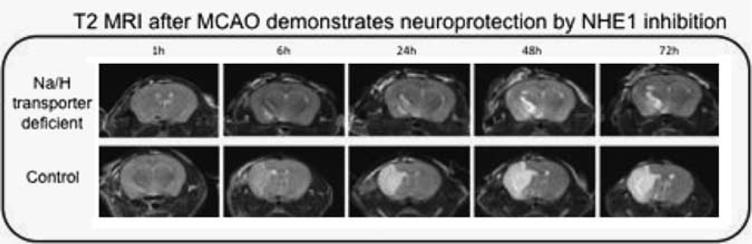
528. Inhibition of the sodium/hydrogen exchanger is neuroprotective after focal cerebral ischemia: a small animal mri study
P. Ferrazzano1, A. Shi2, N. Manhas2, E. Hutchinson3, E. Meyerand3 and D. Sun2
1Pediatrics; 2Neurosurgery; 3Medical Physics, University of Wisconsin School of Medicine and Public Health, Madison, Wisconsin, USA
Objectives: After focal cerebral ischemia, over-stimulation of the Na+/H+ exchanger isoform 1 (NHE1) causes a loss of Na+ ionic homeostasis and contributes to cell swelling and ischemic injury. Recently, inhibition of NHE1 has been shown to be neuroprotective in in vitro and in vivo ischemic injury. However, the therapeutic window for NHE1 inhibition, the time-course of infarct evolution after transporter inhibition, and the neuroprotective effects of NHE1 inhibition on white matter injury and cytotoxic edema formation, are difficult to assess using traditional methods. Small animal MRI provides an ideal means to study this complex physiology following cerebral ischemia and NHE1 transporter inhibition.
Methods: Transient focal cerebral ischemia was induced by a filament occlusion of the middle cerebral artery (MCAO) in wild-type controls (NHE1+/+), NHE1 deficient mice (NHE1+/−), and NHE+/+ mice treated with the NHE1 inhibitor Cariporide (HOE). In the HOE treated animals, 0.5 mg/kg was initially administered IP 30 mins prior to the onset of reperfusion (Rp), and the same dose was repeated at 24 h and 48 h Rp. T2 and diffusion weighted imaging was performed over the 72 h following reperfusion on a Varian 4.7 Tesla small animal MRI scanner. Diffusion-weighted spin-echo images (DWI) and T2-weighted fast spin-echo images were acquired in 12 contiguous axial slices with a field of view of 17 × 17 mm and a slice thickness of 1 mm. High resolution Diffusion Tensor Imaging (DTI) was performed on fixed brain specimens to evaluate white matter injury.
Results: The T2 images demonstrate excellent resolution and anatomic detail. A T2 lesion was first visible at 6 h Rp, and evolved over the following 24 to 72 h. The lesion seen in NHE1+/− mice and HOE treated mice was significantly smaller at all time points, suggesting neuroprotection in these animals. The size and extent of this T2 lesion correlated well with the infarction seen on TTC staining. At 1 h Rp, DWI revealed a lesion that was not visible on conventional T2 images, and that correlated well with the final infarct size seen at 72 h on T2 images and histology. High resolution DTI Mean Diffusivity Maps also demonstrated a smaller lesion in NHE1+/− animals which spared the cortex. Moreover, Fractional Anisotropy Maps revealed injury to the corpus callosum in NHE1+/+ animals but not in the NHE1+/− animals. Taken together, the DTI results suggest white matter integrity is maintained in the NHE+/− animals.
Conclusion: Small animal MRI is a useful measure for evaluating the evolution of injury after cerebral ischemia. T2 images correlate closely with histology, and DWI is a sensitive early marker of ischemic injury. Our MRI studies demonstrate that genetic and chemical inhibition of NHE1 is neuroprotective in this mouse model of transient focal cerebral ischemia.
563. Increased cortical infarct after transient ischaemic stroke in mice lacking the IP receptor for prostacyclin
S. McCann1, C. Roulston1 and G. Dusting1,2
1Bernard O'Brien Institute of Microsurgery; 2Department of Surgery, University of Melbourne, Melbourne, VIC, Australia
Background and aims: Prostacyclin is a vasodilator, platelet anti-aggregatory and cytoprotective prostanoid generated mainly via the cyclooxygenase-2 (COX-2) pathway. Both enzyme and metabolites in the brain are up-regulated following stroke as part of the inflammatory cascade. While inhibition of COX-2 reduces brain damage following ischaemic stroke, prostacyclin treatment also has documented protective actions. The recent withdrawal of selective COX-2 inhibitors rofecoxib and valdecoxib due to an increased risk of adverse cardiovascular events has highlighted the need to re-examine the role of COX-2 products in the search for new therapies. We have investigated whether endogenous prostacyclin plays a role in the brain damage following transient ischaemic stroke.
Methods: Prostacyclin receptor-deficient mice (IP−/−; n = 7) on an apolipoprotein-E-deficient background were compared to control littermates with functional IP receptor (IP+/+; n = 5) after occlusion of the middle cerebral artery (2 h) by intraluminal filament. Cerebral blood flow was monitored during stroke using laser Doppler flowmetry. Infarct area and volume were calculated using MCID image analysis of unstained brain sections. Following stroke, brain oedema was estimated using MCID images and superoxide generation was examined using dihydroethidium (DHE) fluorescence.
Results: Following 24 h reperfusion, infarct volume was increased in the cerebral cortex of IP−/− mice (44.6±9.6 mm3) compared with IP+/+ littermates (10.0±4.4 mm3; P<0.01). There was no difference in infarct size in the striatum. There was no difference between groups in cerebral blood flow changes during stroke. Brain oedema tended to be increased in IP−/− mice but this was not significant. Compared to the appropriate contralateral control and to IP+/+ mice (90%±5%), stroke affected cortical brain regions of IP−/− mice exhibited an increase in DHE-detected superoxide (122%±3%). An increase in DHE fluorescence was detected in the ischaemic penumbra of both genotypes.
Conclusions: The increased infarct size and oxidative stress in IP−/− mice indicates that endogenous prostacyclin signaling exerts a protective effect on cortical neuronal survival after transient stroke, but the mechanisms remain to be clarified.
577. Reduced ischemic injury in HSP110/105 gene knock-out mice after focal cerebral ischemia
T. Marumo1, J. Nakamura2, M. Fujimoto1, K. Nozaki3, K. Nagata2 and Y. Takagi1
1Department of Neurosurgery, Kyoto University Graduate School of Medicine; 2Department of Molecular and Cellular Biology, Institute for Frontier Medical Sciences, Kyoto University, Kyoto City; 3Department of Neurosurgery, Shiga University of Medical Science, Otsu City, Japan
Background and aims: Hsp 110/105 belongs to the HSP110 heat shock protein family, which is a subgroup of the HSP70 family. In mammals, Hsp110/105 is constitutively expressed but exhibits particularly high levels in the brain. It has recently been shown that both Hsp110/105 and Hsp70 are elevated after cerebral ischemia. To study the role of this protein in vivo, we used hsp110/105 knock-out (KO) mice and investigate the effect of reduced Hsp110/105 levels on focal cerebral ischemia.
Methods: There are no histological and morphological differences in Hsp110/105 KO mice and their WT littermates. Hsp110/105 KO and wild-type mice were subjected to 30 mins of transient middle cerebral artery occlusion followed by reperfusion for 24 h. The infarct volume and neurological scores were measured and compared.
Results: The infarction volume and neurological deficit scores were significantly (P< 0.05) reduced in hsp110/105 KO mice compared with wild-type controls.
Conclusions: These results demonstrate that hsp110/105 KO mice are resistant to ischemic injury.
581. Effect of clopidogrel on laser induced thrombus formation in mouse brain microvasculature observed by intravital fluorescence microscopy
T. Fukuoka1, K. Hattori1, N. Araki2 and N. Tanahashi1
1Neurology, Saitama International Medical Center, Saitama Medical University, Hidaka City; 2Neurology, Saitama Medical University, Iruma-gun, Japan
Background and aims: We developed an apparatus of laser induced thrombus formation in murine brain microvasculature instantaneously. The purpose of this study was to observe the effect of clopidogrel on the process of laser induced thrombus formation and platelet behavior in the brain microvasculature of mice using intravital fluorescence microscopy.
Methods: C57 BL/6J mice (N = 13) were anesthetized with chloral hydrate; their heads were fixed with a head holder, and a cranial window was made in the parietal region. Platelets were labeled in vivo by intravenous administration of carboxylfluorescein succinimidylester (CFSE). In six mice, clopidogrel (100 mg/kg) was administered orally for two days before experiment. Seven mice were used as control. Laser irradiation (1000 mA, DPSS laser 532 nm, TS-KL/S2; Sankei) was spotted for 4 sec on pial arteries to induce thrombus formation. Labeled platelets and thrombus were observed continuously with a fluorescence microscope.
Results: After laser irradiation to the pial artery, complete occlusion rate in control group 60%(12/20 vessels) was significantly higher (P<0.01) compared to clopidogrel group 20%(5/25 vessels) (Figure 1). All occluded vessels except one were recannalized within 1 to 2 min. Area of platelet thrombus at 30 mins after laser irradiation in the control group (358±256 mm) was significantly (P<0.05) larger than that of clopidogrel group (209±128 mm) (Figure 2).
Conclusions: Clopidogrel significantly inhibited laser induced thrombus formation in pial arteries of mice.
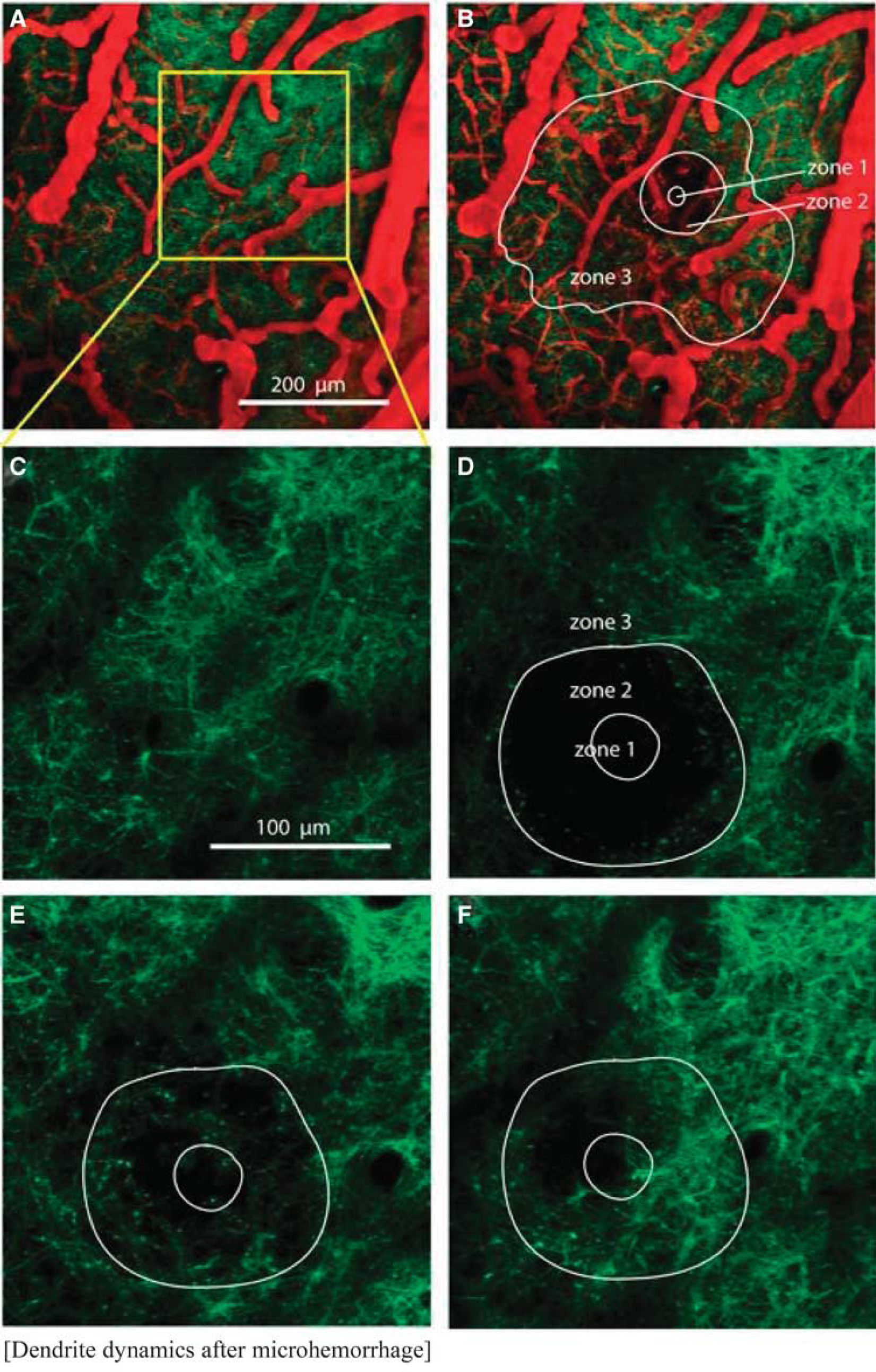
585. Chronic two photon imaging of dendrite dynamics after microhemorrhage of single penetrating arterioles using femtosecond laser ablation
J. Zhou, N. Rosidi, N. Nishimura and C. Schaffer
Biomedical Engineering, Cornell University, Ithaca, New York, USA
Objectives: Clinical evidence indicates that a higher incidence of microhemorrhages is linked to the development of dementia,1 suggesting that small hemorrhages may damage nearby neurons. In addition previous work has shown that occlusions of microvessels can lead to neural pathology such as shriveling of dendrites into nodules (‘blebbing’).2 Here we use two-photon excited fluorescence (2PEF) imaging of mice expressing YFP in neurons to determine whether hemorrhage of cortical penetrating arterioles (PA), without cessation of blood flow, leads to dendrite damage.
Methods: We prepared chronic glass-covered craniotomies in five adult transgenic mice expressing YFP in a subset of cortical pyramidal neurons.3 2PEF microscopy was used to visualize blood vessels labeled by intravenous injection of Texas-red dextran and YFP-labeled neurons (Figure 1a). Microhemorrhages were produced by rupturing individual, specifically targeted PA using tightly-focused femtosecond laser pulses (Figure 1b).4 Image stacks were taken at baseline and immediately, 1 hour, and 1, 3, and 7 days after the microhemorrhage. Dendrite morphologies were classified to identify signs of degeneration.
2PEF imaging of dendrites (green) and blood vessels (red) (A) before and (B) after microchemorrhage. Dendrites (C) before, (D) immediately after, (E) 1 day and (F) 7 days after hemorrhage.
Results: The laser ablation used to cause the hemorrhage consistently caused the immediate blebbing of dendrites in an approximately 40±20 μm (mean±s.d.) diameter region surrounding the target vessel (Zone 1). After triggering vascular rupture, expanding blood plasma and red blood cells pushed the nearby dendrites outward as the targeted vessel bled, forming a sphere of approximately 120±40 μm diameter (Zone 2). Fluorescently-labeled plasma and diffuse red blood cells also penetrated into the tissue beyond this central sphere to a diameter of about 350±60 μm (Zone 3). As red blood cells and plasma are cleared away over days, the damaged and displaced dendrites in Zone 2 returned and blebbing was reduced. We observed no significant changes in dendrite morphology in Zone 3 at any time point in 10 out of 11 hemorrhages (Figure 1d, e, and f).
Conclusions: Chronic two-photon imaging of dendrites reveals that the pressure of blood entering the parenchyma after a microhemorrhage displaces and damages nearby dendrites, with some recovery after a few days. Surprisingly, dendrites further away from the target vessel that are exposed to blood plasma did not show signs of significant dendrite pathology acutely or out to one week post hemorrhage.
588. Laser irradiated thrombus formation and platelet behavior in mouse brain microvasculature observed by intravital fluorescence microscopy
K. Hattori1, T. Fukuoka1, N. Araki2 and N. Tanahashi1
1Neurology, Saitama International Medical Center, Saitama Medical University, Hidaka City; 2Neurology, Saitama Medical University, Iruma-gun, Japan
Background and aims: Platelet behavior and thrombus formation in the microvasculature of the brain has not been fully investigated due to methodological difficulties. Photothrombosis using filtered light and a potent photosensitizing dye was occasionally applied to make vessel occlusions. However, the method is not satisfactory due to reproducibility, weak power etc. We developed an apparatus of laser induced thrombus formation in mouse brain microvasculature.
Purpose: The purpose of this study was to observe the process of laser irradiated thrombus formation and platelet behavior in the brain microvasculature of mice using intravital fluorescence microscopy.
Methods: C57 BL/6J mice (N = 21) were anesthetized with chloral hydrate; their heads were fixed with a head holder, and a cranial window was made in the parietal region. Platelets were labeled in vivo by intravenous injection od carboxylfluorescein succinimidylester (CFSE). Laser irradiation (1250 mA, DPSS laser 532 nm, TS-KL/S2;Sankei) was spotted for 2 to 4 secs on pial microvessels to induce thrombus formation. Labeled platelets and thrombus were observed continuously with a fluorescence microscope.
Results: After laser irradiation to the pial artery, thrombus formed along the vessel wall immediately, and platelets accumulated on the surface of the thrombus leading to vessel occlusion and extended to the lesion of branch artery followed by spontaneous recannalization within 1 to 2 mins (Figure). Fragmentation of thrombus was occasionally seen. Platelet adhesions to irradiated area were seen thereafter.
Laser irradiation to pial veins induced thrombus formation faster than that of pial arteries and occluded pial veins were hardly recannalized.
Conclusions: Laser irradiated thrombus formation is a promising method by which to investigate the pathophysiology of the interaction between platelets and endothelial cells.
601. Sonic hedgehog attenuates brain infarction in rat focal ischemia
D.-I. Yang1 and S.-S. Huang2
1Institute of Brain Science, National Yang-Ming University, Taipei; 2Department of Pharmacology, Chung Shan Medical University, Taichung, Taiwan ROC
Objective: Sonic hedgehog (Shh), a morphogen well known for its critical roles in embryonic development, has recently been shown to carry protective effects against ischemia/reperfusion. Shh gene therapy protected against myocardial ischemia by triggering expression of multiple trophic factors and engendering tissue repair in the adult heart.1 Shh is also a critical factor in the pathophysiology of tissue injury induced by ischemia/reperfusion in liver2 and kidney.3 In this study, we test whether topical application of the biologically active N-terminal domain of sonic hedgehog (Shh-N) may exert protective effects against ischemic insults in a rat model of middle cerebral artery occlusion (MCAO).
Methods: Male Sprague-Dawley rats were subjected to MCAO for 60 min. At the onset of reperfusion, fibrin glues (20 microliter) containing Shh-N and/or its inhibitor cyclopamine (CPM) were applied to the ipsilateral cortices. After reperfusion for 7 days, animals were sacrificed and brains removed for triphenyl tetrazolium chloride (TTC) staining. Data are expressed as Mean±s.e.m. Results shown were derived from combination of at least three independent experiments.
Results: Shh-N at 100, 300, and 1000 ng/ml substantially attenuated brain infarction caused by MCAO. The control animals treated with vehicle alone (0.1% BSA) exhibited infarct volumes of 139.5±23.0 mm3 (N = 7). Application of Shh-N at 30 ng/ml was without effects (150.4±23.2 mm3; N = 6). However, Shh-N at 100 ng/ml (62.94±20.3 mm3; N = 7, P<0.05), 300 ng/ml (75.3±13.4 mm3; N = 8, P<0.05), and 1000 ng/ml (47.5±19.1 mm3; N = 7, P<0.01) significantly reduced the extent of cerebral infarction. Furthermore, while Shh-N at 300 ng/ml reduced infarct volumes (84.0±7.4 mm3 in Shh-N-treated rats, N = 14, versus 136.8±21.0 mm3 in control rats with BSA only, N = 11, P<0.01), co-treatment of its pharmacological inhibitor CPM (10 micromolar) completely abolished this effect (84.0±7.4 mm3 in Shh-N-treated rats, N = 14, versus 170.2±20.1 mm3 in rats with Shh-N plus CPM, N = 7, P<0.001). CPM alone was without effects in attenuating brain infarction (179.6±9.6 mm3, N = 7).
Conclusion: Shh may exert neuroprotective effects in cerebral ischemia in addition to its established roles in neural development. Pharmacological activation of signal transduction pathways downstream of Shh may therefore carry therapeutic potential in ischemic stroke.
605. Effects of cilnidipine on no production and ischemic change of hippocampal CA1 neurons during cerebral ischemia and reperfusion in mice
Y. Kato, T. Ohkubo, Y. Asano, K. Hattori, T. Shimazu, M. Yamazato, H. Nagoya, Y. Ito and N. Araki
Neurology, Saitama Medical University, Saitama, Japan
Introduction: Cilnidipine, an L/N-type Ca2+ channel blocker, has been reported to suppress ischemic brain injury in several animal models. The present study investigates the effects of cilnidipine on nitric oxide (NO) production and ischemic change of hippocampal CA1 neurons during forebrain ischemia and reperfusion.
Methods: Male mice (cilnidipine group; n = 9, control group; n = 17) were anesthetized by inhalation of halothane. NO production was continuously monitored by in vivo microdialysis. Microdialysis probes were inserted into the left striatum and were perfused with Ringer's solution at a constant rate 2μl/min. A laser Doppler probe was placed on the right skull surface. After 2 h equilibrium period, fractions were collected every 10 mins. Forebrain cerebral ischemia was produced by clipping both common carotid arteries using Zen clip for 10 mins. Before 30 mins of ischemia, 50 μg/kg of cilnidipine was intraperitoneally administered. Levels of nitric oxide metabolites, nitrite (NO2−) and nitrate (NO3−), in the dialysate were determined using the Griess reaction. (2) Analysis of hippocampus CA1 neurons: Two hours after the start of reperfusion, animals were perfused with 4% paraformaldehyde. Hippocampal CA1 neurons were analyzed into three phases (severe ischemia, moderate ischemia, survive), and the ratio of the number of surviving neurons was calculated (survival rate). (3) Brain sections were immunostained with an anti-nNOS antibody. To determine the fractional area density of nNOS-immunoreactive pixels to total pixels of the whole field, the captured images were analyzed. Mann-Whitney U test was used for group comparisons.
Results:
Blood Pressure (Figure 1): Cilnidipine group showed significantly higher blood pressure than that of control group before the administration of cilnidipine, but blood pressure of both groups become almost same when ischemia started.
Cerebral Blood Flow (CBF) (Figure 1): There were no significant differences in CBF during ischemia. CBF of cilnidipine group (64.2±14.0) was significantly higher than that of control group (49.0±11.6) in 80 mins after the start of reperfusion.
NO Metabolism: (1) NO2− (% change): The level of NO2− in cilnidipine group was significantly higher than that of control group during ischemia (87.6±35.0 vs. 65.3%±18.7%) and in 10 mins (104.6±51.0 vs. 77.9±19.4) after reperfusion. 2) NO3− (% change): There were no significant differences in the level of NO3− between cilnidipine and control group.
Survival rate in hippocampus CA1 area: There were no significant differences between the both groups.
nNOS activity: The percentage of nNOS-immunoreactive pixels to whole area in cilnidipine group (0.84%±0.16%) was significantly lower than that of the control group (1.96%±0.53%).
Conclusion: These data suggest that cilnidipine may improve CBF by activating eNOS rather than nNOS.
607. Identification of early metabolic markers of various degrees of ischemia in the mouse brain by localized 1H magnetic resonance spectroscopy
C. Berthet1, H. Lei2,3, R. Gruetter2,3 and L. Hirt1
Objectives: Magnetic resonance (MR) imaging and spectroscopy (MRS) allow the establishment of the anatomical evolution and neurochemical profiles of ischemic lesions. The aim of the present study was to identify markers of reversible and irreversible damage by comparing the effects of 10-mins middle cerebral artery occlusion (MCAO), mimicking a transient ischemic attack, with the effects of 30-mins MCAO, inducing a striatal lesion.
Methods: ICR-CD1 mice were subjected to 10-mins (n = 11) or 30-mins (n = 9) endoluminal MCAO by filament technique at 0 h. The regional cerebral blood flow (CBF) was monitored in all animals by laser-Doppler flowmetry with a flexible probe fixed on the skull with < 20% of baseline CBF during ischemia and >70% during reperfusion. All MR studies were carried out in a horizontal 14.1T magnet. Fast spin echo images with T2-weighted parameters were acquired to localize the volume of interest and evaluate the lesion size. Immediately after adjustment of field inhomogeneities, localized 1H MRS was applied to obtain the neurochemical profile from the striatum (6 to 8 microliters). Six animals (sham group) underwent nearly identical procedures without MCAO.
Results: The 10-mins MCAO induced no MR- or histologically detectable lesion in most of the mice and a small lesion in some of them. We thus had two groups with the same duration of ischemia but a different outcome, which could be compared to sham-operated mice and more severe ischemic mice (30-mins MCAO). Lactate increase, a hallmark of ischemic insult, was only detected significantly after 30-mins MCAO, whereas at 3 h post ischemia, glutamine was increased in all ischemic mice independently of duration and outcome. In contrast, glutamate, and even more so, N-acetyl-aspartate, decreased only in those mice exhibiting visible lesions on T2-weighted images at 24 h.
Conclusions: These results suggest that an increased glutamine/glutamate ratio is a sensitive marker indicating the presence of an excitotoxic insult. Glutamate and NAA, on the other hand, appear to predict permanent neuronal damage. In conclusion, as early as 3 h post ischemia, it is possible to identify early metabolic markers manifesting the presence of a mild ischemic insult as well as the lesion outcome at 24 h.
627. Histological and MRI characteristics of exofocal postischemic neuronal death, following middle cerebral artery occlusion in mice
V. Prinz1,2, G. Kronenberg1,2,3, C. Leithner1, K. Gertz1,2, M. Balkaya1,2, S. Mueller2, H. Hoertnagle4 and M. Endres1,2
1Klinik und Poliklinik für Neurologie; 2Center for Stroke Research Berlin (CSB); 3Klinik und Hochschulambulanz für Psychiatrie und Psychotherapie, Campus Benjamin Franklin; 4Institut für Pharmakologie und Toxikologie, Charité – Universitätsmedizin, Berlin, Germany
Objectives: Cerebral ischemia in the territory of the middle cerebral artery (MCA) can induce delayed neuronal death remote to the primary lesion in the ipsilateral substantia nigra (SN). Nigral histopathological changes occur days to weeks after the initial event. However, the mechanisms leading to exofocal postischemic neuronal death (EPND) in the SN are complex and poorly understood. Especially the fate of dopaminergic neurons is discussed controversially.
Using a mouse model of mild stroke, we investigated the time-course and characteristics of EPND using sequential magnetic resonance imaging (MRI) and subsequent immunohistochemistry.
Methods: 129/SV mice were subjected to 30 mins. of filamentous MCA occlusion and followed up by MRI on day 1, 4, 5, 7, 14, 21 and 28 post ischemia. At day 28, animals were sacrificed and immunohistochemistry was performed to investigate histopathological changes in the ipsilateral SN, compared to the contralateral side. Additional groups of animals were exposed to 30 mins. of MCA occlusion and sacrificed 1, 4, 5, 7, 14 and 21 days after reperfusion for histological evaluation of the SN. MRI was performed on the respective day of sacrifice. A subset of animals was exposed to 30 mins. of MCA occlusion and cerebral dopamine levels were measured using high pressure liquid chromatography (HPLC), 90 days post ischemia.
Results: On day 1, T2 weighted imaging showed high signal intensity in the striatum, but no changes in the ipsilateral SN. Likewise, diffusion weighted imaging (DWI) and T2 mapping did not reveal any changes in the SN. On days 4 and 5, T2 weighted imaging showed high signal intensity in the ipsilateral SN, accompanied by an increased T2 value (P<0.05) and a decrease in the apparent diffusion coefficient (P<0.05). These alterations persisted until day 7, but were no longer detectable at later time points.
Compared to the contralateral side, the density of NeuN and tyrosine hydroxylase (TH) positive cells in the ipsilateral SN decreased significantly over time, starting as early as day 7 post ischemia. Consequently, striatal dopamine levels were significantly decreased.
Conclusions: Secondary neuronal degeneration in the SN occurs within days after striatal ischemia and is characterized by a marked neuronal loss, including dopaminergic neurons. In our studies, transient MRI changes in the SN, indicating cellular edema, are of predictive value for the occurrence of EPND, as they are observed prior to histological cell loss.
Together with the striatum the substantia nigra forms a unique dual projection system, which is of high importance for behavioral and motor functions. We speculate that EPND may be responsible for neurological impairment that cannot be correlated to the primary ischemic lesion. In particular, EPND of dopaminergic neurons in the midbrain (SN and ventral tegmental area) may play an important role for impaired motor function and emotional sequelae.
Our findings provide a mouse model to further elucidate mechanisms of EPND that might be targeted for the development of novel therapeutic approaches for the prevention of EPND following stroke.
628. Neuroprotective effects of d-allose, rare sugar, against cerebral ischemia/reperfusion injury in rats
T. Nakamura1, O. Miyamoto2, F. Lu1, N. Okabe1, N. Kawai3, T. Tamiya3 and T. Itano1
1Neurobiology, Kagawa University Faculty of Medicine, Miki; 2Physiology, Kawasaki Medical University, Kurashiki; 3Neurological Surgery, Kagawa University Faculty of Medicine, Miki, Japan
Objectives: Rare sugars have received increasing attention in recent years for a variety of usages such as low calorie carbohydrate sweeteners and bulking agents. D-alllose, an aldo-hexose, is one of the exceptions among rare sugars whose biological function have been suggested. The scavenging activity of D-allose was examined using electron spin resonance.1 The present study investigates anti-oxidative effects of D-allose on ischemic damage.
Methods: Animal protocols were approved by the Animal Committee of Kagawa University Faculty of Medicine. Male Sprague-Dawley rats, each weighing 250 to 350 g, were used for all experiments. Rats were subjected to temporary middle cerebral artery occlusion (MCAO) for 1 h under pentobarbital anesthesia. D-allose was intravenously infused during occlusion and further 1 h after reperfusion (100 or 400 mg/kg). Sections were stained with hematoxylin and eosin, and measured brain damage (infarction and atrophy volume) 1 week after MCAO. In another set of rats, apurnic/apyrimidic abasic sites (AP-sites) and 8-hydroxy-2′-deoxiguanosine (8-OHdG), oxidative stress markers,2 were investigated 24 h after MCAO to clarify anti-oxidative effect of D-allose.
Results: All physiological variables were measured immediately before and 30, 70 mins after ischemia. Blood glucose, pH, pCO2, pO2, and mean arterial blood pressure were within the normal range. These variables were not influenced by D-allose administration. Brain damage was significantly decreased by administration of 400 mg/kg of D-allose compared with vehicle group (P<0.05, Figure 1). However, there were no significant changes in the 100 mg/kg of D-allose treatment group. The DNA oxidation markers, AP-sites and 8-OHdG, were increased in ipsilateral cortex compared with sham ipsilateral cortex 24 h after MCAO (P<0.05). D-allose (400 mg/kg) treatment significantly reduced both DNA oxidation markers (P<0.05).
Bar graphs demonstrating the significant effect of D-allose treatment (400 mg/kg i.v.) on brain damage (infarction atrophy volume) following MCAO (P<0.05, Vehicle versus D-allose, n = 5 in each group.
Conclusions: D-allose suppressed brain damage and the oxidative DNA injury markers in the brain after MCAO. These results suggest that D-allose is a potential therapeutic agent for cerebral ischemia. They may act by reducing the oxidative stress caused by the cerebral ischemia/reperfusion.
637. Neuroprotective effect of ceftrioxone in a model of venous ischemia
A. Heimann1, T. Inui1,2, B. Alessandri1, K. Frauenknecht3, C. Sommer3 and O. Kempski1
1Institute for Neurosurgical Pathophysiology, Universitätsmedizin, Johannes Gutenberg-University, Mainz, Germany; 2Department of Neurosurgery, Nara Medical University, Nara, Japan; 3Department for Neuropathology, Universitätsmedizin, Johannes Gutenberg-University, Mainz, Germany
Background and aims: The glutamate transporter GLT-1 removes glutamate from the extracellular space. Recently it has been shown that the beta-lactam antibiotic Ceftriaxone (CTX) increases the expression of GLT-1. It is controversial, however, whether the upregulation of GLT-1 has an neuroprotective effect in the ischemic penumbra. Thus, the aim of the study was first of all to investigate whether CTX reduces the infarct volume in a rat model of venous ischemia. A second experiment was designed to examine whether CTX pretreatment has an effect on glutamate (NMDA, AMPA) and GABA receptors.
Methods: In the first set of experiments 29 male Wistar rats were randomized into four groups. All animals were pre-treated either with saline (vehicle; n = 7) or with 200 mg/kg/d (CTX; n = 11) for five days. In two further groups GLT-1was inhibited by intraventricular injection of dihydro-kainate 30 mins before induction of focal venous ischemia (CTX+DHK; n = 6; vehicle+DHK; n = 5). The animals were anaesthetized, intubated and ventilated. Blood pressure and blood gases were monitored and regional cerebral blood flow (rCBF) was assessed by laser Doppler scanning on the ipsilateral parietal cortex. Focal ischemia was induced by photochemical occlusion of two adjacent bridging veins (2-VO: see Crit Care Med 2003, 31: 2495–2501). The brains were removed 2 days after 2-VO and infarct volume was assessed by quantitative morphometry (HE-staining). In the second set of experiments 10 native rats (without ischemia) were pretreated with CTX (200 mg/kg/d) or vehicle for 5 days before the brains were removed and immediately frozen for later evaluation for receptor density of NMDA, AMPA and GABA receptors using in-vitro autoradiograhy.
Results: Mean arterial blood pressure, rectal and brain temperature and blood gases remained normal in all groups. There was no difference in rCBF at baseline (42.21±16.4 LD-units), it decreased identically after 2-VO in all groups by approx. 50% (Figure 1A). The infarct volume was 8.21 mm3 in the vehicle group. CTX pre-treatment resulted in a significant infarct reduction to 5.53 mm3 (Figure 1B). After inhibition of GLT-1 transporter by dihydro-kainate the infarct size was 12.3 mm3 in the vehicle+DHK and 9.3 mm3 in the CTX+DHK group, respectively. Thus, both groups had significantly bigger infarcts than CTX treated subjects. In the second set of experiments quantitative in vitro receptor autoradiograpy did not show any differences for NMDA, AMPA and GABA receptors between the CTX/saline pre-treated native rats without ischemia.
Conclusion: CTX has a neuroprotective potential without changing glutamate receptors density. This effect can be abolished by GLT-1 inhibition, indicating that the up-regulation of glutamate transporter GLT-1 is one mechanism of action.
640. No production, hydroxyl radical metabolism and ischemic change of hippocampal CA1 during cerebral ischemia and reperfusion in AT2KO mice
Y. Ito1, T. Ohkubo1, Y. Asano1, K. Hattori1, T. Shimazu1, M. Yamasato1, H. Nagoya1, Y. Kato1, M. Horiuchi2, M. Iwai2 and N. Araki1
1Neurology, Saitama Medical University, Saitama; 2Molecular Cardiovascular Biology and Pharmacology, Ehime University, Graduate School of Medicine, Ehime, Japan
Introduction: It is reported that an angiotensin II type2 (AT2) receptor is up-regulated during cerebral ischemia, and the ischemic area induced by MCA occlusion was significantly larger in AT2 receptor knock out (AT2KO) mice than in wild type.1 It is also suggested that AT2 receptor stimulation may protect brain tissue during cerebral ischemia. The purpose of this study is to investigate the nitric oxide production, hydroxyl radical metabolism and ischemic change of hippocampal CA1 during cerebral ischemia and reperfusion in AT2KO mice.
Methods:
AT2KO mice [n = 5] and control mice (C57BL/6 mice [n = 6]) were anesthetized by halothane. Both NO production and hydroxyl radical metabolism were continuously monitored by in vivo microdialysis. Microdialysis probes were inserted into the bilateral striatum. The in vivo salicylate trapping method was applied for monitoring hydroxyl radical formation via 2,3 dihydroxybenzoic acid (2,3-DHBA), and 2,5 dihydroxybenzoic acid (2,5-DHBA). A Laser Doppler probe was placed the skull surface. Blood pressure, blood gases and temperature were monitored and maintained within normal ranges throughout the procedure. Forebrain cerebral ischemia was produced by occlusion of both common carotid arteries for 10 mins. Levels of nitric oxide metabolites, nitrite (NO2−) and nitrate (NO3−), in the dialysate were determined using the Griess reaction.
CA1 neurons: Hippocampal CA1 neurons were analyzed into three phases (severe ischemia, moderate ischemia, survive), and the ratio of the number of surviving neurons was calculated (survival rate).
Brain sections were immunostained with an anti-nNOS antibody. To determine the fractional area density of nNOS-immunoreactive pixels to total pixels of the whole field, the captured images were analyzed. Mann-Whitney U test was used for group comparisons.
Results:
Blood Pressure: AT2KO group (82.1±10.4 mm Hg; mean±s.e.) showed significantly higher blood pressure than that of the control group (68.0±13.2), 50 to 10 mins before ischemia, and 10 mins after the start of reperfusion (P<0.05).
Cerebral Blood Flow (CBF): No significant differences were obtained between the groups.
Nitric oxide metabolites (NO2− & NO3−): There were no significant differences between the groups.
Hydroxyl radical metabolites: 1) 2,3-DHBA; AT2KO group (99.1%±3.87%) showed significantly lower than that of control group (107.1±7.41) 20 mins after the start of reperfusion (P<0.05) (Figure 1). 2) 2,5-DHBA; There were no significant differences between the groups.
Survival rate in CA1 area: There were no significant differences between the groups.
nNOS activity: There were no significant differences between the groups.
2,3-DHBA.
Conclusion: These in vivo data suggest that AT2 receptor may be closely related to the hydroxyl radical metabolites in striatum following ischemia and repurfusion.
651. Effects of rehabilitation on behavioral recovery and neuronal damage: a pet study with ischemic brain model of monkey
H. Tsukada1,2, D. Fukumoto1, H. Ohba1, S. Nishiyama1 and T. Kakiuchi1
1Central Research Laboratory, Hamamatsu Photonics K.K., Hamamatsu; 2CREST, Saitama, Japan
Objective: Rehabilitation by physiotherapist is one the essential treatments for patients suffered by cortical ischemic insult to recovery from movement inability. The rehabilitation started from acute phase has been well known to be more effective than that started from chronic phase. Although this suggests the existence of ‘critical period’ for the recovery from the ischemic damage, the neurochemical mechanisms have not been clarified yet. In this study, we applied the monkey stroke model with occlusion/reperfusion of middle cerebral artery (MCA)1,2 to assess the effects of start timing of rehabilitation on behavioral recovery and neuronal damage.
Methods: Twenty mail young adult cynomolgus monkeys (Macaca fascicularis) were subjected to the study. Occlusion of right hemisphere MCA with surgical clip was performed under isoflurane anesthesia. After MCA occlusion for 3 hrs, the clip was removed for reperfusion of blood flow through the MCA. The effects of occlusion and reperfusion in MCA was confirmed by PET measurements with [15O]gas. Rehabilitation as well as behavioral performance test was conducted with ‘apple test’, where monkeys had to pick up small pieces of apple by right or left hand separately through small right or left window at the cage front door. Rehabilitation with ‘apple test’ was started 4, 7 and 14 days after ischemic insult, and [11C/18F]Flumazenil-PET was performed before (normal), 1 and 28 days after the insult.
Results: When rehabilitation started 4 and 7 days after ischemic insult, behavioral performance was significantly recovered compared with control monkeys without rehabilitation. In contrast, when rehabilitation started 14 days after the insult, no significant recovery was observed. Neuronal damages determined with [11C/18F]Flumazenil-PET indicated that the ratio of mildly damaged area was significantly reduced when rehabilitation started 4 and 7 days after ischemic insult, while no significant changes in the damaged area when started 14 days after the insult.
Conclusion: We have reported that this stroke monkey model is useful for the assessment of neuroprotective reagent.3,4 These results with behavioral performance task and PET imaging suggest that this animal model is also useful for the clarification of neurochemical mechanisms of rehabilitation, showing that the ‘critical period’ might exist between 7 and 14 days after the ischemic insult in the stroke model of monkeys.
660. Role of brain type fatty acid binding protein in the transient brain ischemia
T. Kato1, H. Yoshioka1, Y. Owada2, M. Sugita1, T. Yagi1, T. Wakai1 and H. Kinouchi1
1Neurosurgery, University of Yamanashi, Chuo; 2Anatomy, Yamaguchi University Graduate School of Medicine, Yamaguchi, Japan
Objectives: It has been generally accepted that hippocampal neurogenesis does occur in matured mammals, especially after various injuries such as ischemia. The mechanism of this phenomenon is not clearly elucidated yet, but the radial glia cells, known as neuronal stem cells, are considerd to be involved. On the other hand, brain-type fatty acid-binding protein (B-FABP), a family of intracellular lipid-binding proteins, has been observed in radial glia cells of the adult animals and it has been considered to grow and protect neurons. Since there were no previous studies concerning to the neuroprotective and neurogenetic properties of the B-FABP, in this study, we investigate the distribution of expression and the extent of delayed neuronal death concomitant with the neurogenesis to determine whether this protein possesses neuroprotective and/or neurogenetic properties in the adult mouse using B-FABP knock-out (BKO) mice (Owada Y Eur J Neurosci. 2006; 25:175) comparing to the wild type.
Materials and methods: The transient forebrain ischemia (20 mins bilateral carotid artery ligation and recirculation) models were prepared using C57BL/6 mice under generalized anesthesia and body temperature were maintained 37°C. We have evaluated and compared the difference between wild type and BKO regarding the expression and distribution of B-FABP together with the extent of the neuronal cell death, which was classified into 4 groups according to the survived cell numbers in the hippocampus: 76% or more; grade 1, 75% to 51%; grade 2, 50% to 26%: grade 3, 25% or less; grade 4, after 3 and 8 days after ischemia. In addition, we have assessed the neurogenesis in the the subgranular zone (SGZ), which is a part of the dentate gyrus of hippocampus by multi-labeled immunohistochemical staining with B-FABP, BrdU, Nestin, NeuN and GFAP.
Results: The upregulation of B-FABP expression was observed only in the wild type 3days after ischemia. Delayed neuronal death of the hippocampal CA2 region was significantly increased in the BKO group (grade 3.1) than the wild type (grade 1.9, P<0.05). With regard to the neurogenesis, most of the BrdU positive cells were copositive to the Nestin and some of them were concurrently positive to the GFAP. The number of the BrdU positive cells in SGZ of the wild type animals significantly increased (584.2±185.1/mm2) in comparison with that of BKO (32.8±92.6/mm2, P<0.05) 8 days after ischemia. Besides, Nestin was detected in the BrdU positive cells after 28 days after ischemia.
Conclusions: This study suggests that the B-FABP has neuroprotective and neurogenetic effect against transient forebrain ischemia. In addition, the specific expressions of B-FABP indicated that it is essential element for proliferation of radial glia and differentiation to the neuronal cells.
662. Assessment of the neuroprotective potential of an isoflavone-enriched diet in ischemic stroke associated with hypertension in rats
G. Torregrosa1,2, M. Castelló-Ruiz1,2, M.C. Burguete2, J.B. Salom1,2, J.V. Gil3, T. Jover-Mengual2, O. Hurtado4, M.A. Moro4 and E. Alborch1,2
1Centro de Investigación, Hospital Universitario La Fe; 2Departamento de Fisiología; 3Departamento de Medicina Preventiva y Salud Pública, Universidad de Valencia, Valencia; 4Departamento de Farmacología, Facultad de Medicina, Universidad Complutense de Madrid, Madrid, Spain
Objectives: We and others have recently demonstrated that high-soy diets improve stroke outcome in rats undergoing either transient or permanent middle cerebral artery occlusion (MCAO).1–3 We have also shown that the pure soy-derived isoflavone, genistein, limits the consequences of ischemic stroke when given at early stages.4 As hypertension is the main ethiopathogenic factor associated with stroke, the present study was carried out to determine whether the neuroprotective action of high-soy diet takes place also in ischemic stroke associated with hypertension in rats.
Methods: Just weaned (by about 50 g and 21-day old) male normotensive (WKY) and hypertensive (SHR) rats were allowed to grow with either soy-based high-isoflavone (468 μg/g diet) (high-IF) or isoflavone-free [IF(0)] diets. Therefore, four experimental groups were established: high-IF WKY (n = 10), IF(0) WKY (n = 8), high-IF SHR (n = 11), and IF(0) SHR (n = 12). Eight weeks later, transient focal cerebral ischemia (90 min) was induced by MCAO following the intraluminal thread technique. Cerebro-cortical laser-Doppler flow (cortical perfusion, CP), arterial blood pressure (ABP), core temperature, PaO2, PaCO2, pH and glycemia were mesured before (basal), during (ischemia) and after (reperfusion) MCAO. Neurological examination and infarct volume measurements (2,3,5-triphenyltetrazolium chloride) were carried out 3 days after the ischemic insult.
Results: CP values at the ischemia stage decreased to 28% to 47% of the basal values. After 90 mins. of MCAO, the thread was removed and CP began to increase to a sustained plateau amounting 112% to 191% of the basal values. Neither intraischemic nor postischemic CP values were significantly different among the four groups. As to ABP, no significant changes were shown neither among the three intervals (basal, ischemia and reperfusion) nor among the four groups. Overall, during the ischemia period both PaO2 and pH decreased, PaCO2 increased, and glucose did not change. During the reperfusion period all the altered values tended towards basal values. Neither basal, ischemia or reperfusion values of the four parameters were significantly different among the four groups. At 3 days reperfusion, neurological scores (median with its minimum and maximum values) were: 2[0–8] (high-IF WKY group), 3[2–4] (IF(0) WKY group), 3[2–5] (high-IF SHR group), and 3[2–5] (IF(0) SHR group). As to the infarct volume (percentage of the whole hemisphere), the corresponding values were: 15.7%±7.7%, 15.5%±6.9%, 23.8%±9.2%, and 25.3%±9.4%. Statistical analysis showed no differences in neurological impairment or infarct volume among the four experimental groups.
Conclusions: Under the reported experimental conditions, the high-isoflavone diet does not reduce the impact of ischemic stroke associated with hypertension, in terms of both neurological impairment and infarct volume. The current results do not confirm the neuroprotective effects of the high-isoflavone diet previously reported in normotensive male Wistar rats, what means that the rat strain could have a great influence in the study of the impact of dietary interventions in experimental stroke.
(Supported by RETICS-RENEVAS and grant PI06/0981, ISCIII)
667. In vivo monitoring of tissue viability and lesion growth in focal cerebral ischemia in rats using 201TlDDC small-animal spect
U.H. Schröder1, O. Großer2, H. Amthauer2, H. Scheich3, K.G. Reymann1 and J. Goldschmidt3
1PG Neuropharmacology, Leibniz-Institut for Neurobiology; 2Clinic for Radiology and Nuclear Medicine, Otto-v.-Guericke University; 3Auditory Learning and Speech, Leibniz-Institute for Neurobiology, Magdeburg, Germany
In vivo monitoring of lesion size and lesion growth in focal cerebral ischemia in rodents is usually done using magnetic resonance imaging. But the diffusion- and perfusion-weighted images obtained with MRI cannot provide direct information on tissue viability in acute cerebral ischemia. In particular, it has remained highly controversial whether or to what degree irreversible damage in the ischemic core can be detected using diffusion weighted imaging.
We here introduce a novel method for in vivo monitoring of tissue viability and lesion growth in focal cerebral ischemia in rodents. The method is based on high-resolution SPECT-imaging of brain potassium metabolism using the thallium isotope 201Tl as a tracer.
We induced cerebral ischemia in rats by endothelin-mediated reversible occlusion of the middle cerebral artery. After induction of ischemia rats were intravenously injected with the lipophilic chelate complex 201thallium diethyldithiocarbamate (201TlDDC) and the time course of the 201Tl distribution was monitored with a dedicated small-animal SPECT/CT-scanner. In addition, we used a histochemical technique—a modified Timm-technique or autometallographic method—for mapping, with cellular resolution, the thallium distribution in the brain.
We provide evidence, using the histochemical technique and in vivo monitoring of 201Tl redistribution, that Tl+ is released from TlDDC into the brain extracellular space, from which neurons and glial cells take up the tracer. Upon MCAO induction we find, with SPECT-imaging as well as with thallium autometallography, a core region, in which Tl+-uptake is reduced to background levels. This core region expands over time and can be monitored with submillimeter spatial resolution in vivo using small-animal multipinhole SPECT-imaging.
We conclude from these findings that 201TlDDC small-animal SPECT can be used for in vivo monitoring of tissue viability and lesion growth in cerebral ischemia. 201Tl has been used for more than three decades as a tracer for SPECT-imaging of myocardial viability in humans. Tl+ is a well-established K+-analogue, and K+-uptake as well as the maintenance of intra- to extracellular K+-gradients are closely linked to cellular viability. The use of 201Tl for imaging brain potassium metabolism, however, was limited due to the poor blood-brain barrier K+-permeability. Application of 201TlDDC, from which 201Tl+ is released after bypassing the blood-brain barrier, makes it possible to use 201Tl for SPECT-imaging of tissue viability in the brain in the same manner as has been done previously in myocardial imaging.
Small-animal multipinhole SPECT-imaging can be performed at higher spatial resolution than small-animal PET-imaging. In addition, due to the long half-life of 201Tl (73 h) the spatiotemporal patterns of 201Tl redistributions can be monitored, depending on the dose injected, over a period of about three days after a single injection of 201TlDDC. 201TlDDC small-animal SPECT is a convenient metabolic imaging technique that offers substantial advantages over both PET and MRI in assessing tissue viability in rodent models of cerebral ischemia in vivo.
669. Anterior cerebral artery occlusion in rats: 1-year follow-up of structural and metabolic alterations using MRI and μPET
H. Endepols, H. Backes, G. Mies and R. Graf
Max Planck Institute for Neurological Research, Cologne, Germany
Objectives: The anterior cerebral artery (ACA) supplies the medial portions of frontal lobes and superior medial parietal lobes in humans. ACA strokes are rare (approx. 3%), but lead to severe cognitive impairments and reduced incentive drive. In this study we evaluated interactions of structural and metabolic loss and recovery of function over one year using a rat model of ACA occlusion (ACAo).
Methods: In eight male Lister hooded rats the vasoconstrictor endothelin-1 (ET-1; 150 pmol in 0.3 μl phosphate buffer) was injected stereotactically near the anterior pericallosal part of the ACA. Two sham operated controls were injected with phosphate buffer. We used 15O-H2O-μPET to measure cerebral blood flow (CBF) before and 45 mins after ACAo; 18F-fluorodeoxyglucose (18F-FDG)-μPET to measure regional brain glucose metabolism before, 1 h, 28 days, 90 days, and 360 days after ACAo, and MRI (T2 maps) to determine structural changes 24 h, 28 days, 90 days, and 360 days after ACAo. A correlation analysis (Pearson test) was performed between 15O-H2O-μPET and metabolic as well as structural data.
Results: Early measurements: 15O-H2O-μPET 45 mins after ET-1 injection showed a massive reduction of cerebral blood flow in areas supplied by the ACA, including anterior cingulate cortex, prelimbic region, olfactory bulb, and septum. This was reflected by edema and ventricular enlargement seen in T2 maps 24 h after ACAo. 18F-FDG-μPET 1 h after ACAo revealed a strong metabolic reduction in the olfactory bulb, but not in the other ACA territory areas. Additionally, metabolic deficits could be seen in the piriform region of three animals. In controls, a reduction of CBF was observed as well, but it did not cause lasting metabolic and structural lesions.
Late measurements: While there was a partial metabolic recovery in the olfactory bulb, metabolism progressively decreased in the anterior cingulate cortex, prelimbic region, and septum. MRI scans revealed further ventricular enlargement and cyst formation in the ACA territory and the piriform region.
15O-H2O-μPET in the ACA territory was inversely correlated to early 18F-FDG-μPET. A positive correlation was found between 15O-H2O-μPET and 18F-FDG-μPET at day 28, but not with later measurements. In contrast; 15O-H2O-μPET correlated positively with structural lesion size (MRI) at 24 h, day 28, and day 90. Metabolic and structural losses in the piriform region were not correlated to 15O-H2O-μPET.
Conclusions: Metabolic lesions in the ACA territory follow different time courses after ET-1 induced ACAo. Metabolic reduction in the olfactory bulb is fast, followed by partial recovery, whereas it progresses over time in prelimbic, cingulate, and septal regions. The inverse correlation between early CBF and early metabolic scans indicates that in regions of mild ischemia, metabolic reduction develops faster than in severely affected areas. Early CBF reduction correlates well with structural changes (edema and cyst formation) for up to day 90, but neither structural nor metabolic outcome after 360 days can be predicted by early CBF measurements. The piriform lesion occurring in three out of eight animals does not seem to be caused by ischemia and might represent secondary degeneration.
676. Behavioral tests to reveal long-term functional benefit of potentially neuroprotective compounds in cerebral ischemia in mouse
V. Beray-Berthat, M. Frechou, B. Coqueran, M. Plotkine and I. Margaill
Pharmacology of Cerebral Circulation EA2510, Paris Descartes University, Paris, France
Objectives: All compounds showed neuroprotective in models of ischemia on the basis of short-term reduction in infarct volume have failed in clinical trials. Therefore a better evaluation of therapeutic strategies including long term behavioral outcome is needed1. In this context, the present study aimed to select sensorimotor tests that show long lasting deficits after cerebral ischemia in mouse.
Methods: Male Swiss mice (27 to 30 g) anesthetized with ketamine and xylazine hydrochloride (50 mg/kg and 6 mg/kg ip respectively) underwent intraluminal occlusion of the left middle cerebral artery (n = 23). Sham-operated mice (n = 11) and non operated mice (n = 7) were also included in the study. Behavioral tests were assessed at day 2 after ischemia and once a week until day 56 after the surgery. The neurological score evaluates sensorimotor reflexes. Beam walking tests assess the capacity of the mouse to walk on beams of different wide (1, 2 and 3 cm) and beam balance tests the ability to balance on a rectangular or a cylindrical beam. The pole test values the capacity of the mouse to go down a vertical wooden pole. For the chimney test, the mouse has to back up a vertical glass tube. The grip test measures the time during which the mouse hang on a horizontal string and the string test evaluates the way the mouse grips and moves on the string. Adhesive removal test measures the time to remove adhesives put on each forepaw. The circle test values the ability and rapidity of the mouse to exit concentric circles of different diameters.
Results: Whatever the time after surgery, the grip test and the beam balance test on the rectangular beam did not reveal any deficit after ischemia. In all the other tests, animals exhibited a deficit on day 2 after ischemia. An early functional recovery (before day 9) was observed for the chimney test, the string test and the balance beam test on the cylindrical beam. The neurological score, the beam walking tests, the pole test, the adhesive removal test and the circle test revealed a deficit up to 56 days.
Conclusions: In conclusion, among our battery, five tests are able to reveal long-term sensorimotor deficits. These tests appear of particular interest to screen strategies relevant for the treatment of clinical stroke.
681. Synaptic dysfunction following recurrent hypoglycemia in diabetic rats may contribute to increased ischemic damage
K. Dave1, I. Saul1, R. Defazio1, A. Raval1, M. Perez-Pinzon1 and A. Pileggi2
1Neurology; 2DRI, University of Miami, Miami, Florida, USA
Objectives: Stroke and heart disease are the most serious complications of diabetes accounting for more than 65% of the mortality among diabetics.1 Hyperglycemia is one of the factors responsible for a worse outcome following stroke in diabetics.2 However, intensive therapy targeted to blood glucose control was able to delay onset and retard the progression of secondary complications of diabetes. The major side effect of intensive therapy in both type 1 and type 2 diabetics is recurrent hypoglycemic episodes (RH). The aims of the present study were:
to determine if RH episodes exacerbate cerebral ischemic damage and
to evaluate synaptic function following RH.
Methods: Streptozotocin (Stz)-induced diabetic rats were used as an animal model. Global cerebral ischemia was induced by tightening the carotid ligatures bilaterally following hypotension for 8 min. We determined the extent of neuronal death at 7 days of reperfusion in CA1 hippocampus following global cerebral ischemia in control, Stz-induced diabetic, insulin treated Stz-diabetic (ITD), and RH (two hypoglycemic episodes/day for 5 days). Hypoglycemia was defined as ∼55 to 65 mg/dl blood glucose. Pre-ischemic synaptic function was measured in acute hippocampal slices. General population measurements of excitatory post-synaptic field potentials (fEPSP) were recorded with NaCl-filled glass micropipettes inserted into the stratum radiatum of the CA1 hippocampal subfield, and the Schaffer collaterals were electrically stimulated with bipolar tungsten electrodes.
Results: Following ischemia, in Stz-diabetic rats only 18% of pyramidal neurons survived as against 62% in control group (P<0.001). Insulin treatment decreased ischemic damage by 41% (P<0.001) as compared to the diabetic group. RH rats had 34% (P<0.05) and 41% (P<0.001) more damage as compared to ITD or control group, respectively. To determine if increased ischemic damage in RH group was due to synaptic dysfunctions, first we measured fEPSP amplitudes at different stimulation intensities as an index of synaptic strength in hippocampal slices. No significant difference was detected between ITD (15 slices from 4 animals) and RH (13 slices from 4 animals) groups. Next we examined paired pulse plasticity as an index of use-dependent synaptic plasticity. We observed that the ratio of fEPSP amplitudes following second pulse and first pulse was lower by 17% (P<0.02) in RH group (ratio 1.69±0.08, 13 slices from 4 animals) as compared to ITD group (ratio 2.03±0.04, 15 slices from 4 animals) when the duration between two pulses was 50 ms. No significant differences were noted when duration was 25, 75, 150, 300 or 400 ms.
Conclusion: We are first to demonstrate that RH in diabetic animals exacerbates cerebral ischemic damage. Results also suggest that synaptic dysfunction following RH may be responsible for increased ischemic damage. These data indicate that recurrent hypoglycemic episodes may be an unexplored but important factor responsible for increased ischemic damage in diabetes.
Support: AHA (0735106N) and S.J. Glaser Research Grant.
682. Aromatase and ABC1 alteration in ischemic hippocampi of megestrol acetate treated-rats
P. Kelicen1, M. Cincioglu1 and S. Ugur2
1Department of Pharmacology, Hacettepe University, Faculty of Pharmacy; 2Department of Vaccine, Serum and Experimental Animals, Refik Saydam Hıfzıssıhha Center, Ankara, Turkey
Objectives: Brain aromatase has been shown to be increased in expression after experimental stroke.1 The present study investigated whether or not brain aromatase and ABC1 expressions were altered by aromatase inhibitor treatment (Megestrol acetate) after an experimental global brain ischemia produced by bilateral carotid artery occlusions plus systemic hypotension (BCAO+ht). Hippocampal cytosolic extracts were subjected to immunoblot analysis using anti-aromatase and anti-ABC-1. Global ischemia after cardiac arrest, intraoperative hypoxia/hypotension, or hemorrhagic shock is one of the causes of brain injury, resulting in severe neurological and neurobehavioral deficit. Since neurodegeneration can be protected by local aromatase expression and estrogen synthesis can be neuroprotective in the ischemia/reperfusion, aromatase may be a potential target to study reperfusion injury after brain ischemia. The expression of aromatase, an essential component of the aromatase cytochrome P450 (P450 arom; CYP 19) protein and the enzyme that catalyzes the biosynthesis of estrogens from androgenic precursors, is increased in the brain after injury or neurodegenerative disease, suggesting that, aromatase may be involved in neuroprotection.2 The blood-brain barrier is a natural diffusion barrier, which expresses active carriers extruding drugs on their way to the brain back into the blood against concentration gradients. Whereas these so-called adenosine triphosphate-binding cassette (ABC) transporters prevent the brain entry of toxic compounds under physiological conditions, they complicate pharmacotherapies in neurological disease. Recent observations in animal models of ischemic stroke showed that some of the the prototypes of ABC transporters are upregulated on brain injury, deactivation of this carrier considerably enhancing the accumulation of neuroprotective compounds. We investigated the expression of aromatase and ABC1 proteins using Western blotting in rat hippocampus after transient global ischemia + hypotension (BCAO+jvht).
Methods: Transient two vessel occlusion global ischemia plus hypotension was produced. Briefly, s.d. male rats (250 to 280 g) were anesthetized and body temperature maintain within the normal range (37.0°C to 37.5°C), throughout the experiment. The femoral artery was exposed and catheterized to allow continuous recording of mean arterial blood pressure (MABP). Bilateral common carotid arteries were temporarily occluded (CAO), and blood was gradually withdrawn from the jugular vein to reduce the MABP to 35 to 45 mm Hg (ht). After 10 mins of ischemia, the arterial clips were removed; the withdrawn warmed shed blood was reinfused to restore normotension. Hippocampi were homogenized and applied to immunoblotting.
Results and conclusion: Protein expressions of aromatase and ABC1 were observed in both control and damaged tissues. Immunoblot analysis demonstrated that aromatase and ABC1 expressions enhanced in the damaged (BCAO+jvht) hippocampi after 1 week reperfusion. Aromatase inhibitor megesetrol acetate treatment (30 days, p.o.; 20 mg/kg/day) decreased both aromatase and ABC1 levels in damaged hippocampi suggesting that ABC1 could be involved in the signal transduction cascade regulated by aromatase.
This work is supported by Hacettepe University Research Foundation Project no: 02 02 301 009; approved by the ethical committee at H.U. (2002/63-5).
699. Platelet activating receptor null mutation renders neurons more sensitive to mcao induced toxicity
S. Whitehead1,2, S. Jiang1, J. Slinn1, A. Aylsworth1, S. Hou1,2 and S. Bennett2
1Institute for Biological Sciences, National Research Council of Canada; 2Biochemistry, Microbiology and Immunology, University of Ottawa, Ottawa, ON, Canada
Objectives: Platelet-activating factor (PAF, 1-O-alkyl-2-acetyl-sn-glycero-3-phosphocholine) is a family of potent lipid second messengers generated through the cPLA2/LPCAT lipid remodeling pathway. PAFs possess a wide range of biological functions including pro-inflammatory and neuromodulatory effects within the CNS.1 Under normal physiological conditions, PAFs play a role in long-term potentiation in learning and memory dependent upon interaction with the G-coupled receptor platelet activating factor receptor (PAFR).2 Increased PAF metabolism is linked to neuronal death in ischemia, seizure, and AIDS dementia. PAF signal transduction, however, can be either neurotoxic or neuroprotective3,4 depending upon the identity and degree of saturation of the sn-1 ether-linked alkyl chains of individual isoforms and the expression pattern of PAFR in target neurons.5,6 Moreover, PAF-induced neuronal apoptosis can be triggered independently of PAFR with PAFR activation protecting neurons from the toxic effects of specific PAF isoforms. It is not known whether neurons are more sensitive to cell death following ischemia in the absence of the PAFR.
Methods: To address this, PAFR+/+ and PAFR−/− mice were subjected to a 1 h middle cerebral artery occlusion (MCAO) followed by 24 h of reperfusion. In vitro studies involved primary cortical neurons and cerebellar cortical neurons (CGNs) from PAFR+/+ and PAFR−/− pups to oxygen glucose deprivation (OGD) followed by reperfusion. Rescue experiments involved the re-expression of PAFR by means of adenoviral infection into the CGNs from PAFR−/− mice.
Results: Here, we show that mice lacking the PAFR developed larger infarct sizes following ischemia-reperfusion injury suggesting a role for PAFR in neuroprotection. To confirm that PAFR null mutation protects neurons in vitro, we subjected primary cortical neurons and CGNs from PAFR+/+ and PAFR−/− pups to OGD followed by reperfusion. Our results demonstrated that neurons from PAFR−/− mice are more sensitive to OGD-induced cell death than cells generated from PAFR+/+ littermates. Restoration of PAFR expression by means of adenoviral infection into the CGNs from PAFR−/− mice rescued the PAFR−/− neurons from OGD-reperfusion induced neurotoxicity.
Conclusions: Overall these results clearly demonstrate the neuroprotective role of PAFR following cerebral ischemia reperfusion injury.
702. Image-based mathematical modelling for ischemic stroke
Q. Li and S. Payne
University of Oxford, Oxford, UK
Objectives: Our aim is to construct a mathematical model of ischemic stroke that can be coupled with imaging data obtained from subjects who are still within the ‘window of opportunity’ for treatment in order that likely stroke outcome can be predicted and clinical decision making improved. We intend to build a new global phenomenological model of ischemic stroke from the cellular level to the voxel level, split into sub-models representing the underlying independent sub-systems.
Methods: The sub-models used here are based on the cell state, necrosis, toxin level, apoptosis, OEF and ATP, similar to the model proposed by1 but including ATP dynamics and fully-coupled flow-metabolism behaviour. The cell can die in either necrotic or apoptotic fashion, the former being dependent upon cell state, the latter upon the energy available and a necrotic threshold. A point-based model approach was used as the physiological variables are regarded as functions of time only: hence any diffusive behaviour is neglected at this stage. Suitable parameter values were chosen, based on experimental data in the literature.2
Results: The temporal behaviour of the six state variables is shown in Figure 1:
CBF has been reduced from 100% to 25% of baseline at a time of hour. The cell state (u) increases gradually from healthy (0) to partially impaired (0.7), as the necrosis state (σ) decreases from 1 to 0; there is only a brief build-up of toxins (θ) during the transient as OEF increases. ATP (a) decreases from 1 to 0.3 and the apoptosis state (φ) does not reach 1. In this case, cell death occurs by necrosis: however, for different fractional cut-offs of CBF, cell death can occur by apoptosis. (Results not shown here).
Conclusions: The model proposed here mimics the different types of cell death occurring in a very simple manner: it is found that there is threshold phenomenon in the model, which affects the behaviour considerably, although the cut-off fraction of CBF can affect the type of cell death occurring. Despite the promising results shown here, there remains much further work to be done in terms of interpreting the cellular level behaviour and improving our modelling of the flow-metabolism coupling. Given the availability of imaging data, which has the potential to give spatially-resolved CBF and OEF, this model has the potential to form a basis for a clinically-based decision making tool.
745. Is cyclooxygenase type 2 a ‘good’ target for stroke treatment?
D. Lerouet, M. Lechaftois, I. Popescu, B. Palmier, B. Coqueran, M. Plotkine and I. Margaill
Pharmacology of Cerebral Circulation, EA 2510, Paris Descartes University, Paris, France
Objectives: Cyclooxygenase type 2 (COX-2) has been demonstrated deleterious in various experimental models of cerebral ischemia, due to the production of prostaglandin E2 (PGE2).1–3 However, the recent reports of an increased risk of cardiovascular events, e.g. ischemic stroke, in patients receiving COX-2 selective anti-inflammatory drugs raised concern about the safety of these inhibitors4. In this context, the present study investigated the relevance of COX-2 as a therapeutic target in stroke.
Methods: Ischemia was induced by intravascular occlusion of the left middle cerebral artery for 1 h in male Swiss mice (25 to 30 g) anesthetized with ketamine/xylazine.
Experiment 1: Animals were euthanized 30 mins, 2, 6, 24 or 48 h after reperfusion. Their brains were removed and cut into 7 coronal sections to measure (1) COX-2 protein expression by western blot, and (2) lesion volume by the triphenyl-tetrazolium chloride (TTC) technique.
Experiment 2: The COX-2 inhibitor nimesulide (12 mg/kg, i.p) was administered just after the onset of ischemia and 4 h later, and brain PGE2 production was measured 6 h after reperfusion by Radio-Immuno Assay (RIA).
Experiment 3: Nimesulide was administered just after the onset of ischemia, then 4 and 8 h later to study its effect on the lesion volume at 24 h.
Results: Compared to sham-operated mice, COX-2 expression was significantly increased 6 h (× 1.7, P<0.01) and 24 h (× 1.4, P<0.05) after reperfusion. PGE2 was also increased at 6 h (163±22 pg/mg of proteins versus 83±12 pg/mg of proteins in sham-operated animals, P<0.05). Brain lesion was 17±6 mm3 after 2 h of reperfusion and 61±7 mm3 at 24 h (P<0.001), and did not increase further at 48 h (65±2 mm3).
Nimesulide totally suppressed post-ischemic PGE2 production at 6 h (P<0.05), but did not modify the brain lesion at 24 h (66±6 mm3 versus 66±4 mm3).
Conclusions: Our results show that COX-2 induction associated with a PGE2 production occurred after cerebral ischemia. However, although the COX-2 inhibitor, nimesulide, suppresses post-ischemic PGE2 production, it has no neuroprotective effect. Thus COX-2 inhibition might not be a relevant strategy in every models of cerebral ischemia.
749. The ‘skimming lesion’ model of cortical photochemical stroke minimizes artifactual edema
R. Defazio and B.D. Watson
Neurology, University of Miami, Miami, Florida, USA
Objectives: Small-vessel cortical models of Type II (rose Bengal, erythrosin B) photochemically induced stroke in rodents have been widely utilized owing to their relative noninvasiveness and high reproducibility. Although the lesion features acute occlusion of cortical vessels specifically by platelets (no fibrin), vasogenic edema due to blood-brain barrier damage can induce occlusion by vascular compression even in the absence of platelet aggregation.1–3 We hypothesized that the contribution of blood-brain barrier breakdown to infarct volume can be minimized by inducing a ‘skimming’ lesion, wherein a laser beam is focused into a line shape directed onto the skull parallel to the parietal convexity. The beam traverses the skull at a shallow grazing angle, effecting photothrombosis of only the pial vessels at the cortical surface.
Methods: We used adult mice of either sex. After anesthesia and femoral artery catheterization, the skull was exposed and coated with mineral oil. A 532 nm Nd:YAG laser beam was applied perpendicular to (standard model), or focused as a line beam parallel to (‘skimming’ model), the skull surface. Both beams were 2 mm in width with a total power of 4 mW. The laser was applied for 1 min upon injection of 20 mg/kg erythrosin B. To investigate the volume of edema, Evans Blue was injected immediately after photo-excitation. Four hours after injury, the mice were cardiac perfused and the cortical surface area of Evans Blue labeling measured. With a separate set of mice, animals were perfusion-fixed at 3 days post-injury and infarct width was quantified using standard hematoxylin and eosin staining.
Results: In both models, Evans Blue revealed an intense staining in the center of the lesion, surrounded by a fainter ring of staining. The skimming lesion greatly reduced the surface area of Evans Blue staining. After the skimming lesion, intense Evans Blue labeling occurred only within the boundaries of the initial stimulation and was 75% less than the extravasation observed using the standard model (2.9 mm2 vs. 12.4 mm2, skimming vs. standard, n = 2; initial photo-excitation region area = 3.1 mm2). Faint Evans Blue extravasation infiltrated an area 63% less than the standard model (9.5 mm2 vs. 25.7 mm2, skimming vs. standard, n = 2). We compared the histological impact of skimming photothrombosis with published reports using the standard model. Skimming photothrombosis resulted in a sharp-edged infarction delimited by boundaries perpendicular to the cortical surface. The width of infarction was 95% of the stimulation line beam width (n = 2). Published results using the standard model have demonstrated bowl-shaped boundaries indicating edematous expansion of the infarction beyond the initial photo-excitation dimensions (>100% spot diameter).
Conclusions: Skimming photothrombosis minimizes the impact of edema yet maintains the powerful advantage of a reproducible infarct volume determined by the pattern of illumination. The skimming lesion results in a state of relatively pure ischemia in the absence of widespread blood-brain-barrier breakdown.
Supported by the UM SJ Glaser Foundation.
791. Spatial match of maximum changes of sodium and potassium in ischemic rat brain
V.E. Yushmanov1, A. Kharlamov1, B. Yanovski1, G. Laverde2, F.E. Boada2 and S.C. Jones1,2,3
1Anesthesiology, Allegheny-Singer Research Institute; 2Radiology, MR Research Center, University of Pittsburgh School of Medicine; 3Neurology, Allegheny-Singer Research Institute, Pittsburgh, Pennsylvania, USA
Background: Regional inhomogeneities in sodium concentration ([Na+]br) increase1,2 and potassium brain concentration ([K+]br) decrease3 in ischemic brain have been observed. The peripheral edge of the infarct is characterized by more prominent edema formation4 and transient BBB disruption5 consistent with higher residual blood flow in these areas than in the infarct center. We hypothesize that the maximum rate of sodium increase occurs in the same peripheral ischemic regions where the decrease of potassium is maximal.
Methods: The rate of sodium increase by 23Na MRI,2,6 [Na+]br and [K+]br by flame photometry,7 and [K+]br spatial distribution by K+ histostaining3,8 were measured on the same rat brain after permanent MCA occlusion.9 The ischemic region was localized by ADC mapping with MRI,2 and by microtubule-associated protein-2 immunohistochemistry and changes in surface reflectivity.10 Flame photometry and K+ histostaining were performed at 4.4 h after ischemia. The images were aligned and analyzed using AMIDE software.
Results: The maximum rate of Na+ increase and maximum decrease of [K+]br were observed in peripheral regions of ischemic core (Figure 1, regions 1 and 3). [Na+]br and [K+]br from flame photometry confirmed normal values in non-ischemic cortex and the differences between the central and peripheral ischemic regions noted in Figure 1: at the infarct edge (regions 1 and 3), [Na+]br was increased to 125% in comparison to the central region, whereas [K+]br was decreased to 56%.
(A) Pseudocolor image of the 23Na slope superimposed over the grayscale 3D reconstruction of the brain from the slices. (B) K+-stained slice. 1—frontoparietal cortex at the dorsal edge of ischemic core; 2—central ischemic core; 3—ischemic region near the ventral edge. The maximum rate of Na+ increase in the peripheral regions of ischemic core (a: regions 1 and 3) corresponds to the maximum decrease of [K+]br in the same regions (B).
Conclusion: Maximum changes in sodium and potassium in the peripheral regions of infarct indicate differences in pathophysiologic processes between the center and periphery of the infarct. The periphery of the infarct requires more attention for better understanding the mechanisms of ischemic pathology.
Support: NIH-NS030839.
803. PPAR-Gamma agonist rosiglitazone is neuroprotective after stroke in type II diabetic rodents
R. Vemuganti1, K. Tureyen1, I. Satriotomo1, K. Bowen1, R. Kapadia1 and D. Feinstein2
1Neurological Surgery, University of Wisconsin, Madison, Wisconsin, USA; 2Dept of Anesthesiology, University of Illinois-Chicago, Chicago, Illinois, USA
Type-2 diabetics show exacerbated brain damage, neurological dysfunction and mortality following stroke. Millions of type-2 diabetics use rosiglitazone (rosi; a potent agonist of the transcription factor PPAR-gamma) to control blood glucose and lipid balance. Rosi was also shown to induce neuroprotection against ischemic neuronal death in rodents. We currently tested the efficacy of rosi in adult db/db mice (a genetic model of type-2 diabetes) with 4 times higher blood sugar level than their normoglycemic littermates (db/+ mice). Following a 2 h transient middle cerebral artery occlusion (MCAO), the mortality rate in the first day of reperfusion was observed to be 75% for the db/db compared to 13% for the db/+ mice (n = 13/group). Following a 45 mins transient MCAO and 3 days reperfusion, rosi treatment (2 mg/Kg at 2 h reperfusion; i.p.) decreased the infarct volume by 54% in the db/+, but only by 21% in the db/db mice compared to vehicle control (P<0.05; n = 7/group). Even pre-treatment (at −4 h and 1 mins reperfusion) with 4 mg/Kg rosi decreased the infarct volume by only 30% in the db/db compared to 68% in db/+ over vehicle control (P<0.05; n = 7/group). The acute pre- or post-MCAO treatment with rosi had no significant effect on the blood glucose levels measured at 6 h, 1 day and 3 days of reperfusion compared to pre-MCAO levels in either genotype. In human type-2 diabetics, there is a lag phase of ∼3 weeks before the beneficial blood glucose lowering effect can be seen after starting rosi treatment. Hence, we fed a cohort of db/db and db/+ mice with rosi-fortified chow for 3 weeks. Rosi chow fed db/db mice showed a 32% decreased blood glucose level at 21 days compared to pretreatment levels. In the db/db (but not db/+) mice, the long-term oral pre-treatment with rosi induced a significantly better neuroprotection (47% decrease in infarct volume over normal chow fed controls; P<0.05; n = 7/group) and decreased mortality than bolus injections. On the other hand db/db mice fed on metformin for 3 weeks show decreased blood sugar level, but not infarction volume following transient MCAO. Both the genotypes (db/db and db/+) fed on control chow showed significantly elevated mRNA expression of the pro-inflammatory transcripts IL6, MCP1, ICAM1 and E-selectin following transient MCAO. However, the post-ischemic fold increases for all the transcripts were significantly higher for the db/db group compared to db/+ group. Feeding mice on rosi chow significantly prevented the post-ischemic induction of all these transcripts by ∼60 to 90% in both the db/db and db/+ mice indicating that prevention of inflammation is a mechanism of the neuroprotection induced by rosi. In addition, the rosi-fed db/db mice showed significantly less neutrophil infiltration and brain water accumulation following transient MCAO. Thus, rosi is a potentially beneficial therapeutic agent that not only decreases blood glucose but also prevents stroke-induced inflammation, edema, brain damage and mortality in type-2 diabetics.
845. Enlarged infarcts in mice expressing the archetypal NOTCH3 R90C cadasil mutation
J.H. Lee1, K. Eikermann-Haerter1, A. Joutel2, M.A. Moskowitz1 and C. Ayata1,3
1Stroke and Neurovascular Regulation Lab, Department of Radiology, Massachusetts General Hospital, Harvard Medical School, Charlestown, Massachusetts, USA; 2Université Paris 7-Denis Diderot, Faculté de Médecine, Site Lariboisière, Paris, France; 3Stroke Service and Neuroscience Intensive Care Unit, Department of Neurology, Massachusetts General Hospital, Harvard Medical School, Charlestown, Massachusetts, USA
Background and aims: Cerebral autosomal dominant arteriopathy with subcortical infarcts and leukoencephalopathy (CADASIL) is the most common inherited small vessel disease responsible for strokes, migraines, mental disorders, and dementia. The disease is caused by highly stereotyped mutations in NOTCH3, resulting in progressive degeneration of cerebrovascular smooth muscle cells. We recently reported increased sensitivity to cerebral ischemia in notch3 knockout mice. However, the impact of CADASIL mutations on focal ischemic outcome has not been tested.1
Methods: All experiments were conducted by a blinded investigator. Transgenic mice overexpressing mutant human NOTCH3 R90C (TgR90C) were compared to those overexpressing wild-type human NOTCH3 (TgWT) and to non-transgenic wild-type (WT) mice (C57BL/6 background). Mice were anesthetized with isoflurane (2% induction, 1.5% maintenance, in 70% N2O/30% O2), and subjected to 1 h transient middle cerebral artery occlusion (MCAO) using an intraluminal filament inserted via the external carotid artery. Regional CBF was monitored using a laser Doppler probed placed over the core MCA territory. Systemic physiological parameters were monitored before and during ischemia, and after reperfusion. Neurological deficits were assessed before sacrifice at 24 h, using five-point grading. Circle of Willis was examined after intracardiac carbon black perfusion. Indirect infarct volumes were calculated using 1 mm-thick TTC-stained coronal sections.
Strain
BW (g)
N
Mortality
CBF post-CCAO (%)
CBF post-MCAO (%)
Deficit
Indirect infarct (mm3)
Edema (mm3)
WT
31±4
12
3
73±16
11±4
2 (1–2)
68±15
14±7
TgWT
31±2
10
3
59±16
10±4
2 (2–3)
74±14
12±6
TgR90C
32±4
9
2
65±13
10±4
3 (2–3)*
93±24*
15±9
Purpose: To test whether NOTCH3 R90C CADASIL mutation worsens outcome after focal ischemia.
Results: Arterial blood pressure, pO2, pCO2 and pH were within normal range in all groups. Regional CBF did not differ among the groups after common carotid occlusion (CCAO), MCAO, and reperfusion. TgR90C mice developed significantly larger infarcts in both cortex and striatum compared to both TgWT and WT mice (*P<0.05). Neurological deficits were significantly more severe in TgR90C mice compared to both TgWT and WT strains. Circle of Willis anatomy and presence of a posterior communicating artery did not differ among groups.
Conclusions: To our knowledge, this is the first demonstration of worse ischemic tissue and neurological outcome associated with a CADASIL mutation. Although R90C NOTCH3 retains Notch3 function, in vivo,2 mutants develop impaired cerebrovascular autoregulation and an age-dependent arteriopathy similar to CADASIL,3,4 suggesting a hemodynamic mechanism for the enlarged infarcts in NOTCH3 R90C mutants.
846. The effect of pre-morbid intravenous administration of dipyridamole in a rabbit model of embolic middle cerebral artery occlusion
C.D. d'esterre1, W.G. Eisert2 and T.Y. Lee1,3
1Medical Biophysics, University of Western Ontario, London, ON, Canada; 2Corp. Dept. Medical Affairs, Boehringer Ingelheim, Ingelheim, Germany; 3Robarts Research Institute, London, ON, Canada
Objective: In twenty-nine trials examining the use of anti-platelet agents in patients with cerebrovascular disease, dipyridamole (DP) reduced the rate of vascular events but not vascular death. Thus, the role of DP to decrease stroke severity remains unclear. We investigated the potential of pre-morbid intravenous DP administration to minimize stroke severity in a rabbit embolic model.
Methods: Fourteen male New Zealand white rabbits (2.7 to 3.6 kg) were randomized to DP (n = 7) or Saline (sham) treatment (n = 7). Animals were infused via the right common jugular vein with DP or saline for 7 days prior to stroke induction. DP plasma concentrations were measured and maintained at >1 μg/ml plasma. The left middle cerebral artery was occluded by injection of an autologous blood clot into the corresponding internal carotid artery.
On day of stroke induction, three computed tomography perfusion (CTP) scans were performed: baseline (pre), 10 and 30 mins (post embolus injection). A neurological assessment, with a 25 point scale, and a CTP scan were performed on days 1,4,7,14,21, and 28 post stroke. On completion of the study, the animals were sacrificed and 2,3,5-tetrazolium chloride (TTC) and H&E stained brain slices were analyzed for viability. For each animal, mean CBF and CBV in the ipsilateral cerebral hemisphere (CBFih, CBVih, respectively) were determined with CT Perfusion 4 software (GE Healthcare) at each time point. An omnibus repeated measures ANOVA and post-hoc Tukey test were used to identify significant differences in the collected data.
Results: The DP and Saline groups survived 15.3±12.7 and 5.6±3.1 days, respectively. 66% of the DP group survived past 28 days, while the Saline group were euthanized by day 7 due to severe neurological deficits. The DP group had more viable ipsilateral hemisphere tissue, shown with TTC staining, than the Saline group, 78.5%±36.4% versus 46.7%±% (P<0.05) at sacrifice. On both day 4 and 7, the DP group had decreased deficit (lower neurological scores) than Saline group, 6.8±5.3 versus 17.9±7.3 (P<0.05).
Mean CBFih between DP and Saline groups were significantly different on day 1 (75.6±24.7 versus 55.1±21.2) and 4 (77.2±19.7 versus 59.3±16.8) post stroke induction; however, mean CBFih between groups were not different on the day of stroke induction. Within the DP group, mean CBFih at day 1 post stroke was 15% higher than that at 30 mins post stroke (P<0.05) but not different from subsequent days. Within the Saline group, day 1 and 4 mean CBFih was not different from that at 30 mins post stroke. An inter-group comparison demonstrated a significant difference in mean CBFih on days 1 and 4. For CBVih, there were no differences between or within groups.
Two non-treated animals presented with small microbleeds within the infarcted tissue, observed on H&E staining.
Conclusion: Pre-morbid dipyridamole administration was shown to aid in the maintenance of cerebral perfusion, and long term recovery during an acute embolic stroke.
852. Quantitative assessment of [18F]-annexin V uptake by micropet imaging as a biomarker of ketamine-induced neuronal death
X. Zhang, G. Newport, T. Patterson, M. Paule, W. Slikker and C. Wang
Neurotoxicology, NCTR/FDA, Jefferson, Aucansas, USA
Ketamine is a dissociative anesthetic that is primarily used for the induction and maintenance of general anesthesia. Recent reports indicate that ketamine anesthesia for 6 to 12 h triggers neuronal apoptosis in postnatal day (PND) 7 rats. In all apoptotic neurons, phosphatidylserine (PS) is rapidly redistributed from the inner to outer surface of the plasma membrane and can be selectively recognized and bound by annexin V. Because the high-resolution positron emission tomography scanner (microPET) provides in vivo molecular imaging at sufficient resolution to resolve neuronal activities in the rat brain, it has been proposed as a minimally invasive method for detecting apoptosis in the brain using the tracer [18F]-labeled annexin V. In this study, the effect of ketamine on the uptake and retention of [18F]-annexin V in the rat brain was investigated using microPET imaging. On PND 7, rat pups in the experimental group were exposed to 6 injections of ketamine (20 mg/kg at 2 h intervals) and control rat pups received 6 injections of saline. On PND 35, [18F]-annexin V (1 mci) was injected into the tail vein of treated and control rats and static microPET images were obtained over 2 h following the injection. Radiolabeled tracer accumulation in the region of interest (ROI) in the frontal cortex and hippocampus were converted into Standard Uptake Values (SUVs). After the injection, radiotracer was quickly distributed into the brains of both ketamine- and saline-treated rats. Compared with the control group, the uptake of [18F]-annexin V was significantly increased in the ROI of ketamine-treated rats. Additionally, the duration for wash-out of the tracer was prolonged in the ketamine-treated animals. This preliminary study demonstrates that microPET imaging is capable of distinguishing differences in retention of [18F]-annexin in different brain regions and suggests that this approach may provide a minimally invasive biomarker of neuronal apoptosis.
875. Endovascular canine transient middle cerebral artery occlusion model with leptomeningeal collateral assessment
G. Christoforidis, C. Rink, R. Koch, M. Kontzialis, A. Abduljalil, Y. Mohammad, V. Bergdall, S. Roy, S. Khanna, A. Slivka, M. Knopp and C. Sen
The Ohio State University, Columbus, Ohio, USA
Objective: In an effort to bridge the translational gap between laboratory and clinical research the goal of this work was to develop a large animal minimally invasive reversible middle cerebral artery occlusion (MCAO) model which also takes into account leptomeningeal collateral formation.
Methods: Endovascular MCAO materials and methods were optimized in 15 mongrel canines. Subsequently, 11 mongrel dogs (20 to 30 kg) received 300 mg clopidogrel and intravenous heparin. Using isofluorane anesthesia and bilateral common femoral artery punctures digital subtraction angiography (OEC 9800) was used to guide a microcatheter (SL10, Boston Scientific) through the third spinal ramus artery to access the middle cerebral artery (MCA) through the circle of Willis and deploy a 3 mm by 20 centimeter coil (Matrix, Boston Scientific) to occlude the middle cerebral artery and carotid terminus for one hour. Repetitive 4 vessel cerebral arteriography confirmed occlusion and reperfusion and allowed assessment of pial collateral formation using an 11-point scale. Infarct volumes were calculated from MRI (3T Achieva, Philips) using a threshold method by two observers on 1 h post reperfusion mean diffusivity (MD) maps and 24 h FLAIR MRI. A ten point canine stroke scale modified from the National Institutes of Health stroke scale score was developed to quantify neurologic assessments following reperfusion.
Results: MCAO was successful in 7 of 11 dogs (63% efficiency). Angiography was able to confirm that external carotid and circle of Willis colaterals were not present. Following coil removal reperfusion occurred in all successful MCAOs. Between two observers, Bland-Altman statistic indicated that infarct volume calculations from MD maps (15.9%) and FLAIR MRI (5.6%) and scoring of pial collateral formation (22.6%) were reproducible. One hour post-reperfusion infarct volumes calculated on MD maps was predictive of 24 h post-reperfusion infarct volumes calculated on FLAIR MRI (linear fit; r2 = 0.970; P = 0.0022). Pial collateral formation was found to be predictive of infarct volumes calculated on 1 h post reperfusion mean diffusivity maps (linear fit; r2 = 0.950; P = 0.0003) and 24 h FLAIR MRI (linear fit; r2 = 0.961; P = 0.0033). The canine stroke scale score correlated with both infarct volume calculated at one hour post-reperfusion and 24 h post reperfusion as well as pial collateral score.
Conclusion: An endovascular approach to transient MCA occlusion which takes into account leptomeningeal collateral formation is feasible and can consistently produce defined infarct lesions in the canine neocortex.
Acknowledgements: Supported by NIH NS42617 (CKS) and Boston Scientific (GAC).
Note: First two authors provided equal effort towards this work.
995. Intravenous administration of human umbilical cord blood cells did not reduce infarct volume and caspase-3-dependent apoptosis
A. Kranz1,2, U.-M. Riegelsberger1,2, C. Poesel1, J. Boltze1,2,3, F. Emmrich1,2,3 and D.-C. Wagner1
1Neurorepair Research Group, Fraunhofer Institute for Cell Therapy and Immunology; 2Institute for Clinical Immunology and Transfusion Medicine, University of Leipzig; 3Translational Centre for Regenerative Medicine, University of Leipzig, Leipzig, Germany
Objectives: Beneficial effects of human umbilical cord blood (HUCBC) administration in animal models of ischemic stroke have been repeatedly described over the past decade. This cell population was predominantly used in intravenous approaches. Originally, the accepted opinion about the mechanism was a homing of transplanted cells into the lesioned brain areas and a subsequent integration and differentiation. Nevertheless, later studies substantiated alternative mechanisms of action, focusing on neuroprotective factors released by transplanted cells. Even the CNS entry of administered cells seemed unnecessary for providing substantial improvements within the readout parameters. A modulation of the inflammatory response, the release of neurotrophic factors and an enhancement of endogenous neurogenesis are imaginable mechanisms for a beneficial HUCBC treatment. In the present experiment, we aimed to investigate the potential neuroprotective and antiapoptotic effects of intravenously transplanted HUCBC within a 96 h time window.
Methods: Forty male spontaneously hypertensive rats were subjected to a permanent occlusion of the right middle cerebral artery. Subsequently, the animals were randomly assigned into control and cell treatment group (n = 20 each). The infusion of 8x10E6 HUCBC per kilogram bodyweight or the administration of vehicle solution was performed 24 h upon stroke onset by an investigator blinded to the group allocation. Five subjects of each group were sacrificed 25 h, 48 h, 72 h and 96 h following the release of brain ischemia. Formalin fixed brain specimen were cut into 20 μm thick coronal slides. Every fifteenth slide was selected for the evaluation of the infarct volume, determined by the absence of MAP-2 immunoreactivity. The slides at bregma +1.6, +0.7, −0.2 and −1.1 were selected for the quantification of caspase-3 positive cells adjacent to the ischemic lesion. An investigation for RNA expression of caspase-3 and the antiapoptotic protein survivin within the cortical infarct border was performed for both experimental groups before MCAO, respectively 6 h, 24 h, 36 h and 48 h thereafter (n = 3 each). All post mortem analyses were conducted by investigators blinded to the experimental groups.
Results: The infarct volume of both experimental groups did not show a significant variation during the experiment. No significant difference between cell treated and control animals was evident at any investigation time point. The amount of caspase+ cells increased from 25 h to 72 h and remained stable between 72 h and 96 h. Again, there was no significant difference between the treatment and the control group. Quantification of caspase-3 mRNA expression increased significantly between 6 h and 24 h and levelled off until 48 h following stroke onset. We measured a cumulation of antiapoptotic protein survivin from 6 h to 48 h following stroke. Once more, we found no significant difference between the experimental groups for both investigated markers.
Conclusions: Regarding those surrogate parameters, we found no evidence for a significant neuroprotective or antiapoptotic effect of the used transplantation setup. The investigation of lesion development showed a constant infarct volume during the experiment despite ascending amounts of apoptotic cells. Possibly, the used therapeutic time point was too late to produce a neuroprotective effect in a permanent occlusion model.
1083. Enduring deficits in neurophysiological and behavioral functions of intact somatosensory cortex following focal injuries to the contralateral somatosensory cortex in adult rats
R. Chaudhary, Z. Darokhan and V. Rema
National Brain Research Centre, Manesar, India
Deleterious influences of brain injury could cause neurological deficits in people of all ages and can lead to significant, long-lasting behavioral and cognitive deficits. The degree of recovery of neurological functions, in most instances would depend on the type, severity, location and extent of injury. Functional impairments attributed to the site of injury are likely to be severe due to tissue loss and therefore limiting the possibilities for interventions. However ongoing secondary reactions in areas anatomically connected to the injured site could aggravate neurophysiological as well as behavioural deficits over time. Interventions using combination of pharmacological and rehabilitative therapy given at the right time could help in reducing deficits arising from secondary reactions. Designing optimal interventions requires in-depth knowledge of response of the brain to injuries. In this regard we addressed two major issues: (i) How do neurons in an intact region respond to focal lesions in a reciprocally connected area? (ii) What are the changes in behavioral functions controlled by an intact cortical region following lesion in a reciprocally connected site? For these studies we removed a part of the primary somatosensory (whisker barrel) cortex unilaterally and examined changes in neuronal activity and behavioral functions of the intact somatosensory cortex in adult rats. Surgical lesions were made by sub-pial aspiration of the whisker barrel cortex under anesthesia. Earlier studies have shown that such lesions caused deficits in activity of neurons in the intact contralesional barrel cortex after long survival times (Rema and Ebner, 2003). We monitored ongoing changes at regular intervals for the first 12 h post-lesion. Our results show early onset of laminar changes in spontaneous and stimulus evoked activities of neurons in the homotopic contralesional hemisphere. Neurons in cortical output layers 2/3 increased their activity whereas neurons in the thalamocortical input layer 4 do not change significantly. Alterations in neuronal functions could affect sensory behavior. To examine deficits in somatosensory behavior we used the whisker-sensitive gap-crossing task (Hutson and Masterton, 1986). The rat uses its sensitive facial whisker to gather tactile information from its environment. The gap-crossing task tests the ability of rats to use sensory information from whiskers to judge and cross a gap between two raised platforms for food reward. The effect of lesion on behavioral functions of the intact barrel cortex in the opposite hemisphere was monitored at regular intervals upto 70 days post-lesion. The rats had all whiskers contralateral to lesion trimmed and all whiskers on the side ipsilateral to lesion were intact. This forced the use of whiskers that project to the intact cortex in the performing the gap-crossing task. Our results show that unilateral barrel cortex lesion produces long-term deficits in somatosensory behavioral functions controlled by intact somatosensory cortex in adult rats. Our preliminary experiments suggest that one of the mechanisms for these long-term impairments in neuronal activities and behavioral functions, of distant anatomically connected intact brain regions following focal cortical injuries is synaptic dysfunction at these regions.
Funding: Wellcome Trust International Senior Research Fellowship: WT066676 to V. Rema.
References
1.
StenmanE. Stroke2002;33(9):2311–6.
2.
AnsarS. Am J Physiol Heart Circ Physiol2007;293(6):H3750–8.
KatzIRStreimJParmeleeP. Prevention of depression, recurrences, and complications in late life. Prev Med1994;23:743–7502.
13.
GottfriesCG. Late life depression. Eur Arch Psychiatry Clin Neurosci2001;251(Suppl 2):II57–613.
14.
FujikawaTYamawakiSTouhoudaY. Incidence of silent cerebral infarction in patients with major depression. Stroke; a Journal of Cerebral Circulation1993;24:1631–44.
15.
YamashitaHFujikawaTYanaiI. Clinical features and treatment response of patients with major depression and silent cerebral infarction. Neuropsychobiology2001;44:176–1825.
16.
YanaiIFujikawaTHoriguchiJ. The 3-year course and outcome of patients with major depression and silent cerebral infarction. Journal of affective disorders1998;47:25–30.
17.
LiPARasquinhaIHeQPSiesjöBKCsiszárKBoydCDMacManusJP. Hyperglycemia enhances DNA fragmentation after transient cerebral ischemia. J Cereb Blood Flow Metab2001;21:568–76.
18.
MuranyiMDingCHeQLinYLiPA. Streptozotocin-induced diabetes causes astrocyte death after ischemia and reperfusion injury. Diabetes2006;55:349–55.
MeunierJMHollandCKLindsellCJShawGJ. Duty cycle dependence of ultrasound enhanced thrombolysis in a human clot model UltrasoundMed Biol2007;33(4):576–83.
27.
MedrihanLTantalakiEAramuniGSargsyanVDudanovaIMisslerMZhangW. Early defects of GABAergic synapses in the brain stem of a MeCP2 mouse model of Rett syndrome. J Neurophysiol2008;99:112–21.
28.
FischerMReuterJGerichFJHildebrandtBHägeleSKatschinskiDMüllerM. Enhanced hypoxia susceptibility in hippocampal slices from a mouse model of Rett syndrome. J Neurophysiol2009 (in press).
29.
Jones. Stroke2006;37:883–8.
30.
Dawson. JCBFM1996;16:170–4.
31.
Kato. Exp Neurol1987;96:118–26.
32.
Kharlamov. JCBFM2007;27(S1):BP53–7W.
33.
Jones. JCBFM2007;27(S1):PBO5–9.
34.
CullenKM. J Cere Blood Flow Metab2005;25:1656.
35.
ZhangS. J Neuro2005;25:5333.
36.
FengG. Neuron2000;28:41.
37.
NishimuraN. Nature Methods2006;3:99.
38.
Kusano. Sonic hedgehog myocardial gene therapy: Tissue repair through transient reconstitution of embryonic signaling. Nat Med2005;11:1197–204.
39.
Tuncer. Interaction of L-arginine-methyl ester and Sonic hedgehog in liver ischemia-reperfusion injury in the rats. World J Gastroenterol2007;13:3841–846.
40.
Ozturk. Nitric oxide regulates expression of Sonic hedgehog and hypoxia-inducible factor-1alpha in an experimental model of kidney ischemia-reperfusion. Ren Fail2007;29:249–56.
41.
Murata. J Biosci Bioeng2003;96:89–91.
42.
Nakamura. Stroke2008;39:463–9.
43.
Iwai. Possible Inhibition of focal cerebral ischemia by angiotensin II type 2 receptor stimulation. Circulation2004;110:843–8.
44.
TakamatsuH. Detection of reperfusion injury using positron emission tomography in a monkey model of cerebral ischemia, J Nucl Med41(2000) 1409–16.
45.
TsukadaH. Transient focal ischemia affects the cAMP second messenger system and coupled dopamine D1 and 5-HT1a receptors in the living monkey brain: A PET study using microdialysis. J Cereb Blood Flow Metab2004;24:898–906.
46.
TakamatsuH. FK506 attenuates early ischemic neuronal death in a monkey model of stroke. J Nucl Med2001;42:1833–40.
47.
UmemuraK. Positron emission tomographic study of the neuroprotective effect of TRA-418, an anti-platelet agent, in a monkey model of stroke. J Nucl Med2005;46:1931–36.
48.
Burguete. Eur J Neurosci2006;23:703–10.
49.
Lovekamp-Swan. Neuroscience2007;148:644–52.
50.
Schreihofer. Am J Physiol Regul Integr Comp Physiol2005;289:R103–8.
51.
Castelló-Ruiz. 2008. Int J Stroke3(Suppl. 1):166.
52.
Stroke Therapy Academic Industry Roundtable (STAIR). Stroke1999;30:2752–8.
GrenierE. A phenomenological model of the growth of the necrotic area in ischemic stroke. Mathematical and Computer Modelling2008 (in press).
65.
DronneM. A modelling approach to explore some hypotheses of the failure of neuroprotective trials in ischemic stroke patients. Progress in Biophysics and Molecular Biology2008;97(1):60–78.
MonetM. The archetypal R90C CADASIL-NOTCH3 mutation retains NOTCH3 function in vivo. Hum Mol Genet2007.
85.
LacombeP. Impaired cerebral vasoreactivity in a transgenic mouse model of cerebral autosomal dominant arteriopathy with subcortical infarcts and leukoencephalopathy arteriopathy. Stroke2005;36(5):1053–8.
86.
RuchouxMM. Transgenic mice expressing mutant Notch3 develop vascular alterations characteristic of cerebral autosomal dominant arteriopathy with subcortical infarcts and leukoencephalopathy. Am J Pathol2003;162(1):329–42.