17. The stem cell marker prominin-1/CD133 on membrane particles in human cerebrospinal fluid offers novel approaches for studying CNS disease
H. Huttner1, M. Köhrmann1, J. Jászai2, C. Nimsky3, E. Jüttler4, S. Schwab1, M. Wilsch-Bräuninger5, A.-M. Marzesco5 and D. Corbeil2
1Neurology; University of Erlangen, Erlangen; 2Tissue Engineering Laboratories (Biotec), University of Dresden, Dresden; 3Neurosurgery, University of Erlangen, Erlangen; 4Neurology, University of Heidelberg, Heidelberg; 5Max-Planck-Institute of Molecular Cell Biology and Genetics, Dresden, Germany
Objective: Cerebrospinal fluid (CSF) is routinely used for diagnosing and monitoring neurological diseases. The CSF proteins used so far for diagnostic purposes (except for those associated with whole cells) are soluble.1 Here we investigated whether CSF contains membrane particles carrying the somatic stem cell marker prominin-1/CD133.
Methods: CSF samples of 61 healthy adult subjects and 14 patients with different stages of glioblastoma were analyzed. Differential and equilibrium centrifugation as well as detergent solubility analyses were performed followed by quantitative immunoblotting for prominin-1/CD133 using the monoclonal antibody (mAb) 80B258.2,3 Caco-2 cells were used as positive control4 and a standard curve (R2 = 0.9892) served to determine the amount of membrane particle-associated prominin-1/CD133.
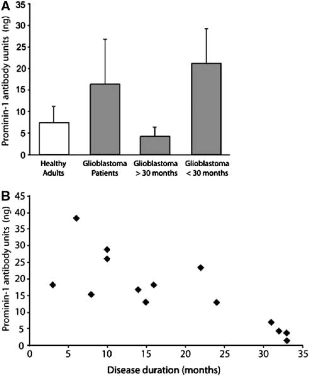
Results: Differential centrifugation of CSF followed by immunoblotting of the resulting fractions revealed the presence of prominin-1 in the 200,000 g (P4) pellet.2 All healthy individuals analyzed showed a narrow range of membrane particle-associated prominin-1/CD133 levels in the CSF (7.4±3.8 ng of bound prominin-1 antibody). The membrane particles were similar in physical properties and microdomain organization to small membrane vesicles previously shown to be released from neural stem cells in the mouse embryo.5 Glioblastoma patients showed elevated levels of membrane particle-associated prominin-1/CD133 (see Figure), which decreased dramatically in the final stage of the disease.2
Conclusions: Our observations represent the first demonstration that a somatic stem cell marker present in central nervous system (CNS) tissue is released into the CSF. This finding strongly suggests that CSF has a greater potential for diagnosis and monitoring of neurological diseases than previously assumed. To analyze the amount of membrane particle-associated prominin-1/CD133 in CSF reflects a novel approach and may open up new avenues for studying CNS disease.2,6
223. Characterisation of neural stem cells for oestrogen in vitro: potential for improving stem cell based therapy for stroke
S. Patkar1, R. Tate1, M. Modo2, R. Plevin1 and H.V.O. Carswell1
1SIPBS, University of Strathclyde, Glasgow; 2Centre for the Cellular Basis of Behaviour, Kings College London, London, UK
Stroke is a problem for the ageing population. The ageing population is increasing but there is no licensed therapy for chronic stages of the disease. Stem cells are already known to have enormous potential to improve stroke outcome in the chronic stages1 but we need to improve their integration in vivo. Evidence also suggests that the female hormone, oestrogen, enhances differentiation of neural stem cells.2 The long-term aim of the present study is to determine whether oestrogen can improve the success of neural stem cell grafting in experimental stroke. We used an immortalised temperature sensitive murine neural stem cell line, the Maudsley hippocampal stem cell line clone 36 (MHP36) because they proliferate only at low temperatures (33°C) in vitro, develop into mature neurons and glia on transplantation into the higher temperature brain (37°C) and cease dividing once matured reducing the chance of producing tumours. The short-term aim was to fully characterise the MHP36 for oestrogen receptors (ER) and the enzyme, aromatase, which synthesizes oestrogen.
The expression of aromatase (gift from J. Hutchison), ERalpha (Novachem) and ERbeta (gift from G. Greene, CO1531) protein in the MHP36 stem cells were investigated by immunofluorescene and Western blotting. In addition, reverse transcriptase PCR (RT-PCR) and the sequencing of amplicons were carried out to confirm the expression of each transcript of interest. Appropriate controls were used in each assay.
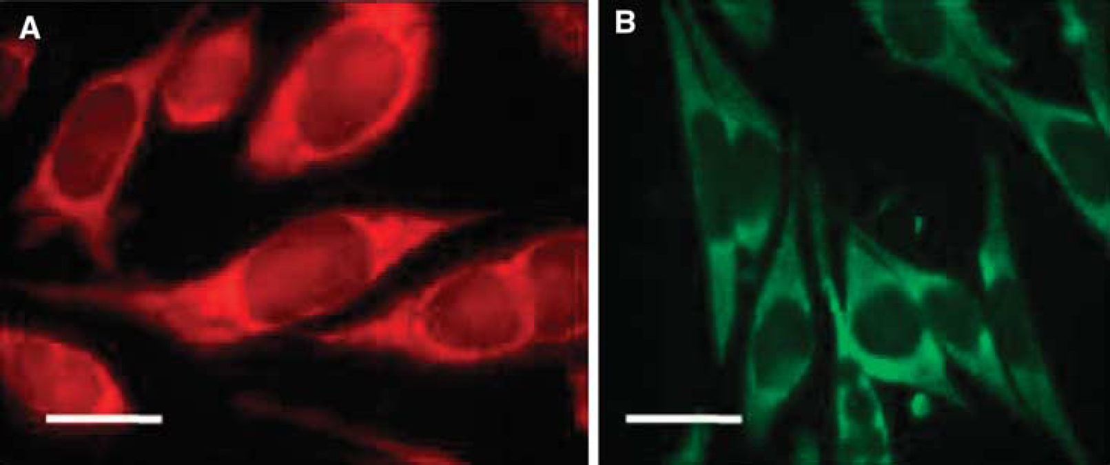
Undifferentiated MHP36 cells were shown to express aromatase using Western blots. Immunofluorescence staining revealed expression of aromatase in the cytoplasm and not in the nucleus (Figure A, MHP36 cells stained for aromatase using Texas red, bar represents 20 μmol/L). Expression was confirmed by semi-nested RT-PCR. ERalpha was absent in the MHP36 stem cells when compared to positive control breast cancer cells MCF-7, which stained the nucleus for ERalpha using immunofluorescence and Western blotting, these results were confirmed by RT-PCR. On the other hand, ERbeta was observed to be expressed in the peri-nuclear membrane of the MHP36 cells using immunofluorescence (Figure B, MHP36 cells stained for ERbeta with FITC, bar represents 20 μmol/L) and expression was confirmed by RT-PCR.
Immunofluorescence staining for Aromatase and ERb.
We have thoroughly characterised the MHP36 cells for oestrogen and demonstrated for the first time that they express ERbeta and aromatase, but do not express ERalpha. This expression of aromatase and ERbeta by the cells is appealing, since the local endogenous production of oestrogen via aromatase by the MHP36 cells may help improve the integration, survival and differentiation of transplanted cells in vivo after experimental stroke, possibly mediating ERbeta activation which is the hypothesis for our next studies.
430. Gene expression in the brain after insonation with ultrasound: a comparison between experimental and diagnostic ultrasound probes
P. Reuter1, A. Fabrizius2, M. Nedelmann3, J. Marx1, B. Alessandri4, O. Kempski4 and T. Hankeln2
1Neurology; 2Molecular Genetics, Johannes Gutenberg-University, Mainz; 3Neurology, Justus-Liebig University, Giessen; 4Insitute of Neurosurgical Pathophysiology, Johannes Gutenberg-University, Mainz, Germany
Background and aims: Latest experimental and clinical studies could show that the use of diagnostic ultrasound (3 MHz) in addition to rtPA treatment successfully enhances thrombolysis cerebral vessel occlusion. However, a potential effect on gene expression is unknown, but should be considered for safety reasons.
The aim of this study was to examine the effects of ultrasound treatment on cerebral gene expression. For this purpose, three ultrasound probes of different frequencies (3 MHz diagnostic probe; 60 and 488 kHz experimental probes) were compared.
Methods: The brain of healthy rats was transcranially insonated with the above probes. For measuring the gene expression we extracted RNA of the cerebral cortex and transcribed the RNA into cDNA. After normalization to reference genes (GAPDH, actin, cyclophillin A, ARP and albumin) we measured expression of several candidate genes from different functional groups (major focus: apoptosis, angiogenesis, NO-metabolism, stress). Gene expression levels were determined by quantitative Real-Time RT-PCR (qPCR) assays.
Results: The measurement of mRNA-expression of selected genes with qPCR showed significant differences between insonated and non-insonated animals. As one example, insonation with the diagnostic ultrasound probe induced a doubled increase of gene expression of the pro-apoptotic gene bcl-2. Most of the regulations were detected after insonation with 488kHz ultrasound. We found an up to 3-fold change after 4 or 24 h for 14 of the 24 studied genes compared to sham-insonated animals. Angiogenesis factors (Vegfa, Egfr, Egr1) showed a decreased expression. Fewest regulations were detected after insonation with the 60 kHz ultrasound probe (3 of 24 measured genes).
Conclusions: The results of this study indicate that not only histological changes can be observed after ultrasound treatment, but also changes of gene expression are detectable. Most of the changes were found after treatment with 488 kHz ultrasound. Further studies have to identify whether these changes have a positive or negative effect on the ischemic lesion. Additionally these findings give reasons to a global gene expression study.
508. Immune-mediated effects of grafted human embryonic stem cells-derived neural stem cells in pediatric hypoxic-ischemic brain injury model
M. Daadi, A. Arac, Z. Li, G. Sun, J. Wu and G. Steinberg
Stanford University School of Medicine, Stanford, California, USA
Ischemic brain injury in newborn infants represents a major cause of cerebral palsy, mental retardation and epilepsy. Currently, there are no effective interventions to improve the chronic sequelae of perinatal asphyxia. Stem cell-based therapy has the potential to replace the necrotic tissue caused by hypoxia-ischemia (HI) and to restore function. Promising preclinical studies in experimental stroke models have demonstrated the efficacy of stem cells derived from various sources, including bone marrow, cord blood and central nervous system. In most of these studies, grafted animals showed some degree of functional recovery. Although, these findings are promising, little is known about how stem cell transplantation therapy achieves these outcomes. It is generally believed that non-neural cells, such as those derived from bone marrow or cord blood, exert a neurotrophic effect on ischemia-injured tissue and do not survive for long-term in the grafted site. While neural stem cells are thought to provide cell replacement and neurotrophic support of the lesioned area. In the present study, we sought to determine how the inflammatory and cellular immune responses are modulated after stroke and transplantation of human neural stem cell progeny (hNSCs), derived from human embryonic stem cells (hESCs) in the rat model of neonatal HI.
The hNSCs were isolated from the hESCs and perpetuated using serum free media supplemented with epidermal growth factor, basic fibroblast growth factor and leukemia inhibitory growth factor. To generate the model of neonatal HI, seven-day-old rats were subjected to permanent ligation of the left carotid artery, followed by one and half hours in a hypoxic chamber (8%O2 and 92%N2, at 37°C). The newborns were divided into HI vehicle and HI transplant groups. Twenty-four hours after the induction of HI, animals were grafted with single cell suspension of hNSCs (4.5 × 10⁵) into 3 sites in the forebrain. The animals were evaluated 5 weeks after transplantation for their sensorimotor skills in the cylinder and in the rotarod tests.
Our results showed that during the fifth week after transplantation, HI transplanted animals significantly improved in their use of the contralateral impeded forelimb (P<0.05). The hNSC grafts significantly ameliorated the locomotor deficits in the rotarod test (P<0.05). To evaluate potential effects on the immune system, the cellular composition of peripheral immune system was evaluated using flow cytometric analysis. Our data demonstrated that stroke induced a 3-fold increase in the CD3+, CD4+ T-cells and no significant changes in the CD8+ and CD11b+ cells in the blood and other peripheral systems of vehicle group. Graft treated animals manifested a significant decrease (P<0.05) in the CD3+, CD4+ T lymphocytes, suggesting an anti-inflammatory effect. Post-mortem histopathological analysis of grafted hNSCs identified with a human-specific nuclear marker, demonstrated good survival, dispersion and differentiation in the stroke-damaged tissue. Interestingly, the transplanted animals showed a 2-fold increase in Iba1+ microglia with no apparent infiltration or reaction against the grafts. These preliminary results suggest that the peripheral immune system is modulated by hNSCs transplants and that microglia could play a neurotrophic role in HI-transplanted brains.
514. A novel adenoviral vector for gene therapy against cerebral ischemia
S. Hou, D. Huang and A. Desbois
Institute for Biological Sciences, National Research Council Canada, Ottawa, ON, Canada
Background and aims: Viral vector-mediated gene therapy represents a promising approach to treat human neurological diseases. Among the many gene delivery vectors, replication defective adenoviral vectors have been widely used as an effective tool to express therapeutic genes in the brains of rodent models of human neurological diseases including cerebral ischemia. Needless to say, the development of an array of selective gene targeting vectors is vital for the future success of any therapeutic attempts aimed at protecting neurons in the diseased brains. However, selective gene expression in neurons is still a challenge. To this end, we have developed a expression vector which is capable of expressing gene products selectively in neurons under the regulation of hypoxia.
Methods: We have developed several expression vectors using a combination of neuron restrictive silencer elements (NRSEs), hypoxia responsive elements (HREs) and CMV minimal promoter (CMVmp). These elements were packaged into replication defective adenovirus to target gene expression selectively in neurons in a hypoxia-regulated manner. Neuronal selectivity and responsiveness to hypoxia of the constructed novel vectors were determined empirically in both neural cell lines, primary cerebellar granule neurons (CGNs) and in neonatal mouse brain under hypoxia.
Results: The construct p5HRE-3NRSE exhibited not only the highest level of reporter gene expression in neuronal cells, but also in an oxygen concentration-dependent manner when compared with all other constructs. As expected, this construct did not elicit reporter gene expression in non-neuronal cells including human HEK293A and HT29 cells, rat NRK cells, mouse 3T6 cells and 3T3 L1 cells. The construct was packaged into a replication defective adenoviral vector (Ad/5HRE-3NRSE). Remarkably, in response to hypoxia, Ad/5HRE-3NRSE showed strong gene expression in primary CGNs (12-fold induction compared with normoxia), but not in glial cells. Double immunostaining with antibodies against the reporter gene product luciferase and a neuron-specific marker protein, such as MAP-2, confirmed the specificity and inducibility of this construct in neurons. Preliminary in vivo studies using these vectors in a neonatal hypoxia mouse brain confirmed selective gene expression in neurons in response to hypoxia.
Conclusions: Taken together, further refinement of this vector may lead to the development of a useful tool for targeting gene delivery and therapy for stroke research and therapeutics.
545. Monitoring of intra-arterially administered bone marrow mononuclear cells in rat transient focal ischemia model using MRI
N. Kamiya1, M. Ueda1, H. Igarashi2, Y. Nishiyama1, S. Suda1 and Y. Katayama1
1Department of Internal Medicine, Division of Neurology, Nippon Medical School, Tokyo; 2Center for Integrated Human Brain Science, Brain Research Institute, University of Niigata, Niigata, Japan
Background and aims: We had reported that transplantation of bone marrow mononuclear cells (BMMCs) via carotid artery, but not via femoral vein immediately after reperfusion had neuroprotective effect in rat transient ischemic model. We revealed the difference of the protective effect determined by the number of the cells in brain at the acute stage of ischemia. To reveal the mechanisms of neuroprotective effect, the present study was designed to monitor the distribution of transplanted BMMCs in brain in process of time on rat transient focal ischemia model. It is impossible to monitor cells histologicaly on one rat in time course, because we need to euthanize rat to make brain slices. Using MRI system, we can scan brain and monitor administrated cells repeatedly.
Methods: 12 Male Sprague-Dawley rats, weighing 250 to 300 g, were used in the study. Bone marrow was obtained autologously from the right femur of each animal, and BMMCs were isolated prior to ischemia using density-gradient centrifugation method. BMMCs were labeled with SPIO (super paramagnetic iron oxide) using electroporation method. Rats were then subjected to transient (90 mins) left middle cerebral artery (MCA) occlusion followed by 1 × 10⁷ BMMCs administration via the ipsilateral carotid artery (IA group) immediately after reperfusion. Control rats underwent the same procedure but received vehicle injection (Vehicle group). At 1 h, 24 h and 7 days after reperfusion, we underwent MRI study (3d gradient T2*) on every rats. After the final MRI, rats were decapitated and brains were carefully removed. Paraffin-embedded brain blocks were cut into 20 μm-thick coronal sections at 2 mm intervals to obtain six slices. Sections were stained with Berlin Blue to identify iron of transplanted cells and with hematoxylin and eosin (HE) to measure infarct volume.
Results: SPIO labeled BMMCs showed negative contrast enhancement in MRI corresponding to histological finding. Administrated cells distributed over the left MCA perfusing lesion at 1 hr (Figure) and decreased in process of time. Infarct volume significantly reduced in IA group than vehicle group. MRI study also revealed that administered cells were distributed within the ipsilateral hemisphere at 1hr, and that the cells were decreased in number thereafter. BMMCs tended to accumulate around ischemic lesion.
Conclusions: The present study established in vivo monitoring of transplanted SPIO-labeled BMMCs using MRI in process of time. The present data confirm significant neuroprotection following transient focal ischemia by autologous BMMCs administered intra-arterially, immediately after reperfusion in rats.
582. Overexpression of human down syndrome candidate region 1 (DSCR1) gene improves outcome following cerebral ischemia-reperfusion in mice
C.G. Sobey1, K.R. Martin2, M.A. Pritchard2 and V.H. Brait1
1Department of Pharmacology; 2Department of Biochemistry, Monash University, Clayton, VIC, Australia
Background and aims: Down syndrome results from the trisomy of chromosome 21. The Down syndrome candidate region 1 (DSCR1) gene, recently renamed RCAN1, is found on chromosome 21. DSCR1 protein is highly expressed in the brain,1 and following middle cerebral artery occlusion (MCAO) in mice DSCR1 mRNA and protein was found to increase in the peri-infarct region.2 The aim of this study was to investigate the effects of overexpression of DSCR1 on outcome following stroke.
Methods: DSCR1 transgenic (Tg) mice were produced by the overexpression of the human DSCR1 gene, only in cells that normally express DSCR1, on a C57Bl6/J X CBA background. Cerebral ischemia was induced by MCAO for 0.5 h followed by reperfusion for 23.5 h (ischemia-reperfusion; I-R) in age-matched groups of 8 to 14 week old male DSCR1 Tg (n = 14) and wild-type (Wt; n = 9) mice. MCAO was confirmed by a sudden drop in regional cerebral blood flow (rCBF; ∼75%), measured by a laser-doppler flow probe positioned over the region perfused by the MCA. Control mice were subjected to sham surgery. After 24 h, neurological impairment was assessed (neurological score and hanging wire test) and %rCBF re-measured. Brain infarct and edema volume were then measured in thionin-stained coronal sections.
Results: After I-R, Tg mice had significantly less neurological impairment than Wt mice, with a lower neurological score (2.4±0.3 versus 3.9±0.1; P<0.05) and a longer hanging-wire time (38±5 versus 9±3 seconds; P<0.0001). %rCBF at 24 h was significantly higher in Tg than Wt mice (73±10% versus 33±7% pre-ischemia; P<0.01). After I-R, Tg mice had a significantly smaller subcortical infarct volume than Wt mice (15±2 versus 32±5 mm3; P<0.01). In addition, total infarct volume tended to be smaller in Tg than Wt mice (26±5 versus 36±6 mm3, n = 9 to 14; P = 0.26). Furthermore, edema volume was smaller in Tg than Wt mice (17±3 versus 31±6 mm3; P<0.05).
Conclusions: Thus, overexpression of DSCR1 in mice improves outcome following cerebral I-R. Further studies are required to establish the mechanisms behind this improved recovery, and the relevance for individuals with Down syndrome.
589. Neural induction of pericyte progenitor cells from human peripheral blood mononuclear cell cultures
K.-H. Jung, K. Chu, S.-T. Lee, J.-S. Sunwoo, J.-J. Bahn, H.-K. Park, M. Kim, S.K. Lee and J.-K. Roh
Seoul National University Hospital, Seoul, South Korea
Background: Pericytes are located around vessels and contribute to vascular maintenance and vascular regeneration. It has long been suggested that pericytes may have stem/precursor cell potential. Our recent study has identified neural progenitor cells in peripheral blood mononuclear cells (PBMNCs) of acute stroke patients. However, there are still unsolved issues about the nature of circulating neural progenitors. Given that pericytes respond to injury, and regenerate the tissue, we hypothesized that neural progenitor cells outgrown from in vitro culture of PBMNCs might be tissue or bone marrow-derived pericyte progenitor cells (PPCs).
Methods: PBMNCs were isolated from the peripheral blood of 32 acute stroke patients, and 35 controls with risk factor-only. The fraction of pericytes in PBMNCs was measured using flow cytometry with anti-PDGFRβ or anti-NG2 antibodies. We investigated the association between characteristics of circulating PPCs and stroke phenotypes. We also developed the efficient protocols for mobilization, isolation, expansion, and neural induction of circulating PPCs.
Results: The fraction of circulating PPCs in PBMNCs was higher in acute stroke patients than control. These progenitor cells could be more efficiently isolated from acute stroke patients with greater NIHSS scores than risk factor-only group, suggesting that the circulating progenitor cells might be one marker of systemic resilience to ongoing tissue damage. PPCs could be efficiently mobilized by pharmacological stimulation, expanded ex vivo under hypoxia condition, and differentiate into a variety of neural cells under appropriate media conditions.
Conclusion: The feasibility of extracting and culturing PPCs in large numbers suggests that such autologous cells will be useful for applications ranging from basic research to cell-based therapy. Neural induction of PPCs from autologous peripheral blood makes them valuable candidates for transplantation therapy.
638. Intravenous coadministration of smooth muscle and endothelial progenitor cells enhances angiogenesis in focal permanent cerebral ischemia in mice
N. Deroide1, L.R. Nih1, C. Déan2, M. Clergue1, B.I. Lévy1, J.-S. Silvestre1, G. Tobelem2 and N. Kubis1
Objectives: Extensive evidences indicate that angiogenesis plays a key role in stroke recovery. Strong cooperation between endothelial and smooth muscle cells is required for the formation of a mature functional vascular network. Therefore, we assessed whether therapy based on coadministration of smooth muscle progenitor cells (SMPCs) and endothelial progenitor cells (EPCs) isolated from human cord blood enhances angiogenesis and neurogenesis, in a murine model of focal permanent cerebral ischemia.
Methods: Ischemia was induced in adult male C57Bl6 mice by the thermocoagulation of the middle cerebral artery (MCA). SMPCs (0.5 × 10⁶), EPCs (0.5 × 10⁶), SMPCs+EPCs (0.25 × 10⁶+0.25 × 10⁶) or phosphate-buffered saline (PBS) were intravenously administered 24 h post-MCA occlusion (n = 6 to 8/group). To assess proliferation, BrdU (100 mg/kg, i.p.) was injected 4 and 2 h before sacrifice on day 7 post-MCAo. Infarct volume was measured by histomorphological analysis of Cresyl Violet stained sections. Blood-brain barrier (BBB) permeability was quantified using Evans Blue extravasation. Vascular area, cell proliferation and proliferating cell phenotype were determined in ischemic and peri-ischemic areas, using co-immunostaining with antibodies against doublecortin, laminin, CD31 and BrdU.
Results: Infarct volumes were not significantly different (7.09±2.97, 6.83±3.16, 6.70±2.48 mm3) for SMPCs, EPCs, SMPCs+EPCs-treated groups respectively, compared to the PBS group (6.01±2.39 mm3). However, the vascular area in the infarct was significantly increased in all treated groups with SMPCs, EPCs or EPCs+SMPCs, in the same range, compared to controls (+17.4%±8%, +19.4%±5.2% and +17.3%±3.6%, respectively, P<0.05). Phenotypical vascular differences were evidenced between groups. SMPCs-treated mice showed an immature disorganized and dilated vasculature, EPCs-treated mice showed more numerous small vessels, whereas SMPCs+ EPCs-co-treated mice showed more mature and numerous vessels. The number of co-labelled BrdU-CD31 positive cells was significantly increased in all treated groups, compared to PBS but more markedly for the EPCs- and SMPCs+EPCs co-treated mice: 14.7±1.2/ROI (controls), 22.7±1.5/ROI (SMPCs), 33.3±3.2/ROI (EPCs) and 35.0±1.4/ROI (SMPCs+EPCs) (P<0.002), indicating angiogenesis in this area of intense remodelling. In addition, the number of peri-infarct BrdU-positive cells was higher only in the SMPCs+EPCs-treated group (3-fold) compared to the three other groups (P<0.002), but no neurogenesis was evidenced in this group. Despite vascular remodelling, we found no significant difference in BBB permeability at day 7 (111.67%±39.8%, 111.39%±31.1%, 114%±19%) for SMPCs, EPCs, SMPCs+EPCs-treated group respectively, compared to the PBS group.
Conclusions: Co-administration of human cord blood smooth muscle and endothelial progenitor cells triggers vascular remodelling and angiogenesis in the infarct and intensive cell proliferation in the peri-ischemic area in cerebral ischemia in mice. Whether, neurological deficit is improved remains to be determined.
645. Intravenous administration of autologous mesenchymal stem cells derived from bone marrow into stroke patients
K. Houkin1, O. Honmou1, T. Matsunaga2, S. Ishiai3, S. Waxman4 and J. Kocsis4
1Neurosurgery; 2Fourth Department of Internal Medicien; 3Rehabilitation, Sapporo Medical University, Sapporo, Japan; 4Neurology, Yale University School of Medicine, New Haven, Connecticut, USA
Background: Intravenous injection of mesenchymal stem cells (MSCs) prepared from adult bone marrow has been reported to ameliorate functional deficits in several CNS diseases in experimental animal models. Bone marrow cells can be enriched in MSCs by selecting for plastic-adherent cells. MSCs will grow to confluency in appropriate culture conditions as flattened fibroblast-like cells. Although MSCs may be present in different proportions in the stromal cell fraction of various species, MSCs have a distinct cell surface antigen pattern including SH2+, SH3+, and CD34−, and methodologies have been established to culture human MSCs in very high purity. Although human MSCs have been clinically used for several diseases, it is still uncertain whether MSCs may have therapeutic benefits on stroke patients.
Objectives: The objectives of this study were to examine feasibility and safety of cell therapy using culture-expanded autologous MSCs in twelve stroke patients. This study was a phase I clinical trial.
Methods: Twelve (male and female) patients aged ≥40 years with stroke were enrolled. Bone marrows from the stroke patients were obtained by aspiration from the posterior iliac crest after informed consent was obtained; the subject's consent was obtained according to the Declaration of Helsinki, and this study was approved by the Institutional Review Board at Sapporo Med. Sch. where the cells were isolated and transplanted. Bone marrow was plated in plastic tissue culture flasks, and the adherent cells were cultured in appropriate medium in a humidified atmosphere of 5% CO2 at 37°C. After reaching confluency, they were harvested and cryopreserved until use. On the day of infusion cryopreserved units were thawed at the bedside in a 37°C water bath and injected intravenously into patients over 30 mins. All patients were monitored closely during and within 48 h of MSC injections. Oxygen saturation, temperature, blood pressure, pulse and respiratory rate were carefully monitored before and after injection. Patients also had chest films before and after MSC injection. Patients had carefully been followed for 12 months.
Results: Human bone marrow-derived MSCs were successfully isolated from bone marrow aspirate from all 12 stroke patients, and all were successfully culture-expanded. Serial evaluations showed no adverse cell-related, serological, or imaging-defined effects.
Conclusions: In patients with cerebral infarcts, the intravenous administration of autologous MSCs appears to be feasible and safe, and merits further study as a therapy that may improve functional recovery.
732. MRI-guided transplantation and monitoring of a microparticle scaffold system for tissue repair and regeneration in stroke
E. Bible1, D. Chau2, M. Alexander2, J. Price3, K. Shakesheff2 and M. Modo1
1King's College London, London; 2University of Nottingham; 3King's College London, Nottingham, UK
Introduction: An alternative to pharmacological intervention in stroke therapy is cell replacement via NSC (neural stem cell) transplantation. As yet, however, neither pharmacological intervention, nor stem cell transplantation has tackled the problem of the large cystic cavity that remains post-lesion. In the present study, MRI-derived stereotactic co-ordinates were used to transplant neural stem cells attached to a microparticle scaffold into the lesion core to facilitate tissue regeneration. T2-weighted images obtained before and after transplantation allowed confirmation of the correct targeting of microparticles and progressive monitoring of scaffold integration with surrounding parenchyma, indicating successful engraftment.
Methods: Cells from the MHP36 mouse hippocampal cell line were attached to PGLA (polyglycolic lactic acid) microparticles that had previously undergone surface chemistry modification by allylamine plasma polymerisation coating (ppAam) and surface adsorption with fibronectin. Animals were lesioned by 60′ MCAo (middle cerebral artery occlusion) and subjected to MRI 2 weeks after stroke to select animals with striatal lesions and transplantation co-ordinates were derived from these images. A 30 ml volume of microparticles with attached cells suspended in N-acetyl-cysteine was injected directly into the lesion cavity. Animals were scanned on a 7.0 T horizontal bore magnet (Varian, Palo Alto, CA, USA) using a custom built head coil with a 40 mm field of view and 128 X 128 data matrix. T2-weighted images were acquired at 4 days pre-transplantation, 1 day post-transplantation and 7 days post-transplantation. A multi-echo, multi-slice, spin-echo pulse sequence (MEMSP: TR = 4200 ms, TE = 10, 20, 30, 40, 50, 60, 70, 80 ms) was used to acquire images. T2 maps and T2-weighted images were generated using VnmrJ software (v2.3, Varian, Palo Alto, CA, USA).
Results & discussion: Conventionally NSCs have been transplanted in suspension into the contralateral non-lesioned side or into intact parenchyma adjacent to the stroke lesion. Transplantation of NSCs on PGLA microparticles using MRI-derived stereotactic co-ordinates allowed for direct transplantation of cells into the lesion. Microparticles could be visualized on MRI and were observed to fill the lesion cavity and to induce a change in signal intensity over time. Changes in the relaxivity of T2-weighted images, provides a means to monitor microparticle engraftment with surrounding tissue non-invasively over time. Immunofluoresent staining of brain sections confirmed MRI findings of scaffold engraftment within the lesion cavity. MHP36 cells could be found still attached to microparticles, but also migrating into the surrounding tissue matrix. Transplanted cells successfully differentiated into neuronal and glial cell populations.
Conclusions: PGLA microparticles can be used to inject NSCs directly into stroke lesions using MRI-derived co-ordinates. MRI is essential for the correct targeting of scaffolds to the lesion and can be used to monitor the extent of engraftment over time.
821. Intraarterial neural stem cell delivery does not reduce cerebral blood flow
J.Y. Chua, X. Gaeta, N. Wang, R.H. Andres, A.V. Pendharkar and R. Guzman
Neurosurgery, Stanford University School of Medicine, Stanford, California, USA
Introduction: Stem cells have been recognized as a potential therapy for stroke and various methods have been developed to deliver these cells. One of these methods, the intraarterial (IA) delivery of cells, provides several benefits such as direct targeting of ischemic tissue, circumventing systemic cell trapping and feasibility in translating into clinical practice. However, it has been reported recently that the IA delivery potentially poses the risk of vascular occlusion. In this study, we compared the cerebral blood flow (CBF) effects of two IA delivery methods – a high volume catheter injection with permanent Common Carotid Artery (CCA) ligation and a low volume needle injection with CCA flow.
Methods: 24 h after undergoing 2 h of middle cerebral artery occlusion (MCAO) with intraluminal suture, adult female Wistar rats had a Laser Doppler probe placed on a thinned portion of the skull above the corpus striatum to monitor CBF. For transplantation, special care was taken to obtain single cell suspensions of Murine C17 cells in phosphate-buffered saline (PBS). In Group 1, the ipsilateral internal carotid artery (ICA) was cannulated with a catheter followed by the infusion of 1 × 10⁶ C17 cells in 1 mL PBS. The Group 1 control animal underwent the same procedure but was infused with 1 ml PBS. In Group 2 animals, 1 × 10⁶ C17 cells in 5 μL and 0.5 × 10⁶ C17 cells in 5 μL were injected into the CCA lumen via the External Carotid Artery. CBF measurements were recorded up to 15 mins after IA delivery.
Results: IA delivery in Group 1 animals resulted in a sharp drop in CBF to about 10% of baseline within the first minute and a recovery to near baseline within 2 mins. 1 animal in Group 1 did not show a recovery in CBF after 1 mins and was hemiplegic before dying. This is consistent with the variability in CBF and mortality reported for this method. The control animal experienced the same drop and recovery in CBF.
The IA delivery in Group 2 animals for both cell concentrations experienced a 20% spike in CBF after CCA reperfusion and maintained its CBF slightly above baseline for 15 mins. The overall comparison between group 1 and 2 demonstrated a statistically significant difference (P<0.05).
Conclusions: The lack of drop in CBF of Group 2 indicates that the reported risk of vascular occlusion and mortality may be confined to the high volume and the CCA occlusion catheter method of IA delivery. Furthermore, the drop in Laser Doppler values during the first minute of the control indicates that the initial drop is an artifact of PBS infusion and is most likely not an indicator of reduced CBF due to an occlusion. Depending on the method, IA delivery of therapeutic cells does not pose a clear risk of vascular occlusion.
824. Creatine modulates survival, migration and differentiation in neural stem cells
R.H. Andres1, A. Pendharkar1, R. Guzman1, A. De2, T.M. Bliss1, E. MacMillan3, C. Svendsen3, S.S. Gambhir2, H. Widmer4 and T. Wallimann5
1Department of Neurosurgery; 2Molecular Imaging Program at Stanford, Stanford University School of Medicine, Stanford, California; 3The Waisman Center, University of Wisconsin-Madison, Madison, Wisconsin, USA; 4Department of Neurosurgery, University of Berne, Bern; 5Department of Cell Biology, Swiss Federal Institute of Technology (ETH), Zurich, Switzerland
Objectives: Creatine kinase (CK) catalyzes the reversible transphosphorylation of creatine (Cr) by ATP. CK isoenzymes are specifically localized at strategic sites of ATP consumption to efficiently regenerate ATP in situ via phosphocreatine (PCr) or at sites of ATP generation to build-up a PCr pool. Accordingly, the CK/PCr system plays a key role in cellular energy buffering and energy transport, particularly in cells with high and fluctuating energy requirements like neurons, and exogenous Cr supplementation results in higher cellular ATP reserves and improved metabolic channeling. Given the high expression of CKs in the developing CNS, we hypothesize that Cr supplementation might be effective in improving the metabolic state of neural stem cells (NSCs). In the present study, we investigated the effects of Cr on survival, migration, and differentiation of mouse NSCs isolated from the subventricular zone (mNSCs) and NSCs derived from the human fetal cortex (hNSCs).
Methods and results: We found both the brain-specific cytosolic (BB-CK) and the ubiquitous mitochondrial (uMt-CK) isoform of CK expressed in mNSC and hNSC, as well as the key enzymes for endogenous Cr synthesis, AGAT and GAMT, and the specific transporter for cellular Cr uptake, CRT. Cr supplementation at 5 mmol/L for 7 days resulted in higher ATP levels in mNSCs and hNSCs, as compared to untreated controls (P<0.01).
Chronic Cr exposure of cultured mNSCs and hNSCs resulted in a dose-dependent increase in neurosphere size and total cell numbers with a maximal effect at 5 mmol/L after 7 days in vitro (P<0.05). Analysis of BrdU incorporation did not reveal a significant increase in proliferation activity, but Cr-treated cultures contained less cells with immunoreactivity for active Caspase-3 (P<0.05), pointing towards an antiapoptotic effect of Cr.
In a modified Boyden chamber assay, both Cr-pretreated mNSCs and hNSCs demonstrated an improved migratory potential (P<0.01) after 60 mins, as compared to untreated controls. In line with this finding, we found that Cr-pretreated, GFP-labeled mNSCs showed improved migration to the ischemic brain area after intrastriatal transplantation in a distal MCA occlusion stroke model in C57/Bl6 mice (P<0.05).
Next we examined the effects of Cr on differentiation of NSCs. Chronic Cr exposure at 5 mmol/L resulted in significantly higher neuronal cell numbers (NeuN+, P<0.05) at the expense of the glial fate (GFAP+, P<0.05) after differentiation conditions in vitro for 5 days. In addition, we found disproportionally higher numbers of GABA-immunoreactive cells in the Cr-treated groups (P<0.05), suggesting that Cr acts as a differentiation factor for specific neuronal subpopulations.
Conclusion: Our findings suggest that the CK/PCr system is critically involved in maintaining the energy metabolism of NSCs. Chronic Cr supplementation resulted in increased cellular ATP levels, inhibited apoptosis during in vitro expansion, and improved NSC migration in vitro and in vivo. Cr exposure also promoted the differentiation of NSCs towards the neuronal lineage, particularly supporting the GABAergic phenotype. Cr pretreatment of NSCs might therefore offer new ways for improving cell replacement approaches for stroke and other diseases of the nervous system.
859. Dual sensitivity of early and late embryonic stem cell-derived neural progenitors to the ischemic environment
C. Seminatore1, J. Polentes1, D. Ellman2, N. Kozubenko3, S. Tine1, L. Tritschler4, J. Blondeau1, E. Guidou4, M. Luillhier4, A. Bugi4, L. Aubry4, A. Perrier4, P. Jendelova3, E. Sykova3, B. Finsen2 and B. Onteniente1
1I-STEM, INSERM UMR894/861, Evry Cedex, France; 2Medical Biotechnology Center, University of Southern Denmark, Odense, Denmark; 3Institute of Experimental Medicine, Prague, Czech Republic; 4I-STEM, INSERM UMR861, Evry Cedex, France
Objectives: Teratoma/tumor formation is a major obstacle to embryonic stem cell (ESC) therapy. However, while it has been described in models of neurodegenerative diseases, teratoma/tumor formation has not been reported for stroke after transplantation of neural progenitors and/or neuronal precursors. This study aimed at understanding the reasons of this discrepancy.
Methods: Neural induction of the SA01 hESC (Cellartis AB, Sweden) line was performed using the rosette protocol with a minimal culture medium. qPCR expression of differentiation and proliferation markers identified 4 maturation stages, named early-, mid-, late-hES-NP, and neuronal precursors (NP). hES-NP and NP were transplanted into rats with no, small or large ischemic lesions. The fate of transplanted hES-NP and NP and their functional effects were investigated 1 to 4 months after transplantation.
Results: The lesion size significantly impacted graft survival of early-hES-NP. This influence was lost with mid- and late hES-NP and with NP. Similar relationships to the lesion were observed with the derivation of non-neuroectodermal cell types. Although teratoma with derivatives of the 3 germ layers were rarely observed (1 out of 12), early-hES-NP generated non-neural cell types of the ectodermal (epidermal cells) and endodermal (cartilage) lineages. The expression of frizzled-3, an inducer of the neural crest, suggested that non-neural formations from early-hES-NP were derived from neural crest-like structures. The sensitivity to the ischemic lesion was lost after the appearance of neuronal markers. Late progenitors and NP nicely integrated into the host brain, underwent a correct neurogenesis sequence by producing neurons then astrocytes, and contained patches of DARPP-32 neurons innervated by host TH afferents. The integration of transplanted cells and the benefit to the host were confirmed by reversal of ischemia-induced rotation behaviour after apomorphin administration.
Conclusions: The influence of the ischemic environment on hESC-NP is limited to the early stages of differentiation and is lost after neural commitment, confirming that intracerebral transplantation of hESC-NP is a safe and reliable approach for stroke.
877. Targeted delivery of ferumoxide labeled human neural stem cells using a neodymium magnet in focal cerebral ischemic rats
M. Song1,2, Y.-J. Kim1, Y.-H. Kim1, J. Roh1 and B.-W. Yoon1,2
1Department of Neurology, Clinical Research Institute, Seoul National University Hospital; 2Medical Research Center, Seoul National University, Seoul, South Korea
Objectives: It was reported that human neural stem cells (hNSCs) can be used to treat damaged tissues in various brain diseases. Injected hNSCs have limitations of delivery to target areas, so more effective methods are required. In our previous study, we established effective labeling of human neural stem cells with ferumoxide, FDA-approved iron oxide nanoparticle, complexed to transfection agent, poly-L-lysine (PLL). We hypothesized that ferumoxides labeled human neural stem cells can be delivered more effectively targeting to an area of interest by magnetic fields in vitro and in vivo.
Methods: hNSCs (HB1.F3 cells) were incubated for 24 h in cell culture media with ferumoxides and PLL or without (control). Cellular iron uptake was identified by Prussian blue staining and iron contents of the cell were analyzed using iron reagents by a 560 nm UV/VIS spectrophotometer. hNSCs were incubated in a culture dish, under which a neodymium magnet was attached. Proliferation, viability and differentiation of the cells were evaluated using an increasing of cell number, trypan blue dye exclusion test, and immunofluorescence cytochemistry with cell-type specific makers, respectively. To investigate the targeted delivery, rats were allocated into three groups (i.e. ischemia only, cell injection with magnet and without magnet). After middle cerebral artery occlusion (MCAo), human NSCs were injected via tail vein 24 h after. In histological sections, transplanted cells were identified by Prussian blue and anti-human nuclei fluorescence staining. Infarct size was analyzed by Nissl staining and the iron contents of rat brain were determined.
Results: Magnetic fields appeared not to affect cell proliferation, viability and differentiation. Cellular iron was detected in hNSCs by Prussian blue stain and mean iron contents was 261±9.8 pg/cell (n = 3). An increased number of labeled hNSCs retained in the area of magnet field. Histological analysis of cell injection with magnet group at 7 days after transplantation revealed significantly reduced infarction volume and significantly increased hNSCs in rat brains comparing with other groups.
Conclusions: Our study suggests that this targeting system may be a useful tool for the cell therapy.
935. Genetically modified mesenchymal stem cells are more effective than unmodified in reducing infarct volume in rodent MCAo models: a meta-analysis
N. Karlupia1, M. Prasad2 and K. Prasad1
1Neurology, All India Institute of Medical Sciences, Delhi; 2Pt. B. D. Sharma PG Institute of Medical Sciences, Rohtak, India
Objective: To determine through meta-analysis the magnitude and consistency of effects on infarct volume with the use of genetically modified Mesenchymal stem cell (MSC) transplantation as compared to primary MSCs (control group), in rodent models of stroke.
Methods: Stem cell intervention studies were identified through a high sensitivity search of Pubmed. We used multiple search strategies to identify the studies. Hits obtained with different key terms (written in parenthesis) were 117{[human stem cells] AND [stroke in rats]}, 24{[human mesenchymal stem cells] AND [stroke in rats]}, 11{[hMSCs] AND [stroke in rats], upto 2008; 44{[MCAo] AND [stem cells]} from 2003 to 2008. The quality assessment of the studies was done independently by two reviewers with an overall agreement of 98.4%. As there was significant heterogeneity, relevant data were synthesized using random effect model of DerSimonian and Laird in Revman 4.2.7 software, to obtain standardized mean difference and 95% confidence interval (CI) for volume of infarction.
Results: We scanned the titles of 117 studies and abstracts of 79 studies to select eight studies for the meta-analysis in rodent MCAo models (five permanent and three transient). Studies were excluded if the primary outcome was not infarct volume or if the treatment group was not of genetically modified mesenchymal stem cells. One study (Kurozumi et al, 2004) was excluded because the effect of primary stem cell group on infarct volume was not measured. The number of rats per group varied from 5 to 46. The cell dose ranged from 0.5 to 10 million cells. The time between occlusion and cell administration was three hours (two studies), six hours (two studies), 12 h (one study) and 24 h (two studies). Treatment with genetically modified mesenchymal stem cells resulted in statistically significant reduction in infarct volume (standardized mean difference −3.06 [−4.70 −1.42]) as compared to primary mesenchymal stem cells, but there was substantial heterogeneity (P<0.00001, I2 = 82.1%).
Conclusion: Pre-clinical studies in rodent stroke models lead to reduction in infarct volume with the use of modified stem cells as compared to primary MSCs, but the results are heterogenous. More studies are required to investigate the factors responsible for the heterogeneity.
References
1.
HuhmerAF. Protein analysis in human cerebrospinal fluid: Physiological aspects, current progress and future challenges. Dis Markers2006;22:3–26.
2.
HuttnerHB. The stem cell marker prominin-1/CD133 on membrane particles in human cerebrospinal fluid offers novel approaches for studying central nervous system disease. Stem Cells2008;26(3):698–705.
3.
FlorekM. Prominin-1/CD133, a neural and hematopoietic stem cell marker, is expressed in adult human differentiated cells and certain types of kidney cancer. Cell Tissue Res2005;319:15–26.
4.
CorbeilD. The human AC133 hematopoietic stem cell antigen is also expressed in epithelial cells and targeted to plasma membrane protrusions. J Biol Chem2000;275:5512–20.
5.
MarzescoAM. Release of extracellular membrane particles carrying the stem cell marker prominin-1 (CD133) from neural progenitors and other epithelial cells. J Cell Sci2005;118:2849–58.
6.
BaoS. Glioma stem cells promote radio-resistance by preferential activation of the DNA damage response. Nature2006;444:756–760.
7.
MillerRH. Brain Research2006;1091(1):258.
8.
BrannvallKKorhonenLLindholmD. Molecular and Cellular Neurosciences2002;21(3):512–20.
9.
ErmakGMorganTEDaviesKJ. Chronic overexpression of the calcineurin inhibitory gene DSCR1 (Adapt78) is associated with Alzheimer's disease. J. Biol. Chem,2001;276:38787–94.
10.
ChoK-OKimYSChoY-JKimSY. Upregulation of DSCR1 (RCAN1 or Adapt78) in the peri-infarct cortex after experimental stroke. Experimental Neurology2008;212:85–92.