
Abstract
The precise biologic mechanisms involved in functional recovery processes in response to stroke such as dopaminergic neurotransmission are still largely unknown. For this purpose, we performed in parallel in vivo magnetic resonance imaging and positron emission tomography (PET) with [18F]fluorodeoxyglucose ([18F]FDG) and [11C]raclopride at 1, 3, 7, 14, 21, and 28 days after middle cerebral artery occlusion in rats. In the ischemic territory, PET [18F]FDG showed a initial decrease in cerebral metabolism followed by a time-dependent recovery to quasi-normal values at day 14 after ischemia. The PET with [11C]raclopride, a ligand for dopamine D2 receptor, showed a sustained binding during the first week after ischemia that declined dramatically from day 14 to day 28. Interestingly, a slight increase in [11C]raclopride binding was observed at days 1 to 3 followed by the uppermost binding at day 7 in the contralateral territory. Likewise, in vitro autoradiography using [3H]raclopride confirmed these in vivo results. Finally, the neurologic test showed major neurologic impairment at day 1 followed by a recovery of the cerebral function at day 28 after cerebral ischemia. Taken together, these results might suggest that dopamine D2 receptor changes in the contralateral hemisphere could have a key role in functional recovery after cerebral ischemia.
Keywords
INTRODUCTION
Stroke, a leading cause of mortality and serious long-term disability in adults, 1 is the result of a prolonged interruption of blood flow in a cerebral vessel. Brain activity is dramatically sensitive to the decrease in blood supply leading to a loss of function in the vascular territory. Brain dysfunction underlying stroke may be triggered by well-known pathophysiological mechanisms such as excitotoxicity, 2 inhibition of protein synthesis, 3 oxidative stress, 4 apoptosis, 5 brain barrier breakdown, 6 and inflammation. 7 Despite the loss of brain function early after stroke, a behavioral recovery in response to brain injury has been described after stroke in both humans 8 and animals. 9 Some of the mechanisms suggested for the functional recovery include neo-formation of brain vessel, brain plasticity, and functional reorganization through recruitment of new brain regions and neuromodulation to the opposite hemisphere. 10 Recently, postischemic thalamic hyperperfusion has also been proposed as a novel type of remote plasticity, which might support removal of necrotic brain tissue and aid functional recovery. 11 Supporting these findings, Martin et al 12 also found long-term hyperperfusion in both the ischemic and remote areas to infarction, which is tightly coupled to neurorestorative processes including neurogenesis and synaptogenesis. 13 Restoration of function after brain damage has also been related to changes in the neurotransmitter system after cerebral ischemia. Modulation of neurotransmission by using levodopa,14,15 amphetamine, 16 and reboxetine 17 in combination with physiotherapy 15 improved sensorimotor function and produced beneficial effects on motor and verbal skills after stroke. Experimental data on experimental models of cerebral ischemia have suggested a marked activation of the nigrostriatal dopaminergic system 18 that might contribute to the protection against ischemia-induced neurodegeneration. 19
Positron emission tomography (PET) with [ 11 C]raclopride has been widely used to investigate the availability of striatal dopamine D2 receptors 20 related to several neurologic and psychiatric disorders. This imaging method has been proven to be helpful in elucidating the role of dopaminergic mechanisms in pathologies such as schizophrenia, 21 Huntington's disease, 22 and Parkinson's disease. 23 Nevertheless, the role of dopaminergic neurotransmission in stroke evolution using PET imaging has been scarcely explored to date.
The purpose of the present study was to investigate the late postischemic dopamine D2 receptor changes in the rat brain after transient cerebral ischemia using PET with [ 11 C]raclopride. In particular, we were interested in clarifying the relationship of the dopaminergic system with (1) cerebral glucose metabolic activity and (2) the recovery of long-term brain function underlying experimental stroke. The results reported here might have significant practical importance as they could provide novel information about the neurotransmitters involved in the recovery of brain function and may ultimately contribute to the development of new treatments and imaging biomarkers for the evaluation of brain plasticity after stroke.
MATERIALS AND METHODS
Animals and Surgery
Adult male Sprague-Dawley rats (300 g body weight; Janvier, France) (n = 48) were used. Animal studies were approved by the animal ethics committee of CIC biomaGUNE and conducted in accordance with Directive 2010/63/EU of the European Parliament. Transient focal ischemia was produced by a 2-hour intraluminal occlusion of the middle cerebral artery (MCA) followed by reperfusion as described elsewhere. 24 Briefly, rats were anesthetized with 4% isoflurane in 100% O2 and a 2.6-cm length of 4-0 monofilament nylon suture was introduced into the right external carotid artery up to the level where the MCA branches out. The ipsilateral common carotid artery was also ligated to reduce the collateral blood flow and animals were sutured and placed in their cages with free access to water and food. After 2 hours, the animals were reanesthetized, the filament was removed and the clip on the common carotid artery was released to allow reperfusion. The contralateral common carotid artery was not occluded during ischemic process and no damage was observed in the contralateral hemisphere after reperfusion. Sham-operated animals were subjected to the same surgical process, without the occlusion of the MCA (MCAO). Rats were repeatedly examined at 1, 3, 7, 14, 21, and 28 days after ischemia (n = 36) or sham operation (n = 5). Nonoperated (n = 7) animals were also included in the study as controls. Sixteen animals (ischemic = 7, sham operated = 5, and control = 4) were subjected to PET studies and thirty-two animals were killed to perform ex vivo autoradiography and immunohistochemistry at control (n = 3), day 1 (n = 5), day 3 (n = 4), day 7 (n = 5), day 14 (n = 5), day 21 (n = 5), and day 28 (n = 5). Six rats died out of forty-two subjected to MCAO during the first 24 hours after reperfusion. None of these animals was included in the present study.
Magnetic Resonance Imaging
Magnetic resonance imaging (MRI) experiments were performed to examine the extent of brain infarction in those animals included in the nuclear studies. T2-weighted (T2W) MRI scans were performed in ischemic animals (n = 7) at 24 hours after reperfusion. Before the scans, anesthesia was induced with 4% isoflurane and maintained by 2% to 2.5% of isoflurane in 100% O2 during the scan. Animals were placed into a rat holder compatible with MRI acquisition systems and maintained normothermia using a water-based heating blanket at 37°C. Measurements were performed by using a 11.7-T Bruker Biospec system (Bruker, Ettlingen, Germany) with a 72-mm volumetric quadrature coil for excitation and a 20-mm surface coil for reception. Acquisition parameters for the T2W spin echo images were repetition time/echo time = 3,300/30 ms, field of view = 1.8 × 1.8 cm2, matrix = 200 × 200, number of excitations = 3, and slice thickness = 0.8 mm. Contiguous slices covering all the infarcted volume were acquired and fat suppression was used.
Magnetic Resonance Imaging Image Analysis
The T2W images were used to calculate the lesion volume. Regions of interest were manually defined using the Open Source software 3D Slicer image analysis software (Version 3.6.3; www.slicer.org) for each rat on the region of increased signal in the ipsilateral hemisphere. The volume of the infarcted region in a slide was calculated by multiplying the area of the delimited region by the slice thickness. The total lesion volume was calculated by summing the volume of the infarcted region of all slices affected by the lesion.
Radiochemistry
For the production of [ 11 C]raclopride, [ 11 C]CH4 was directly generated in an IBA Cyclone 18/9 cyclotron and transferred to a TRACERlab FXC Pro synthesis module (GE Healthcare, Waukesha, WI, USA) where [ 11 C]methyl iodide was generated. At the end of the process, [ 11 C]CH3I was distilled under continuous helium flow (20 mL/min) and introduced in a 2-mL stainless steel reaction loop, precharged with a solution of O-desmethylraclopride (free base, 1 mg, ABX) in dimethylsulfoxide (80 μL) and aqueous 5 mol/L sodium hydroxide solution (3 μL). The reaction mixture was purified by means of high-performance liquid chromatography. The collected fraction was formulated by retention on a C-18 cartridge (Sep-Pak Light, Waters, Milford, MA, USA) and elution with ethanol (1 mL) and saline (9 mL). Double filtration through 0.22 μm sterile filters yielded the final [ 11 C]raclopride solution. Typical radiochemical yields and specific activities were 51.3 ± 11.2% (end of bombardment) and 109 ± 20 GBq/μmol (end of synthesis), respectively. Radiochemical purity was >95% in all cases. [ 18 F]-2-fluoro-2-deoxy-D-glucose ([ 18 F]FDG) was produced as described earlier 25 and provided by IBA Molypharma (San Sebastian, Spain).
Positron Emission Tomography Scans and Data Acquisition
The PET scans were repeatedly performed in ischemic (n = 7), sham-operated (n = 5), and nonoperated (control) (n = 4) rats at 1, 3, 7, 14, 21, and 28 days after reperfusion using a General Electric eXplore Vista CT camera (GE Healthcare). Scans were performed in rats anesthetized with 4% isoflurane and maintained by 2% to 2.5% of isoflurane in 100% O2. The tail vein was catheterized with a 24-gauge catheter for intravenous administration of the radiotracer. Animals were placed into a rat holder compatible with PET acquisition systems and maintained normothermic using a water-based heating blanket. Animals were subjected to two PET scans to assess dopamine D2 receptor binding ([ 11 C]raclopride) and glucose metabolism ([ 18 F]FDG) for every time point and condition (ischemic, nonischemic, and control). First, around 20 MBq of [ 11 C]raclopride was injected concomitantly with the start of the PET acquisition. Brain dynamic images were acquired (18 frames: 4 × 5, 2 × 30, 2 × 60, 2 × 150, 2 × 250, 2 × 450, 2 × 600 seconds) in the 400 to 700 keV energetic window, with a total acquisition time of 63 minutes, providing 0.387 mm thick 175 transaxial and 0.775 mm thick 61 axial slices for 63 minutes. Second, after at least 140 minutes (7 half-lives of 11 C) animals received an intravenous bolus injection of [ 18 F]FDG (around 10 MBq). After an uptake period of 30 minutes, the animals were reanesthetized and placed on the PET for a brain static acquisition during 30 minutes. After each PET scan, CT acquisitions were also performed (140 μA intensity, 40 kV voltage), providing anatomic information of each animal as well as the attenuation map for the later image reconstruction. Dynamic acquisitions were reconstructed (decay and CT-based attenuation corrected) with filtered back projection using a Ramp filter with a cutoff frequency of 1 Hz.
Positron Emission Tomography Image Analysis
The PET images were analyzed using PMOD image analysis software (PMOD Technologies Ltd, Zürich, Switzerland). To verify the anatomic location of the signal, PET images were coregistered to the anatomic data of an MRI rat brain template. Two type of volumes of interest (VOIs) were established as follows: (1) A first set of VOIs was defined to study the evolution of metabolism by using [ 18 F]FDG. A VOI was manually defined in the entire ipsilateral hemisphere that contained the cortical and striatal regions and then mirrored to the homologous contralateral hemisphere using an MRI (T2W) rat brain template. The mean value and the standard deviation (mean ± standard deviation) of each VOI corresponding to the uptake at 30 minutes for [ 18 F]FDG was expressed as percentage of injected dose per body weight (%ID/g). The % of ID/g for both ischemic (ipsilateral) and the homologous contralateral region, as well as the ipsi-to-contralateral ratio was considered. (2) A second set of VOIs was automatically generated in striatums and cerebellum, by using the regions proposed by the PMOD rat brain template, to study the evolution of [ 11 C]raclopride binding potential (BP) after cerebral ischemia. The simplified reference-tissue model 26 from the PMOD software was used to assess BP of the tracer to the tissue in both ipsilateral and contralateral striatums. This model relies on a two-tissue reversible compartment for a target region (here ipsilateral or contralateral VOI) and a single-tissue compartment for a reference region (here cerebellum). The BP values calculated by the simplified reference-tissue model are referred as BPND (nondisplaceable), which is the BP of the tracer to the tissue and refers to ratio of specifically bound radioligand to that of nondisplaceable radioligand in tissue at equilibrium.
[ 3 H]raclopride Binding
A group of 32 rats were used at control (n = 3), day 1 (n = 5), day 3 (n = 4), day 7 (n = 5), day 14 (n = 5), day 21 (n = 5), and day 28 (n = 5) after ischemia. The animals were terminally anesthetized and killed by decapitation. The brain was removed from the skull, immediately frozen, and sliced in 20-μm coronal sections, which were used immediately for autoradiographic studies of [ 3 H]raclopride (100 MBq). Sections were incubated for 20 minutes in Tris Buffer (TRIZMA pre-set Crystals (Sigma, Saint Quentin Fallavier, France) adjusted at pH 7.4 at 4°C, 50 mM, with NaCl 120 mM) and then rinsed for 2 minutes with cold buffer, followed by a quick wash in cold distilled water (Sigma) and washed (3 × 2 minutes) with cold buffer and distilled water. Sections were then placed in a BetaImager 2000 (Biospacelab, Paris, France). Autoradiograms were analyzed using the M3Vision software (Biospacelab, Paris, France). Regions of interest were manually defined for each rat brain section on both ipsilateral and contralateral striatums. The mean value and the standard deviation of each region of interest in autoradiograms were expressed as counts per minute per square millimeter (c.p.m./mm2).
Immunohistochemistry
Immunohistochemical staining was performed for control and at day 7 after ischemia. Animals were terminally anesthetized and killed by decapitation, and the brain was removed from the skull. The brains were frozen and 5-μm-thick frozen sections were generated in a cryostat. Rat brain sections was fixed in paraformaldehyde 4% for 15 minutes and paraformaldehyde blockage was performed with NH4Cl (50 mmol/L) in phosphate-buffered saline (PBS) for 5 minutes. Brain slices were permeabilized with methanol-acetone 1:1 (–20°C), saturated with a solution of BSA 5%/Tween 0.5% in PBS for 15 minutes at room temperature and incubated for 1 hour at room temperature with primary antibodies, rabbit anti-rat tyrosine hydroxylase (TH) (1:500; Merck Millipore, Billerica, MA, USA) and mouse anti-rat NeuN (1/100; Merck Millipore) in BSA 5%/Tween 0.5% in PBS. The sections were then washed (3 × 10 minutes) in PBS and incubated for 1 hour at room temperature with secondary antibodies, AlexaFluor 488 nm goat anti-rabbit IgG and AlexaFluor 594 nm goat anti-mouse IgG (1/1,000; Molecular Probes, Invitrogen, Barcelona, Spain) in BSA 5%/Tween 0.5% in PBS, and then washed again (3 × 10 minutes) in PBS. Finally, the ProLong gold antifade reagent with 4′-6′-diamidino-2-phenylindole (DAPI) (purchased from Molecular Probes, Invitrogen) was added to stain the cell nuclei and mount the slide. Standardized images acquisition was performed with an Axio Observer Z1 (Zeiss, Barcelona, Spain) equipped with a motorized stage.
Assessment of Histologic Damage
Brain damage was evaluated using Hematoxyline-eosine (H-E) and 2,3,5-triphenyltetrazolium chloride (TTC) staining at 1 and 7 days, respectively. For H-E stain, rat brains were frozen and 10-μm-thick frozen sections were generated in a cryostat and then stained with H-E. Standardized images acquisition was performed with an Axio Observer Z1 (Zeiss) equipped with a motorized stage. For TTC (Sigma-Aldrich, Saint Quentin Fallavier, France) stain, the brain was cut into 2-mm-thick sections that were stained with TTC for 10 minutes at 37°C. The TTC staining after animal death shows deep red staining of normal brain tissue and white nonstaining of the infarct tissue.
Neurologic Assessment
The assessment of deficits and neurofunctional recovery induced by cerebral ischemia was based on previously reported tests. 27 Four consecutive tests were performed on every ischemic animal at 1, 3, 7, 14, 21, and 28 days after MCAO and at control (nonoperated) rats as follows: (1) spontaneous activity (moving and exploring = 0, moving without exploring = 1, and no moving or moving only when pulled by the tail = 2); (2) left drifting during displacement (none = 0, drifting only when elevated by the tail and pushed or pulled = 1, spontaneous drifting = 3, circling without displacement, or spinning = 4), (3) parachute reflex (symmetrical = 0, asymmetrical = 1, and contralateral forelimb retracted = 2), and (4) resistance to left forepaw stretching (stretching not allowed = 0, stretching allowed after some attempts = 1, and no resistance = 2). Total score could range from a 0- (normal) to a 9- (highest handicap) point scale.
Statistical Analyses
Behavioral data have been compared with Mann–Whitney U-tests. Comparison by group and time point was performed with two-way analysis of variance (ANOVA). Comparisons within ischemic group were made with one-way ANOVA. A Bonferroni multiple comparisons test was used for post hoc analysis. Statistical analyses were performed with GraphPad Prism software.
RESULTS
Glucose metabolism and dopamine D2 receptor binding were explored by PET imaging after a 2-hour MCAO in rats. All the images were quantified in standard units, i.e., percent of the injected dose per volume tissue for [ 18 F]FDG and BP for [ 11 C]raclopride PET. Hence, the measurements made at different time points in the VOIs of the ischemic area and the mirror contralateral area can be compared directly with the measurements made in control animals. The images with normalized color scales illustrate the evolution of the signal over time (Figure 1).
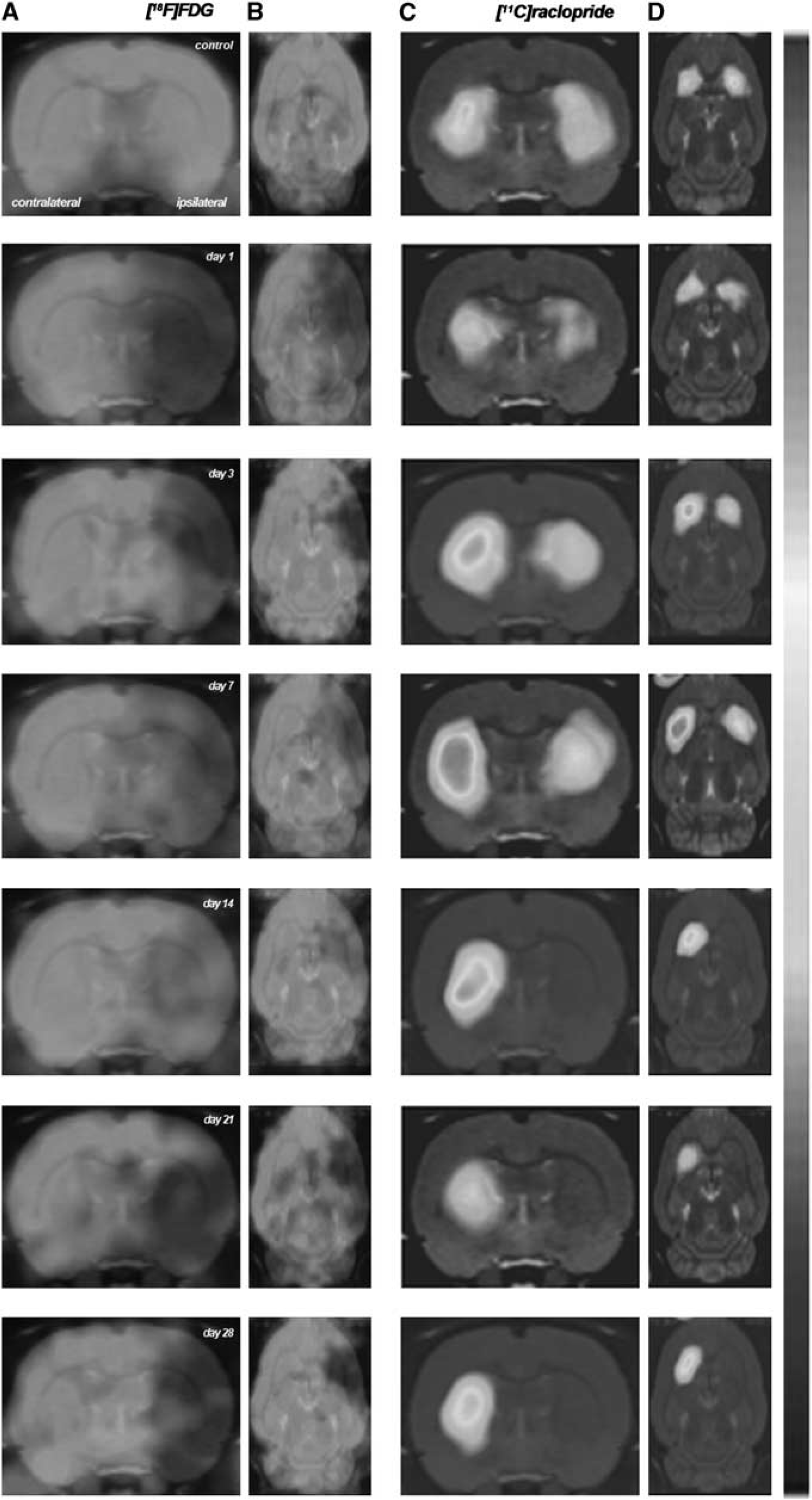
Serial images of [
18
F]fluorodeoxyglucose ([
18
F]FDG) and [
11
C]raclopride positron emission tomography (PET) at control, day 1, day 3, day 7, day 14, day 21, and day 28 after middle cerebral artery occlusion (MCAO). Normalized axial and coronal PET images of [
18
F]FDG (
Brain Damage Assessment After Cerebral Ischemia
The extent of brain damage after cerebral ischemia was assessed using T2W MRI at 1 day after ischemia onset. Hyperintensities of T2W images showed similar infarct extents as well as locations affected. All ischemic rats subjected to nuclear studies showed corticostriatal MRI alterations (mean ± s.d. = 290 ± 55 mm3).
[ 18 F]fluorodeoxyglucose Positron Emission Tomography After Middle Cerebral Artery Occlusion
The time course of cerebral glucose metabolism was evaluated using [ 18 F]FDG PET at 1, 3, 7, 14, 21, and 28 days after MCAO (Figure 2). The [ 18 F]FDG PET signals were significantly lower (–50%) in the cerebral territory of the occluded MCA than in the same region of control animals at day 1 after reperfusion. This was followed by a slight increase in metabolism to 25% of day 1 at days 7 to 14 that did not reach control values. Subsequently, [ 18 F]FDG PET signal decreased again from day 21 to 28 after MCAO (P < 0.05). In the contralateral hemisphere, the [ 18 F]FDG PET signal showed quasi-normal values after cerebral ischemia (Figure 2A).
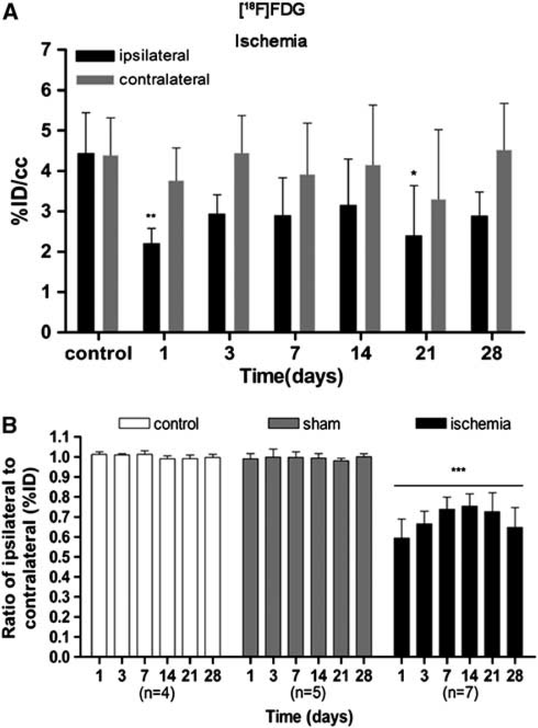
Time course of the progression of the [
18
F]fluorodeoxyglucose ([
18
F]FDG) positron emission tomography (PET) signal after cerebral ischemia. The percentage of injected dose per cubic centimeter of [
18
F]FDG (%ID/cc, mean ± s.d.) was quantified in two volumes of interest (VOIs) defined for each animal over the entire ipsilateral (containing the infarcted area) and the contralateral hemispheres. Rats were repeatedly examined by using PET at 1, 3, 7, 14, 21, and 28 days after ischemia or sham operation. Nonoperated rats were also repeatedly scanned at the same time points and were used as controls. The ipsilateral and contralateral PET values within the ischemic group are shown in (
The ratio of metabolic values in the ipsilateral (occluded MCA) over contralateral (nonoccluded MCA) showed a statistically significant decrease in the signal in the ischemic group in relation to control groups (nonoperated and sham-operated rats) (P < 0.001). The ratio of metabolism values varied from 0.6 at day 1 to 0.7 from days 7 to 14 after MCAO. Subsequently, [ 18 F]FDG PET ratios evidenced a progressive decline from days 21 to 28. The [ 18 F]FDG PET ratio values in control (non and sham-operated) animals showed a ratio of one over time (Figure 2B).
Time course of [ 11 C]raclopride Positron Emission Tomography after Middle Cerebral Artery Occlusion
The time course of striatal dopamine D2 receptor was evaluated using [ 11 C]raclopride PET at 1, 3, 7, 14, 21, and 28 days after MCAO (Figure 3A). The BPND of [ 11 C]raclopride showed quasi-normal values from day 1 to day 7 after reperfusion in the striatum occluded by the MCA. After 7 days, however, [ 11 C]raclopride PET signal decreased sharply from 75% at day 14 to ca. 80% at days 21 to 28 of the control values (P < 0.001). Non and sham-operated animals did not show any significant change over time (Figure 3A).

Time course of the progression of the [
11
C]raclopride positron emission tomography (PET) signal after cerebral ischemia. The binding potential (BP) nondisplaceable of [
11
C]raclopride (BPNP, mean ± s.d.) was quantified in two volumes of interest (VOIs) defined automatically for each animal over the entire ipsilateral (
Interestingly, changes in [ 11 C]raclopride PET signal were also observed in the contralateral, nonischemic area. Contralateral BP of [ 11 C]raclopride showed control values at 1 day after reperfusion, then PET signals rose steadily at day 3, and overshoot to ca. 40% of control values at 7 days after MCAO (P < 0.001). Subsequently, [ 11 C]raclopride signals showed a progressive decrease, reaching control values from day 14 to day 28 (Figure 3B).
Accordingly, the ratio of BPND values in the ipsilateral (occluded MCA) over contralateral (nonoccluded MCA) areas varied from 0.7 at days 1 and 3 to 0.6 at day 7 after MCAO. By day 14, [ 11 C]raclopride PET signal ratios decreased again from 0.2 to 0.1 at day 14 to day 21 (P < 0.001; Figure 3C). At these latter times, the [ 11 C]raclopride signal further decreased in the lesioned striatum in relation to the contralateral hemisphere.
Autoradiography
The time course of [ 3 H]raclopride binding tissue was performed with brain sections of rats killed at 1, 3, 7, 14, 21, and 28 days after the induction of cerebral ischemia, to verify the results obtained in vivo with PET. In the ipsilateral hemisphere, [ 3 H]raclopride binding showed quasi-normal values from day 1 to day 7 after MCAO. After day 14, [ 3 H]raclopride signal showed a fourfold signal decrease in relation to control (nonoperated) brains. The
[ 3 H]raclopride signal in the ischemic striatum considerably decreased at days 14, 21, and 28 with respect to control (P < 0.001; Figure 4A). In the contralateral striatum, [ 3 H]raclopride signal showed a nonsignificant increase from days 1 to 3 followed by the uppermost binding at day 7 (P < 0.001 with respect to control values; Figure 4B) and a return to quasi-normal values from days 21 to 28 after reperfusion.
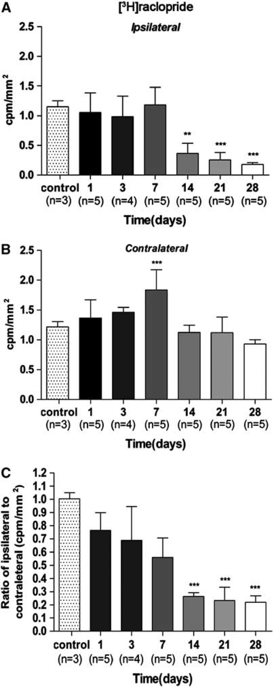
Time course of the progression of [
3
H]raclopride binding after ischemia. The [
3
H]raclopride binding (c.p.m./mm2, mean ± s.d.) was quantified in two volumes of interest (VOIs) defined manually for each animal over the entire ipsilateral (
As a consequence, the ratio of [ 11 H]raclopride signal ratio evidenced a trend of progressive decrease from day 1 to day 7 followed by a further decrease at day 14 and onwards. The ratio of [ 3 H]raclopride signal showed a significant decrease versus that in the controls at days 14, 21, and 28 after ischemia (P < 0.001; Figure 4C).
Time Course of Neurologic Score After Experimental Stroke
Ischemic animals showed the major neurologic impairment at 1 day after MCAO in relation to control animals. After day 3, rats showed a trend to a progressive functional recovery over time. The neurologic impairment showed significant increase versus that in the controls at day 1, 3, 7, 14, 21, and 28 (P < 0.001). After day 3, the score decreased at days 7 (P < 0.05), 14 (P < 0.01), 21, and 28 (P < 0.001) in relation to day 1 after ischemia (Figure 5).
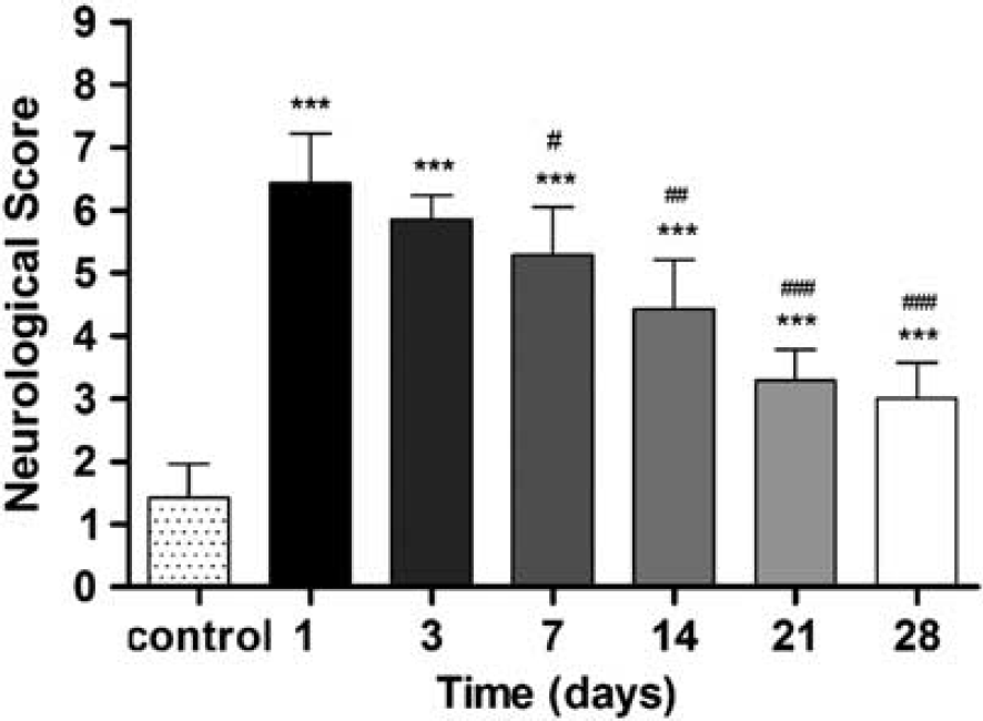
Neurologic outcome after cerebral ischemia. The neurologic score shows an improvement over time. Statistical analyses were performed by using Mann–Whitney U-tests. Statistically different from control: ***P < 0.001. Statistically different from day 1: #P < 0.05, ##P < 0.01, and ###P < 0.001.
Evolution of Brain Lesion After Experimental Stroke
At 1 day after MCAO, T2W MRI evidenced hyperintensities in the territory irrigated by the MCA (Figure 6A). Cortical and striatal MRI alterations coincided with the presence of histopathologic features at 24 hours after ischemia. Histopathologic outcome using H-E staining showed a massive necrosis in the area affected by the blood interruption (Figure 6B). The TTC staining showed infarction evolution (pale area) over the first week after reperfusion, evidencing the hemorrhagic transformation of brain infarction at day 7 after MCAO (Figure 6C). Immunohistochemistry for TH and neuron nuclear antigen (NeuN) illustrated neurons expressing TH in the striatum at control animals (Figure 6D). Surprisingly, in the territory irrigated by MCA, TH and NeuN showed a dramatic decrease at day 7 after ischemia onset. Despite these findings, in the contralateral striatum TH staining did not show an apparent change of the signal at 7 days after ischemia in relation to control rat brain (Figure 6E).

Brain lesion evolution after cerebral ischemia. magnetic resonance imaging (MRI) alterations in T2-weighted (T2W) signal (
DISCUSSION
Clinical observations using PET with [ 11 C]raclopride, a selective striatal dopamine D2 receptor antagonist, have provided more insight into the role that dopaminergic system has in the pathophysiology of major neurologic and psychiatric disorders. 28–31 The modulation of these receptors using pharmacological agents has also provided benefits on learning and motor formation memory in both the subacute state after clinical stroke and chronic period, when less therapeutic options are available.15,32 Despite these findings, PET imaging of dopaminergic receptor density using [ 11 C]raclopride has not been explored neither experimental nor clinical stroke to date. Because of this, we evaluated in parallel in vivo metabolic changes and dopamine receptor density using imaging procedures in combination with ex vivo autoradiography and immunohistochemistry, after transient MCAO in rats.
Glucose Metabolic Changes After Cerebral Ischemia
Positron emission tomography with FDG is a well-validated technique used for the assessment of glucose metabolism that is commonly used in clinical imaging for oncologic, cardiac, and neurologic applications. 33 The PET imaging with [ 18 F]FDG has been shown to be sensitive to metabolic changes that facilitate the detection of functional impairment after focal cerebral ischemia in rats. 34 The area irrigated by MCA showed a dramatic decrease in [ 18 F]FDG uptake at 1 day after 2-hour MCAO that ran parallel to the T2W-MRI hyperintensities observed at this same time point after ischemia. This reduction was followed by a slight recovery of the signal from day 3 to day 7, reaching maximum uptake values at day 14 after ischemia onset evidencing and increase in metabolic activity in the injured territory. Our results are in good perspective with those of Schroeter et al 35 who reported an increase in [ 18 F]FDG uptake 7 days after transient MCAO in rats that colocalized with the activation of neuroinflammatory processes. Thus, these results might suggest that the partial recovery from the baseline physiologic glucose consumption observed in the territory irrigated by MCA after cerebral ischemia maybe related to the neuroinflammation underlying ischemic stroke. Supporting all these findings, Martin et al 12 have also observed a long-term increase in cerebral blood flow and brain perfusion by using PET and Single-Photon Emission Computed Tomography imaging at 7 days after cerebral ischemia. Subsequently, [ 18 F]FDG signal decreased again at days 21 to 28 after reperfusion. The contralateral to the MCAO territory did not show significant differences in relation to control values over time. Accordingly, the ratio of [ 18 F]FDG signals in the ischemic to contralateral side varied from 0.6 at day 1 to 0.8 at day 14. Control and sham-operated animals did not show any difference in the [ 18 F]FDG uptake over time.
In Vivo and Ex Vivo Dopaminergic Changes After Cerebral Ischemia
Experimental studies have shown that dopaminergic neurotransmission is strongly affected by cerebral ischemia; 36 and the modulation of this neurotransmission system has stressed a recovery of movement after stroke in both animals 19 and human.32,37 Likewise, the striatal transplantation of embryogenic stem cells has shown an improvement of rat dopaminergic dysfunction after focal cerebral ischemia in rats. 38 Thus, since these findings suggest a key role of dopaminergic neurotransmission in neural plasticity and functional recovery after stroke, we sought to evaluate for the first time the striatal dopamine D2 receptor evolution using in vivo [ 11 C]raclopride after focal cerebral ischemia in rats.
In the ischemic territory, [ 11 C]raclopride PET showed a (1) nonsignificant decrease in BP at 1 day, (2) followed by a slight recovery, reaching pseudo-control values, and (3) a decrease again to roughly one-third of control values from day 14 to day 28 after ischemia. The [ 11 C]raclopride binding uptake from day 1 to day 7 may be because of the presence of nondegraded dopamine D2 receptors present in necrotic neurons, which showed a particular resistance to proteolytic processes after cerebral ischemia. These events run in parallel with a progressive [ 11 C]raclopride binding increase in the contralateral hemisphere to the lesion at 7 days after cerebral ischemia. Interestingly, the contralateral area to the MCAO showed a time-dependent increase in the BPND of [ 11 C]raclopride from day 1 to day 3, followed by a peak of [ 11 C]raclopride binding at day 7 after MCAO. This result was followed by a slight decrease from day 14 to day 28, reaching again control values. Therefore, these results evidenced an effect of cerebral ischemia over dopamine D2 receptors not only in the impaired tissue, but also in regions remote to the lesion. These results are in agreement with the dopamine D1 receptor increase observed in the contralateral side to the lesion at days 4 and 7 after focal cerebral ischemia in rats. 36 Accordingly, the ratio of the BPND in the lesion over the contralateral hemisphere varied from roughly 0.75 to 0.6 from day 1 to day 7 and followed by a reduction to 0.1 at days 21 to 28 after cerebral ischemia. Control and sham-operated animals did not show significant differences over the different time points, evidencing a minimum contribution of the stress caused by the surgical process underlying MCAO in the density of dopamine D2 receptors.
In vitro autoradiography using [ 3 H]raclopride confirmed the results obtained by in vivo PET [ 11 C]raclopride. In the injured territory, [ 3 H]raclopride binding remained unchanged until the day 7 and then dramatically decreased from day 14 to day 28 after ischemia. In the contralateral hemisphere, D2 receptor binding evidenced a time-dependent signal increase from day 1 to day 7. Accordingly, the ratio of ipsilateral to contralateral to the lesion using in vitro [ 3 H]raclopride showed practically identical results to those obtained by PET [ 11 C]raclopride.
Simplified reference-tissue model is a model that simplifies the experimental setting needed for full kinetic modeling at the cost of assuming certain conditions as follows: (1) the distribution volume is the same for the tissue of interest and the reference tissue and (2) the kinetics in the receptor-rich tissue of interest is such that it is difficult to distinguish between the specific and the free/nonspecific compartment. Therefore, it may seem unrealistic to assume such conditions after cerebral ischemia where the vascularization, neuron viability, and blood–brain barrier are hardly compromised. Nevertheless, the results obtained by in vitro autoradiography using [ 3 H]raclopride validated the use of simplified reference-tissue model to quantify BPND of [ 11 C]raclopride after experimental cerebral ischemia. These findings also showed that in vivo [ 11 C]raclopride binding after MCAO was not influenced by both hemorrhagic transformation that could introduce physical distorsions in PET measurements and long-term changes in brain perfusion, particularly in the remote regions to the infarction after experimental stroke as has been recently described. 12
Immunolabelling showed an evident decrease in TH, the enzyme responsible for catalyzing the conversion of L-tyronise to L-DOPA (a precursor for dopamine) in the injured territory 7 days after ischemia. This decrease evidenced the loss of terminal presynaptic dopaminergic terminals in the striatum. Furthermore, this result supported the posterior loss of postsynaptic dopamine neurons observed by the in vivo and ex vivo decline of raclopride binding. Additionally, the neuronal marker NeuN showed neuronal depletion at day 7 after MCAO showing that brain infarction affected both cortex and striatum. Taken together, these results show the loss of presynaptic and postsynaptic dopamine neuronal population in the rat striatum after 2 hour MCAO.
Likewise, the decrease in TH could be responsible for the decrease in dopamine levels which might as well be the reason for the nonsignificant increase in [ 11 C]raclopride PET binding observed in the infarcted striatum 7 days after ischemia.
Functional Recovery After Stroke
There is clinical evidence showing that optimal functional recovery might appear around 6 months after stroke depending on the effectiveness of neural plasticity and both lesion topography and size. 8 In the present study, ischemic animals showed the highest functional impairment at 24 hours after focal cerebral ischemia followed by a progressive functional recovery over time. At 4 weeks after stroke, animals experienced an optimal recovery of the neurologic function showing similar skills to that presented by control animals. Despite this, the precise mechanisms and biologic basis involved in the recovery after cerebral ischemia are still largely unknown. Clinical and preclinical studies have revealed a major contribution of the hemisphere homolog to the lesion in the compensations of impaired functions after stroke.9,39,40 Likewise, long-term increase in microglial activation and cerebral blood flow have been observed in remote regions to the infarction after MCAO in rats.7,12 Supporting these findings, we observed a significant increase in dopamine D2 receptor density in the contralateral striatum at 7 days after occlusion. Despite these changes, cerebral glucose metabolism did not experience significant variations in the territory homolog to the lesion over the entire study. Therefore, the fact that the increase in dopamine D2 receptor density is not accompanied by an increase in neuronal/astrocytic metabolism in the same region supports the specificity of dopaminergic changes after cerebral ischemia. Therefore, based on previous studies showing that modulation of dopaminergic systems using levodopa (1) improved sensorimotor function and (2) enhanced brain plasticity in human15,37 and also in rat brain after stroke, 14 it is tempting to hypothesize that dopamine D2 receptor might contribute to the recovery of brain function after cerebral ischemia.
Summary and Conclusions
In summary, we report here for the first time the PET imaging of dopaminergic system using [ 11 C]raclopride and its relationship with glucose metabolism and functional recovery after transient focal ischemia in rats. These results should contribute to better understand the role of dopaminergic neurotransmission on stroke evolution and to establish an effective clinical treatment targeted on the healthy hemisphere for the enhancement of the recovery of impaired functions and to provide patients with a better quality of life.
DISCLOSURE/CONFLICT OF INTEREST
The authors declare no conflict of interest.
Footnotes
ACKNOWLEDGEMENTS
The authors would like to thank M González and A Leukona for technical support in the radiosynthesis; A Arrieta and M Puigivila for technical assistance in the PET studies, and the Departamento de Educación, Universidades e Investigación del Gobierno Vasco (PO2011-3) and the Departamento de Industria, Comercio y Turismo Vasco for financial support.