




Abstract
INTRODUCTION
Neurotoxicity mediated by the activation of N-methyl-
In addition to glutamate, the NMDA receptor requires a coagonist at a second site. Although glycine was initially thought to be a ligand for this site, subsequent evidence has revealed that
In terms of cerebral ischemia, the deletion of serine racemase (SRR), which synthesizes
MATERIALS AND METHODS
Animals
All animal procedures were performed in accordance with The Animal Experimentation Guidelines of Keio University School of Medicine and were approved by the Laboratory Animal Care and Use Committee of Keio University. Experiments were performed in 6- to 8-week-old male SRR+/+ or SRR - / - mice (weight: 20 to 24 g). An SRR knockout (SRR - / -) mouse strain was produced as described previously. 12 The mice were obtained from house colonies and were backcrossed onto the C57BL/6 background to generate homozygous animals free of background effects on phenotypes. Wild-type (WT) siblings were used as WT controls.
Timed-pregnant Sprague-Dawley rats were purchased from Japan SLC, Inc. (Hamamatsu, Japan).
Chemicals
All chemicals and materials were obtained from the following sources: Dulbecco's modified Eagle's medium (DMEM) with or without glucose, penicillin, and streptomycin was obtained from Life Technologies (Grand Island, NY, USA); defined fetal bovine serum was obtained from HyClone Laboratories (Logan, UT, USA); Tissue-Tek O.C.T. Compound was obtained from Sakura Finetek Japan (Tokyo, Japan); an anti-SRR antibody was obtained from BD Biosciences (Franklin Lakes, NJ, USA); an antibody for Nissl was obtained from Millipore (Bedford, MA, USA); an antibody for glial fibrillary acidic protein (GFAP) was obtained from Dako (Glostrup, Denmark); secondary antibodies labeled with fluorescein or Texas-Red were obtained from Jackson Immunoresearch Laboratories (West Grove, PA, USA); and the derivatizing reagent 4-fluoro-7-nitro-2,1,3-benzoxadiazole (NBD-F) was obtained from Tokyo Chemical Industry (Tokyo, Japan). Water was purified using a Milli-Q gradient A 10 system (Millipore); Alamar Blue was obtained from Invitrogen Life Technologies (Carlsbad, CA, USA); the LDH assay kit was obtained from Wako Chemicals (Osaka, Japan); all other chemicals were obtained from Sigma (St Louis, MO, USA).
Preparation of Cultured Neurons and Astroglia
A primary culture of neurons was prepared from the cortex and striatum of fetal rats on embryonic day 16 as described previously.
13
Mechanically dissociated cells were counted, and viable cells (1.5 × 10⁶ cells/mL) without trypan blue were cultured in 12-well culture plates (0.8 mL/well, respectively) (Nalge Nunc., Rochester, NY, USA) coated with poly-L-lysine (5 μg/mL). The cells were cultured in a glucose-containing medium (final concentration, 12 mmol/L of
Experimental Protocol for D/L-Serine Release
To assess the effect of hypoxia on D/
Middle Cerebral Artery Occlusion
The procedures used for transient MCAO were identical to those previously described and are only summarized here.15–17 Mice were anesthetized with isoflurane (1.5% to 2%). A fiber optic probe was glued to the parietal bone and then connected to a laser Doppler flow meter (ALF 21R; Advance Co., Ltd., Tokyo, Japan) to monitor the cerebral blood flow (CBF). A heatblunted 6.0 monofilament surgical suture was inserted into the external carotid artery, advanced into the internal carotid artery, and wedged into the circle of Willis to obstruct the origin of the middle cerebral artery. The filament was left in place for 40 minutes and then withdrawn. Only animals that exhibited an 85% reduction in CBF during MCAO and in which the CBF had recovered by 80% after 10 minutes of reperfusion were included in the present study.16,18,19 This protocol for MCAO led to reliable infarcts comparable in size and distribution (cortex and striatum) to those reported by others.20,21 In this study, one mouse for the stroke volume experiment was excluded by the blood flow criteria. In all the mice, the rectal temperature was maintained at 37.0 ± 0.5 °C during surgery and during the recovery period until the animals regained consciousness.
Measurement of Infarct Volume
One day after ischemia, the mice were anesthetized and were perfused transcardially with 2% triphenyltetrazolium chloride. The brains were removed, frozen, and sectioned (thickness, 30 μm) using a cryostat. The brain sections were collected at 600-μm intervals, and the infarct volume was presented as a percentage relative to the entire right hemisphere volume.
Double-Label Immunohistochemistry
To identify cell types expressing SRR and 3-PGDH, mice were anesthetized with sodium pentobarbital (120 mg/kg) and perfused transcardially with 4% paraformaldehyde. The brains were then removed, cryoprotected in 20% sucrose phosphate buffer saline (PBS), frozen in Tissue-Tek O.C.T. Compound (Sakura Finetek Japan), sectioned through the parietal cortex, and stored at −80°C until use. Sections were rinsed in PBS (pH 7.4), permeabilized with 0.3% Triton X-100 for 10 minutes, and incubated at room temperature for 3 hours with a blocking solution containing appropriate normal serum (1:60; Vector Laboratories, Burlingame, CA, USA). Subsequently, the sections were incubated at 4°C for 12 to 72 hours in appropriate primary antibodies (anti-SRR antibody, 1:100, BD Biosciences; anti-3-PGDH, 1:300, a kind gift from Dr Shigeki Furuya; GFAP, 1:1,000, Dako) diluted in PBS containing blocking serum. Then, the sections were incubated at room temperature for 40 minutes in a mixture of the neuronal marker Nissl (1:100; Millipore) and/or species-specific secondary antibodies labeled with fluorescein or Texas-Red (Jackson Immunoresearch Laboratories). The sections were washed again and were mounted using ProLong Gold Antifade Reagent with DAPI (Invitrogen, Carlsbad, CA, USA). Images of double-labeled neocortex were sequentially acquired using a Zeiss LSM 510 confocal microscope (Carl Zeiss, Oberkochen, Germany).
Two-Dimensional High Performance Liquid Chromatography
The amino-acid analysis was conducted using a previously described protocol12,22,23 with a small modification. Brain tissue was freeze-dried for 24 hours using a freeze dryer and was homogenized in a 19.5-fold volume (w/v) of methanol. For the cell cultures, cultured media were mixed with a 10-fold volume (v/v) of methanol. These homogenates were then centrifuged at 20,400 g for 10 minutes, and the supernatants were spindried using Savant SpeedVac (Thermo Scientific, Waltham, MA, USA). Then, the dried residues were resuspended in 200 mmol/L sodium borate. After the addition of 40 mmol/L NBD-F (Tokyo Chemical Industry), the reaction mixture was incubated at 60°C for 2 minutes. The reaction of fluorescent derivatization was stopped by adding 2% trifluoroacetate. The NBD-Fderivatives were subjected to 2D-HPLC (NANOSPACE SI-2 series; Shiseido), separated into each amino acid using a reversed-phase column, a monolithic-ODS column (750mm × 0.53mm ID, 454 prepared in a fused silica capillary, provided by Shiseido), and further separated into enantiomers using an enantioselective column (Sumichiral OA-2500S; Sumika Chemical Analysis Service, Kyoto, Japan).12,22 The fluorescence intensity was detected at 530 nm with excitation at 470 nm.
Evaluation of Cell Damage Using Alamar Blue Reduction Activity and Lactate Dehydrogenase Release
Cellular damage was evaluated quantitatively using the amount of lactate dehydrogenase (LDH) released into the culture media and was confirmed using a colorimetric assay based on Alamar Blue (Invitrogen Life Technologies) reduction.
The LDH release assay, a standard cell death/viability assay reflecting cellular membrane integrity, was applied. Three hundred microliters out of 0.5 mL of culture medium was collected from a 12-well culture plate and was used for the LDH activity assay after 24 hours of hypoxia (1% O2). The remaining cells were used for the Alamar Blue assay, as described below. The LDH activities in the sampled medium were measured using an LDH assay kit (Wako Chemicals) based on the conversion of lactate and NAD+ into pyruvate and NADH catalyzed by LDH. The produced NADH was measured (340 nm), and the LDH concentrations in the samples were calculated using the LDH standard solution. Six wells of appropriate sister culture cells were treated with 0.5 mL of 1% Triton-X 100 in PBS and the average LDH activity was considered as the maximal LDH release. The released LDH activity from each well exposed to hypoxia (n = 6) was divided by the maximal LDH release and was considered as the percent of cell death in each well after 24 hours of exposure to hypoxia (1% O2).
Alamar Blue is a redox indicator that exhibits both fluorescence and colorimetric changes in response to metabolic activity and cellular viability.24,25 After the removal of nutrient medium from the 12-well culture plate, the cells were washed once with PBS, and 0.5 mL of DBSS containing 2 mmol/L glucose and Alamar Blue (10% final volume) was added. The cells were incubated for 60 minutes in a CO2 incubator (21% O2) for color development. The fluorescence level was measured at 0 and 60 minutes using a fluorescence microplate reader (Infinite F200 PRO; TECAN Japan, Kanagawa, Japan) with excitation at 535 nm and emission at 590 nm. As the fluorescent signals increased linearly for up to 60 minutes (data not shown), Alamar Blue reduction (cell viability) was expressed as the percent increase in the fluorescent signal at 60 minutes compared with that at 0 minute. Cell viability after 24 hours of exposure to hypoxia (1% O2) was evaluated using the percent of Alamar Blue reduction activity in the control cells (21% O2).
Western Blotting
The MCAO mice were anesthetized with diethylether and euthanized by the perfusion of PBS. Each brain was then dissected into the right/left parietal or temporal region. The central area of infarction was the right temporal region. Each tissue was homogenized in lysis buffer (50 mmol/L Tris-HCl, pH 7.4, 15 mmol/L NaCl, 20 mmol/L EDTA, 1% Triton X-100, protease inhibitor cocktail) and centrifuged at 20,400 g for 10 minutes. The protein concentration of the supernatant was then analyzed using a BCA assay (Thermo Scientific), and 20 μg of protein was mixed with the sample buffer (320 mmol/L Tris-HCl (pH 6.8), 3.6% SDS, 18% glycerol, and 15% β- mercaptoethanol). The denatured samples were subjected to SDS-PAGE and transferred onto a PVDF membrane. Then, the membrane was immunoblotted with a mouse monoclonal antibody to SRR (BD Transduction Laboratories, Franklin Lake, NJ, USA), a rabbit monoclonal antibody to GAPDH (Cell Signaling Technology, Danvers, MA, USA), and a rabbit polyclonal antibody to 3-PGDH (a kind gift from Dr Shigeki Furuya).
In Vivo D-Serine Production Assay
The ischemic core region of the brain was homogenized in 2 mL of in vivo assay buffer (20 mmol/L Tris-HCl, 4 mmol/L MgCl2, 0.5 mmol/L sodium benzoate, 0.1% Triton X-100) and the debris was removed by centrifugation at 20,400 g for 10 minutes. Then, the small molecules were removed from the homogenate using the Amicon Ultra-4 centrifugal filter device 3000 NMWL (Merck KGaA, Darmstadt, Germany), and the homogenate was concentrated. The protein concentration in the homogenate was measured using a BCA assay, and 600 μg of protein in 75 μL of the assay buffer was mixed with 5 μL of 10 mmol/L ATP'S (Wako, Tokyo, Japan). Then, 20 μL of 50 mmol/L
Statistical Analysis
Data are presented as the mean ± s.e.m. Comparisons between two groups were statistically evaluated using the Student's t-test. Multiple comparisons were evaluated using an analysis of variance (ANOVA) followed by the Newman-Keuls multiple comparison test. Differences were considered as significant at P<0.05.
RESULTS
Serine Racemase - / - Mice have Smaller Infarcts after Middle Cerebral Artery Occlusion
First, we examined the effects of SRR on brain injury at 24 hours after transient MCAO. The infarct volumes after 40 minutes of MCAO were 32.5% and 19.3% of the whole hemisphere in WT and SRR - / - mice, respectively (Figure 1A). The infarct volume in SRR - / - mice was significantly smaller than that in WT mice (Figures 1A and 1C). The reduction in the infarct volume could not be attributed to the differences in the degree of ischemia because the CBF reduction in the center of the ischemic territory was comparable in the WT and SRR - / - mice (Figure 1B).
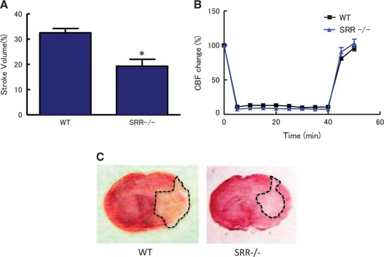
Reduced infarct volumes in serine racemase (SRR) - / - mice. (
Temporal Profile of D-/L-Serine Contents after Middle Cerebral Artery Occlusion
Next, we measured the contents of
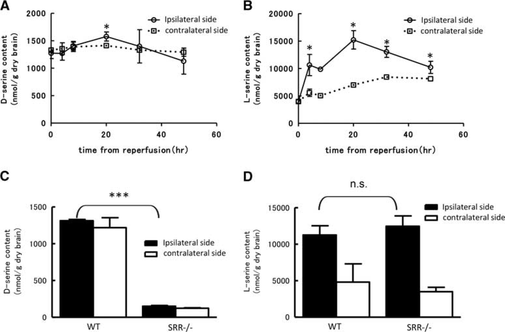
Temporal profile of
Determination of D-/L-Serine Release from Cultured Cells during Experimental Ischemia
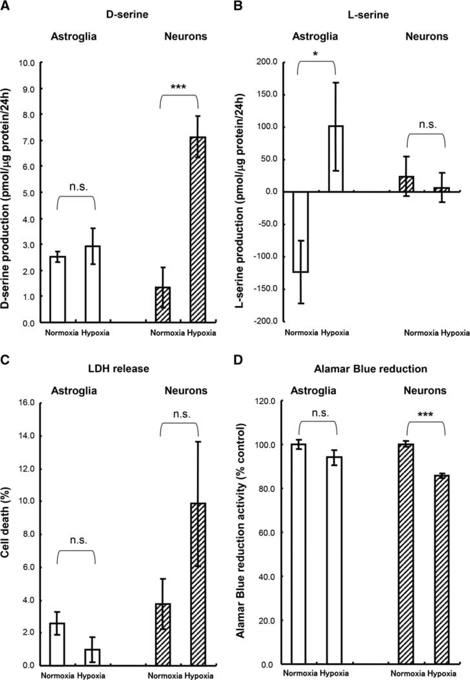
Next, we used cultured cells to determine which cell type can be ascribed to the increase in
Expression of Serine Racemase and 3-Phosphoglycerate Dehydrogenase and Distribution of D-/L-Serine in Tissues after Cerebral Ischemia
Next, we tried to investigate the cellular localization of key enzymes for

Cellular localization of serine racemase (SRR) and 3-phosphoglycerate dehydrogenase (3-PGDH) in the brain after middle cerebral artery occlusion (MCAO). The squares in the ipsilateral and contralateral cortex show the lesion where the pictures were taken (
Western Blots for Serine Racemase and 3-Phosphoglycerate Dehydrogenase after Cerebral Ischemia and
We also attempted to quantify the expressions of SRR and 3-PGDH after cerebral ischemia using a western blot analysis. Samples were separately collected from the temporal region (peri-infarct area) and the parietal region (ischemic core) at 20 hours after reperfusion (Figure 5A). In the peri-infarct lesion tissue, the expression of SRR was almost the same as that in the corresponding area in the contralateral hemisphere; however, the expression was markedly decreased in the ischemic core (Figure 5B). Although immunohistochemistry showed the upregulation of SRR in a small lesion of peri-infarct area, the expression of SRR was not increased overall. The 3-PGDH expression level was slightly decreased in the ischemic core; in the peri-ischemic area, the level was almost the same as that on the contralateral side (Figure 5B). Furthermore, the
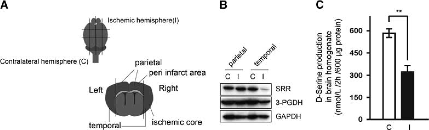
Expressions of serine racemase (SRR) and 3-phosphoglycerate dehydrogenase (3-PGDH) in the peri-infarct area and ischemic core. (
DISCUSSION
The present study showed that SRR - / - mice have a smaller infarct volume than WT mice. In addition, the temporal profiles of D-/
Serine racemase - / - mice were previously reported to have a smaller infarct than WT mice after MCAO.
11
Neuroprotection in SRR - / - mice is expected to be due to a reduction in
The reduction in
Our findings are consistent with a recent report showing that SRR exists in neurons and that 3-PGDH is located in astrocytes.
29
On the basis of these findings, it could be hypothesized that
Cultured neuronal cells that had been exposed to hypoxia (1% O2) for 24 hours were somewhat unhealthy but were not damaged severely, since their axons and dendrites were thinner than those of the control cells but were not torn or cleaved. In fact, LDH release, which reflects membrane lysis and integrity, was not significantly elevated after 24 hours of hypoxic insult. Therefore, an elevation in
The 3-PGDH expression level was not increased overall in the ischemic hemisphere in our western blot analysis. The increase in the
The function of
Footnotes
The authors declare no conflict of interest.
ACKNOWLEDGMENTS
The authors thank Shiseido Co., Ltd., for essential technical support in performing the 2D-HPLC experiments. This work was supported by JSPS KAKENHI Grant Number 26461279.

References
Supplementary Material
Please find the following supplemental material available below.
For Open Access articles published under a Creative Commons License, all supplemental material carries the same license as the article it is associated with.
For non-Open Access articles published, all supplemental material carries a non-exclusive license, and permission requests for re-use of supplemental material or any part of supplemental material shall be sent directly to the copyright owner as specified in the copyright notice associated with the article.
