
Abstract
Abstract
We suggest a straightforward procedure for biofabrication and initial characterization of tissue spheroids with optimal controllable parameters prepared from four different cell types using nonadhesive technology. Applying different immortalized and primary cells, namely HEK293, primary human fibroblasts (HF), primary sheep chondrocytes, and primary sheep osteoblasts, we have demonstrated the reproducibility and scalability of spheroid generation, the strong dependency of ultimate spheroid diameter on initial cell seeding density, and cell type. In addition, the spheroid viability is governed by cell derivation. In this study, we suggest a decision procedure to apply for any cell type one starts to work with to prepare a new type of tissue spheroids with predictable controllable optimal features suitable for high-quality standards in biofabrication and drug discovery.
Introduction
T
Various techniques for spheroid production exist. Classical one is a hanging drop method. 5 In this approach cells are placed in hanging drop culture and incubated under physiological conditions until 3D structures are formed. When just developed the method did not require any special equipment or reagents and was suitable for production of tissue spheroids in low scale. Now adapted for a robotic production, this technique produces spheroids with a considerable variation in diameter, with satellites, which is objectionable for bioprinting and drug discovery.
Nonadhesive hydrogel molding technology and production of spheroids in nonadhesive microplates have a great potential for robotization and automation and generate spheroids that are both consistent in size and perfectly size-controllable. In these techniques cells fall to the bottom of wells because of gravity, nonadhesive hydrogel disables the attachment to the bottom, and cell-to-cell interactions drive the formation of a single spheroid in each well.
The latest techniques such as microfluidic chips, acoustic levitation, and magnetic levitation achieved better efficiency and the easier spheroid manipulations, but they require complex specific equipment such as magnetic levitation equipment, instrumentation for acoustic wave generation, and microfluidic devices which aren't upgraded well for the high-scale production.6,7
Tissue spheroids can be prepared from different types of cells, primary and immortalized ones, from benign cells, cancer cell lines, primary tumor cells, and cancer tissues depending on further application of produced structures. Tissue spheroids imitate the architectural and functional characteristics of native tissue. Cardiomyocyte spheroids beat with heart-like rhythm, hepatocyte spheroids have liver-like functionality, chondrospheres have high-hypoxia tolerance and show collagen II and aggrecan production, and human endothelial cells vascularize fibroblast microtissues.8–11
Until now the comprehensive and generally accepted standardized protocol for quantitative characterization of tissue spheroids biofabricated from different cell types is absent. In this study, we proposed a protocol for assessment of tissue spheroids and tested it on four cell types: HEK293, primary human fibroblasts, primary sheep chondrocytes, and primary sheep osteoblasts. The biofabricated tissue spheroids differ in diameter, roundness, viability, and surface characteristics depending on the cell types, as well as initial cell seeding density. The used protocol is necessary and sufficient for initial step of tissue spheroid characterization.
Materials and Methods
Nonadhesive hydrogel and cell medium
Agarose was purchased from Helicon (Russian Federation, Cat No: Am-0710-0.1). Dulbecco's modified Eagle's medium (DMEM, Cat No: 12491-015), fetal bovine serum (FBS, Cat No: 16000-044), Dulbecco's phosphate-buffered saline (PBS, Cat No: 18912-014), antibiotic–antimycotic, and trypsin/ethylenediaminetetraacetic acid (EDTA) (Cat No: 15240-062) were obtained from Gibco (USA). Versen (Cat No: R080) and L-glutamine (Cat No: F032) were obtained from Paneco (Russian Federation). Glutaraldehyde (Cat No: G5882) was obtained from Sigma-Aldrich (USA). CellTox Green Kit (Cat No: G8742) was purchased from Promega (USA).
Cell culture
Human fibroblasts (HF) were obtained from Lonza (Cat No: CC-2511). HEK293 cell line expressing red fluorescent protein was purchased from Evrogen (Russian Federation). Primary sheep osteoblasts and primary sheep chondrocytes are a generous gift from Dr. N.P. Omelianenko (Central Research Institute of Traumatology and Orthopaedics of N.N. Priorov, Moscow).
Cells were grown in DMEM (Gibco, Cat No: 12491-015) containing 10% FBS (Gibco, Cat No: 16000-044), supplemented with antibiotic/antimycotic (Gibco, Cat No: 15240-062) and 1 mM L-glutamine (Paneco, Cat No: F032). The cells were incubated at 37°C in a humidified atmosphere with 5% carbon dioxide (CO2) and routinely split at 85–95% confluence. Cell transfer and preparation of single-cell suspensions were performed using mild enzymatic dissociation with a 0.25% trypsin/0.53 mM EDTA solution (Gibco, Cat No: 25200-114). Cells were free of mycoplasmal contamination as verified using Hoechst 33258 (Sigma, Cat No: 861405) staining protocol.
Formation of tissue spheroids using MicroTissues 3D Petri dishes
The tissue spheroids were formed using MicroTissues 3D Petri dish micromolds (Sigma Aldrich, Cat Nos: Z764000-6EA, 256 circular wells 400 × 800 μm and Z764019-6EA, 81 circular wells 800 × 800 μm) according to the manufacturing protocol. Briefly, 2% agarose solution in PBS was prepared using microwave oven. Then 450 μL of molten agarose were pipetted into 12-series micromolds for casting MicroTissues 3D Petri dishes. After the agarose had gelled (∼4 min), the micromolds were carefully flexed to remove MicroTissues 3D Petri dishes. To equilibrate MicroTissues 3D Petri dishes, cell culture medium was added (2.5 mL/well for 12-well plate) and incubated for 20 min or longer. Monolayer cells with 95% confluence were rinsed by Versen's solution (Paneco, Cat No: R080), harvested from the culture flasks by 0.25% trypsin/0.53 mM EDTA (Gibco, Cat No: 25200-114), and then suspended in cell culture medium. The concentrations of the HEK293 and HF cells were 2.6 × 10⁶, 1.3 × 10⁶, 5.7 × 10⁵ (for HEK293) or 6.7 × 10⁵ (for HF), 1.7 × 10⁵, and 2 × 10⁴ per milliliter. The concentrations of chondrocytes and osteoblasts were 1.2 × 10⁷, 6.8 × 10⁶, 3.4 × 10⁶, 1.4 × 10⁶, and 4.3 × 10⁵ per milliliter. Excessive culture medium was removed carefully, and 190 μL of cell suspensions were seeded into MicroTissues 3D Petri dishes. For example, when 2.6 × 10⁶ cells per milliliter suspension was used for spheroid formation, 5 × 10⁵ cells got into each MicroTissues 3D Petri dish, containing 256 wells, which resulted in a final concentration of 2000 cells per well/spheroid. Each spheroid is formed in individual well of MicroTissues 3D Petri dish. Forty minutes later an additional culture medium was added into each well of 12-well plate from the outside of MicroTissues 3D Petri dishes. The 12-well plate containing 12 MicroTissues 3D Petri dishes was incubated at 37°C in a humidified atmosphere with 5% CO2 for 9 days.
Formation of tissue spheroids using Corning spheroid microplates
The tissue spheroids were formed using Corning spheroid microplates (Corning, Cat No: 4520) according to the manufacturing protocol. Briefly, monolayer cells with 95% confluence were rinsed by Versen's solution (Paneco, Cat No: R080), harvested from the culture flasks by 0.25% trypsin/0.53 mM EDTA (Gibco, Cat No: 25200-114), and then suspended in cell culture medium. The concentrations of the cells were 2 × 10⁴, 1 × 10⁴, 5 × 10³, and 2.5 × 10³ per milliliter. Hundred microliters of cell suspensions were dispensed to the wells of Corning spheroid microplates. Corning spheroid microplates were incubated at 37°C in a humidified atmosphere with 5% CO2 for 9 days.
Determination of spheroid growth kinetics
Cells were seeded at different concentrations and cultured for 9 days, as described previously. To examine the spheroid growth over time, 80 spheroids for each cell seeding concentration were captured every day using light microscopy (Nikon Eclipse Ti-E, Japan). Spheroid diameters and roundness were measured using ImageJ 1.48v software (NIH, Bethesda, MD). Briefly, all original grayscale images were converted to simplified threshold images under the same converting condition, and the edges of the spheroids were automatically detected. Feret's diameters of the detected spheroid edges were measured initially as pixels and converted to micrometers by comparing to a reference length. Roundness is ImageJ 1.48v shape descriptor and was calculated as 4*area/(π*major axis2).12,13 The effect of cell type and initial cell concentration on spheroid diameter and roundness was analyzed using GraphPad Prism software (GraphPad Software, Inc., La Jolla, CA).
Estimation of tissue spheroid viability
The viability of tissue spheroids was assessed using the CellTox Green Kit (Promega, Cat No: G8742) according to the manufacturing protocol. Briefly, the cell suspensions for each cell type were prepared. CellTox Green Dye was mixed with a part of cell suspensions in a ratio 1:500. The CellTox Green Dye/cells mix and the unlabeled cell suspensions were seeded to the Corning spheroid microplates (100 μL per well). Four microliter of lysis solution were added into eight wells for determination of 0% viability signal. In 24 h tissue spheroids for all cell types were formed. Tissue spheroids labeled with CellTox Green Dye were used for determination of viability during 4 days in culture. The manufacturer guarantees stability of the fluorescent dye for 72 h, but we did not observe the decrease of the signal in the control wells (0% viability) with a highest possible assay signal over the 96 h. On the fifth day after biofabrication, eight unlabeled tissue spheroids of each cell type were treated with CellTox Green Dye and used for determination of viability during next 4 days in culture. Fluorescence was measured during 8 days in culture using VICTOR X3 Multilabel Plate Reader (PerkinElmer, USA). Tissue spheroid viability plots were analyzed using GraphPad Prism software (GraphPad Software, Inc.).
Results
Formation of uniform-sized spheroids using MicroTissues 3D Petri dishes and Corning spheroid microplates
Tissue spheroids have been generated by two alternative methods: using MicroTissues 3D Petri dishes (Sigma), agarose scaffolds with highly-ordered wells (Fig. 1A, B), and using Corning Spheroid Microplates, 96-well plates with nonadhesive coating (Fig. 1C, D). In both methods the nonadhesive surfaces prevented cell adhesion, the bottom of single wells acted as points of cell–cell adhesion, interaction for self-assembly, and subsequent spheroid formation. We have challenged four different cell cultures for their ability to form spheroids applying nonadhesive technology. It was shown that all cell types, immortalized HEK293, primary human fibroblasts, primary sheep chondrocytes, and primary sheep osteoblasts, form tissue spheroids within 24 h of incubation (Fig. 2). To demonstrate reproducibility and scalability of spheroid formation by these methods, the dependency of spheroid diameter on initial cell seeding density was examined. As shown in Figure 2, regardless of the method, spheroids of a homogenous size were reproducibly obtained. Diameters of spheroids increased with the increase of cell seeding concentration.
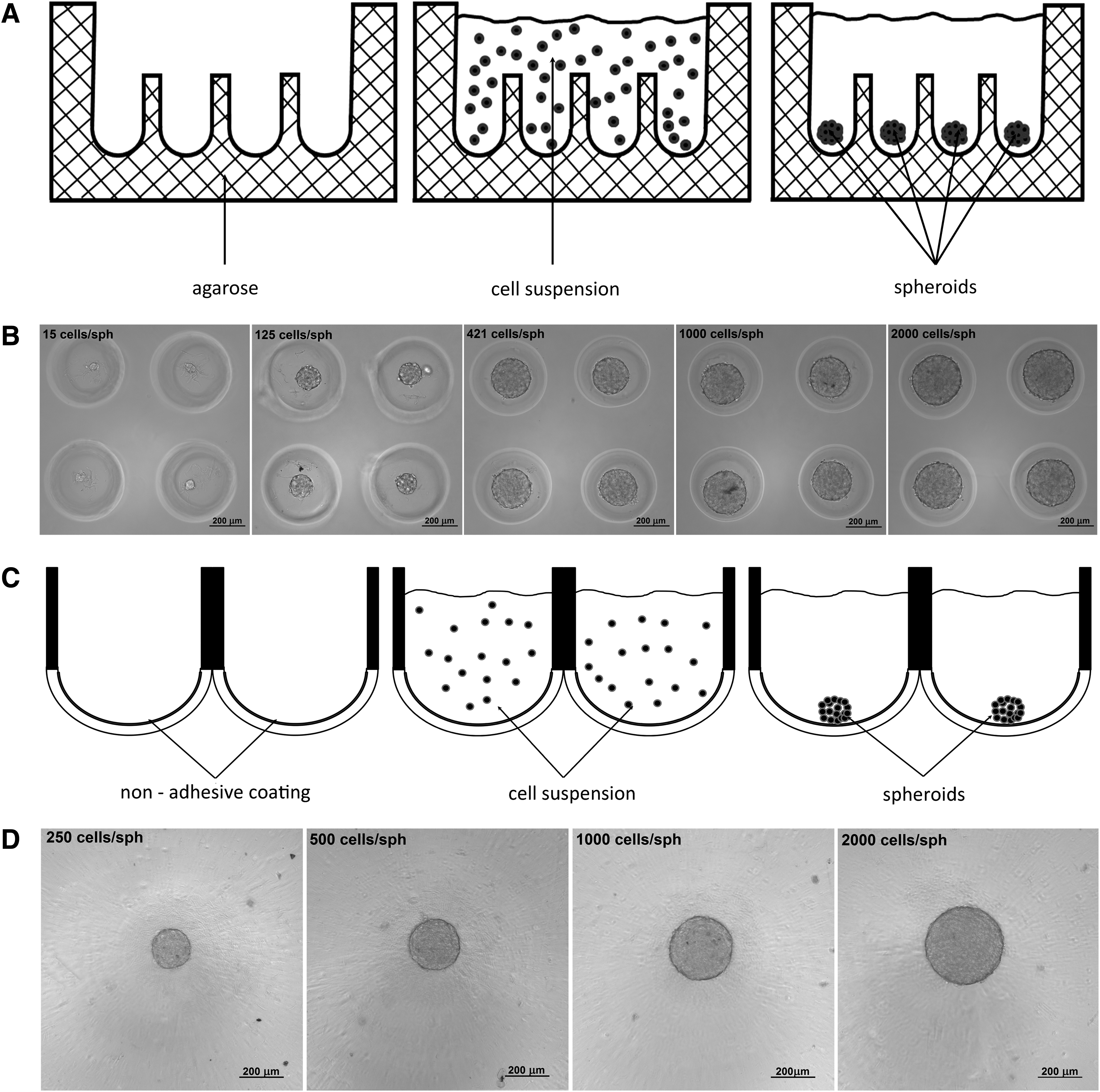
Tissue spheroid biofabrication using
The ratio between initial cell seeding density and resulted diameter of tissue spheroids biofabricated from HEK293 cells, primary HF, primary chondrocytes, and osteoblasts using MicroTissues 3D Petri dishes
Spheroid growth pattern
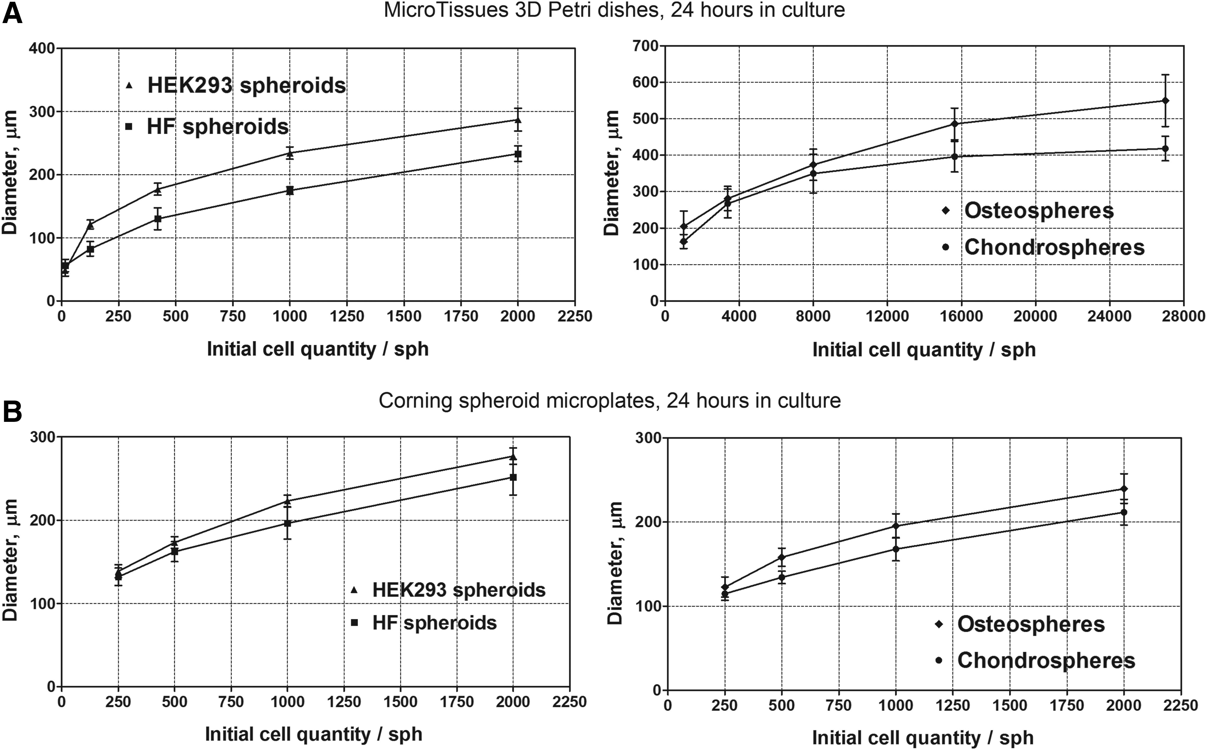
To monitor the growth of tissue spheroids with different initial cell seeding density the changes in spheroid diameter and roundness were investigated (Fig. 3 and 4).

The kinetics of spheroid diameter change.
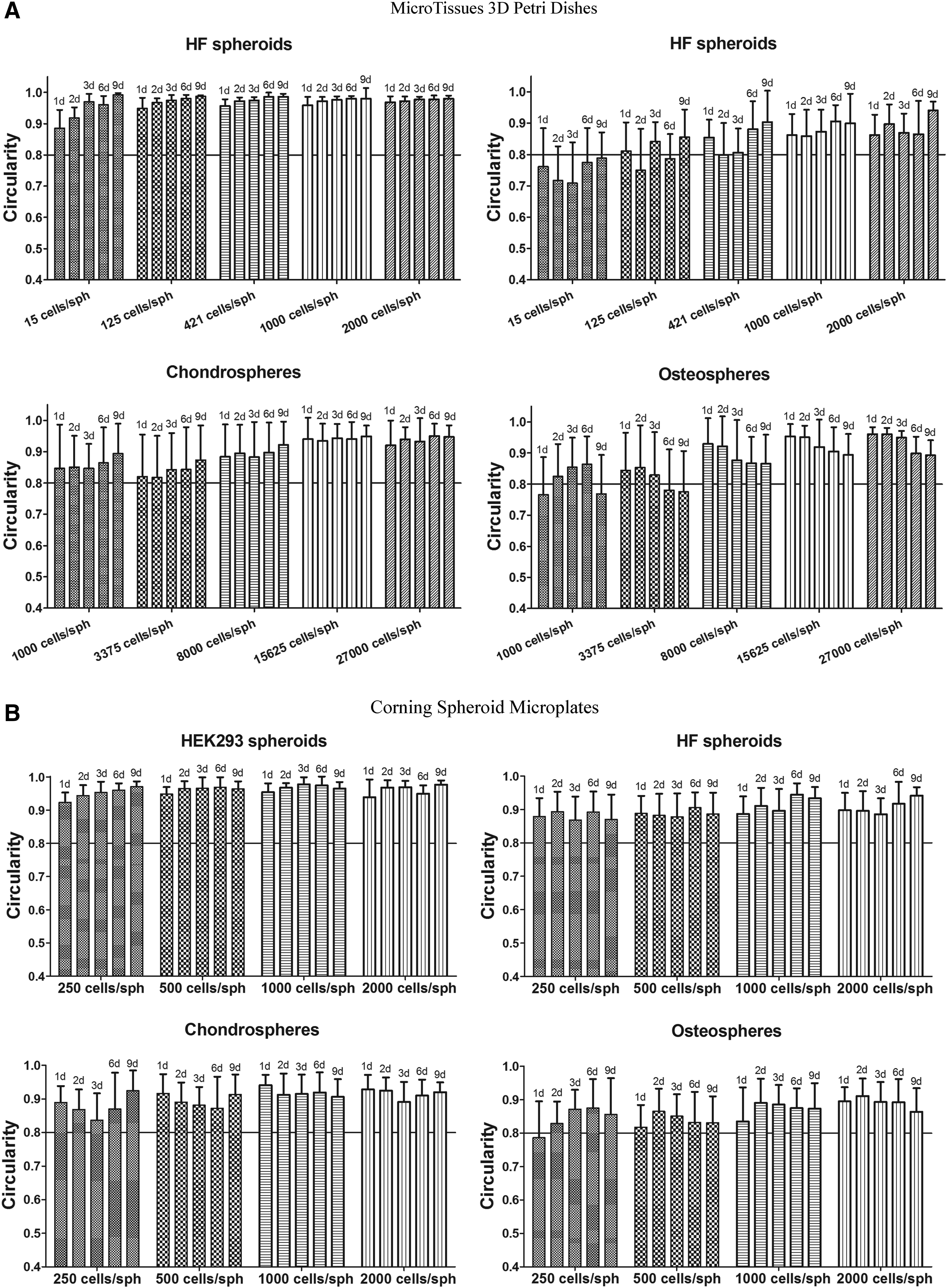
The kinetics of spheroid roundness change.
HEK293 spheroids
HEK293 cells in MicroTissues 3D Petri dishes were tested in concentrations recommended by manufacturer for catalog numbers 12-256: 15, 125, 421, 1000, and 2000 cells per spheroid. HEK293 cells gradually proliferated within tissue spheroids resulting in permanent increase of spheroid diameters. When cultured in MicroTissues 3D Petri dishes, diameter of spheroids increased sequentially and reached plateau, because the largest possible spheroid diameter was determined by the size of the microwell, 400 μm. The rate of diameter increase depended on the initial seeding concentration. The growth rate for smaller spheroids was higher compared to larger ones (Fig. 3A). When cultured in Corning spheroid microplates, HEK 293 cells were tested in concentrations 250, 500, 1000, and 2000 cells per spheroid. Spheroid diameter increased permanently and sequentially up to 1000 μm at ninth day in culture, because no well diameter limitation existed in these conditions. The growth rate for spheroids was similar regardless of the initial seeding concentration (Fig. 3B). The roundness of HEK293 spheroids was quite high (>0.9) irrespective of the initial seeding concentration and biofabrication method. In the case of HEK293 spheroids with very low initial concentration of 15 cells per spheroid, the increase of roundness was observed during the first 3 days (Fig. 4A). The roundness of HEK293 spheroids with the other initial cell densities fluctuated slightly without noticeable tendency to decrease or increase within the time course (Fig. 4A, B).
Chondrospheres
Chondrocytes in MicroTissues 3D Petri dishes were tested in concentrations recommended by manufacturer for catalog numbers 12-81: 1000, 3375, 8000, 15625, and 27000 cells per spheroid. When cultured in MicroTissues 3D Petri dishes, the chondrosphere diameter decreased sequentially irrespective of the initial seeding density from the first day, although well diameter limitation was 800 μm in large microwells. The maximal diameter for highest initial concentration did not exceed 418 μm at first day in culture. When cultured in Corning spheroid microplates, low initial cell concentrations from 250 to 2000 cells per spheroid were tested. The growth of smaller spheroids was observed, whereas the diameter of larger ones actually remained constant. The maximal spheroid diameter did not exceed 215 μm, although diameter limitation for Corning spheroid microplate wells was 2.82 mm. The roundness of all spheroids was high (>0.8). However, at the same initial seeding density (1000 cells per spheroid), the chondrospheres cultured in Corning spheroid microplates had higher roundness compared to ones cultured in MicroTissues 3D Petri dishes (Fig. 4A, B).
HF spheroids and osteospheres
Human primary fibroblasts and sheep primary osteoblasts formed tissue spheroids with the similar growth pattern. For osteospheres the spheroid diameter decreased sequentially from the first day in culture and reached plateau at sixth day for all spheroids regardless of the initial seeding density and biofabrication method (Fig. 3A, B). The reduction rate for osteospheres was higher compared to fibrospheres (Fig. 3A, B). The roundness of HF spheroids and osteospheres cultured in Corning spheroid microplates was high (>0.8) and almost invariable. For osteospheres cultured in MicroTissues 3D Petri dishes, the tendency to roundness reduction by ninth day in culture was observed. The roundness of HF spheroids cultured in MicroTissues 3D Petri dishes changed randomly within the limits of standard deviation.
The viability of tissue spheroids
The viability of tissue spheroids was analyzed using the CellTox Green Kit. The assay system uses asymmetric cyanine dye that is excluded from viable cells but stains the DNA from dead cells. Tissue spheroids were biofabricated from 4 different cell types using Corning spheroid microplates. The average diameters of spheroids were as follows: 204 ± 10 μm (HEK293 spheroids), 251 ± 24 μm (spheroids from HF), 212 ± 15 μm (chondrospheres), and 244 ± 17 μm (osteospheres). The oxygen and nutrient diffusion limit in tissues is around 100–150 μm, so the diameter of tissue spheroids of 200–300 μm is optimal. 14 The time-dependent viability of spheroids was tested. Figure 5 showed that the viability of cells in chondrospheres, HEK293 spheroids, and osteospheres was ≥80% during 7 days (Fig. 5). HF spheroids were less viable. The percentage of living cells was about 70% at fourth day of culture and then dramatically decreased to 50% at day 7.
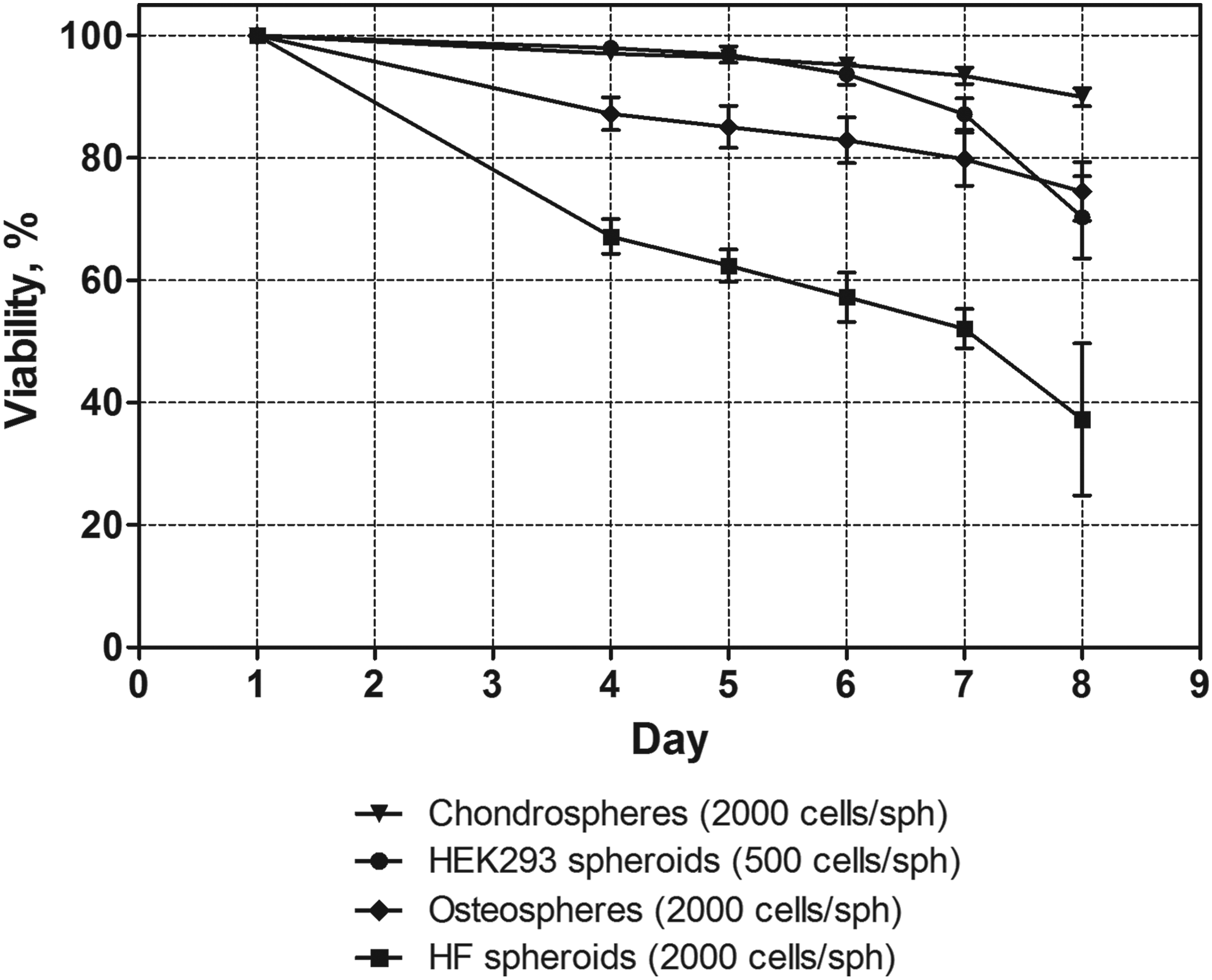
Viability of tissue spheroids biofabricated from HEK293 cells, primary HF, primary chondrocytes, and osteoblasts using spheroid microplates. Data represent the mean ± SD (n = 8).
Discussion
The development of reproducible optimal protocol for scalable and standardized biofabrication of tissue spheroids is necessary requisition for their application in drug discovery and in 3D bioprinting. To accomplish this goal we have tested two different methods for scalable tissue spheroid biofabrication applying four different cell types. In both cases to initiate biofabrication of tissue spheroids nonadhesive substrates have been used. The resulted tissue phenotype is an outcome of competition between cell-to-substrate and cell-to-cell interactions. 15 In case of using nonadhesive substrates the tissue spheroids are usually formed as a direct result of weak cell-to-substrate interactions and relatively strong cell-to-cell contacts. The reproducible scalable biofabrication of tissue spheroids was demonstrated by both methods applied. However, application of MicroTissues 3D Petri dishes-based method involves the cross talk between individual wells which is definitively affecting the accuracy of the initial cell density per well and will result in increased variation in tissue spheroid diameter. The classic hanging drop technology for biofabrication of tissue spheroids has been already automated by companies InSphero (Switzerland) and 3D Biomatrix (USA). Attempts to automate tissue spheroid biofabrication technology based on using micromolded nonadhesive agarose hydrogel have been also reported. 16 However, MicroTissues 3D Petri dishes-based technology does not guarantee a minimization of variation in tissue spheroid diameter or desirable level of standardization for tissue spheroid biofabrication technology. Moreover, the harvesting of tissue spheroids from MicroTissues 3D Petri dishes is still challenging. Thus, the application of standard Corning spheroid microplates with nonadhesive coating is more suitable for automated robotic scalable biofabrication of tissue spheroids with standard diameter and shape. In addition, this technique enables more efficient harvesting of biofabricated tissue spheroids.
The other important accomplishment in development of desirable standard and reproducible protocol of scalable tissue spheroid biofabrication is the demonstration that initial cell density controls the resulted diameter of tissue spheroids. It was shown that the correlation between initial cell density and resulted diameter of tissue spheroids is not linear but cell type dependent. For example, epithelial HEK293 cells possessed stable and prolonged growth within spheroids meanwhile proliferation of other cell types almost completely discontinued after spheroid formation. To generate tissue spheroids with desirable predictable diameter one has to perform the preliminary analysis of correlation between initial cell density and resulted diameter of tissue spheroids for every new cell type applied.
The shape of tissue spheroids is another critical parameter of their standardization. The ideal tissue spheroids must look like a ball or sphere because sphere is the most optimal shape for processing during tissue spheroid-based bioprinting.1,17 Our data demonstrate that biofabrication of standardized tissue spheroids which look like spheres is an achievable goal. However, shape of tissue spheroids is both cell type and time dependent.
The optimal diameter of tissue spheroids is closely related to their viability. Tissue spheroids are living structures, and cells forming the tissue spheroids undergo both proliferation and apoptosis, which can be manifested in changes of tissue spheroid diameter within time course. In this context it has been very important to estimate diameter of tissue spheroids as a function of time.
The viability is a critical issue in the usage of tissue spheroids in terms of in vitro tissue model, as well as building blocks for bioprinting. It was demonstrated that viability of tissue spheroid gradually reduced with time. Moreover, it is also cell type dependent. Some types of biofabricated tissue spheroids, for example, chondrospheres, maintain their viability quite long, whereas tissue spheroids biofabricated from fibroblasts reduce their viability much faster. Thus, it is preferable to use tissue spheroids for bioprinting or preclinical drug analysis during first 3–4 days.
The used protocol for systematic characterization and analysis of biofabricated tissue spheroid with defined quantitative parameters represents a practical approach for optimization of scalable biofabrication of tissue spheroids from any cell types with predictable and controllable, desirable standardized properties. Proposed protocol includes following necessary criteria: (1) estimation of correlation of tissue spheroid size with initial cell seeding density, (2) estimation of diameter and roundness as a function of time, (3) estimation of viability as a function of time, and (4) estimation of diameter and viability as a function of cell type. We suppose that described protocol could be used for development of automated image analysis systems of tissue spheroids.
Conclusion
In this study, we report a protocol to apply for any cell line one starts to work with to prepare a new type of tissue spheroids with predictable controllable optimal features. For scalable and standardized robotic spheroid production we suggest nonadhesive technology applying coated microplates.
Footnotes
Acknowledgment
This work was supported by grant number 15-15-00173 from February 6, 2015 of Russian Science Foundation.
Author Disclosure Statement
No competing financial interests exist.