
Abstract
Abstract
The biocompatibility of photopolymers in additive manufacturing (AM) often referred to as 3D printing (3DP) is an issue of concern due to, among other things, the unique parameters of the manufacturing process, which can influence the physical, chemical, and biological properties of AM-produced devices. The quality of AM-produced devices may consequently vary when identical parts are built using different 3D printers or even when the same 3D printer, parameters, process steps, and materials are used. In this novel study, representative materials built with stereolithography and material jetting processes were subjected to biological evaluation using the Organization for Economic Cooperation and Development (OECD) fish embryo test designed to determine acute toxicity of chemicals on embryonic stages of fish. The study demonstrates that the AM materials are toxic in zebrafish assays; however, the adverse effects of toxicity in some materials were reduced significantly after treatment with ethanol. Within the limitations of the study, it is evident that material composition and cleaning method are significant parameters by which the biological risks of photopolymers in 3DP can be assessed. Furthermore, the zebrafish biocompatibility assay is a reliable assessment tool for quantifying the toxicity of leachates in AM materials.

Introduction
I
In this study, representative materials built with stereolithography (SLA) and material jetting (MJ) processes are subjected to biological evaluation using the OECD fish embryo test designed to determine acute toxicity of chemicals on embryonic stages of fish. 15 In a typical SLA system, layers of liquid photopolymer resin from a vat are selectively cured layer-by-layer with a UV laser beam to form a solid polymer. In MJ, the liquid photopolymer resin is selectively squirted through multiple jet heads, and then cured with a passing of UV light as each layer is deposited. Unlike SLA, postcuring is not required in MJ. 16 In designing this study, the hypothesis that material composition and/or postprocessing methods will influence toxicity was proposed.
Materials and Methods
Specimen preparation
Disk-shaped 15 × 3 mm samples were built from CAD models sent to Stratasys (C-BONS International Center 108, Hong Kong, China) and 3D Systems (3D Systems, Rock Hill, SC) from materials listed in Table 1. Based on the composition of the materials,13,14,17,18 a cross-linked polymethylmethacrylate (PMMA) material 19 produced by injection molding was included in the test for comparison purpose. The PMMA material was tested in “as-built” (ASB) form, whereas the AM materials were tested in both “ASB” and treated (Rx) forms. “ASB” specimens were rinsed with ultrapure water (complying with grade 1 of ISO 3696) and air-dried for 30 min. “Rx” specimens were soaked in ethanol absolute for analysis (purity ≥99.9%; Merck KGaA, Darmstadt, Germany) for 3 min, rinsed five times with ultrapure water, and air-dried for 30 min.
PMMA, polymethylmethacrylate; MED; MJ, material jetting; SLA, stereolithography; USP, United States Pharmacopeia.
Test procedures
Test procedures and toxicological analyses in this study were informed by stringent OECD test guidelines 15 that require newly fertilized zebrafish eggs exposed to test chemicals for a maximum period of 96 h. Ethical approval (MARP/2015/094) for the study was issued by the Animal Ethics Committee in Monash University, Australia.
Embryos for the experiment were obtained by pair-wise mating and natural spawning from 5 to 7 months old wild-type (AB/Tü strain) zebrafish maintained at 28°C, pH 7.2, and 14 h light/10 h dark photoperiod (FishCore, Australian Regenerative Medicine Institute, Monash University). In the preliminary test (Test 1), five randomly selected 1.5-h postfertilized (hpf) embryos were placed in test chambers containing specimens and 1.2 mL of transparent E3 medium, and incubated at 28.5°C in Heracell CO2 incubator (Thermo Fisher Scientific, Inc.). Control assays comprised only embryos in E3 medium. Each test was performed in triplicates and repeated for reliability. Phenotype assessment was carried out at 24-h intervals using Olympus MVX10 Research Macro Zoom Microscope, Olympus DP 72 digital color microscope camera, and cellSens imaging software (Olympus Soft Imaging Solutions GmbH) to identify lethal endpoints: coagulation of the embryo, nondetachment of the tail bud, lack of somite formation, and lack of heartbeat. These apical endpoints indicate acute toxicity and, consequently, death of the embryos. 15 Selected sublethal developmental endpoints (development of eyes, spontaneous movement, heartbeat/blood circulation, pigmentation, and formation of edema) and teratogenic effects (malformation of the head, malformation of tail, yolk deformation, and general growth retardation) in Nagel 20 were also assessed and recorded. At the end of the test, larvae were euthanized in 0.4% anesthetic tricaine mesylate solution. In this study, “embryo” denotes 24–72 hpf fish, whereas “larva” denotes 96 hpf fish. 21
Results
Toxicity data for preliminary test
In addition to embryo deaths recorded in some Visijet assays on day 1, surviving embryos in “ASB” Visijet Clear (60%), “Rx” Visijet Clear (13%), “ASB” Visijet Crystal (67%), and “Rx” Visijet Crystal (100%) recorded severe sublethal and teratogenic effects (Fig. 1) on day 2 and test was discontinued. Representative larvae in Visijet assays are shown in Supplementary Figure S1 (Supplementary Data are available online at www.liebertpub.com/3dp). All embryos in “ASB” MED assays coagulated on day 1, in contrast to normal embryonic developments in “Rx” MED assays. However, sublethal and teratogenic effects were observed in these assays from day 2. On day 3, a 100% hatching rate in all assays was recorded, in addition to minor behavior perturbations, yolk sac edema, pericardial edema, and tail malformations. These endpoints were seemingly pronounced by day 4, preceding larvae mortality in MED610 (≈15%) and MED620 (≈30%) assays. Figure 2 shows a comparison of 96 h representative larvae in MED620 and control assays.
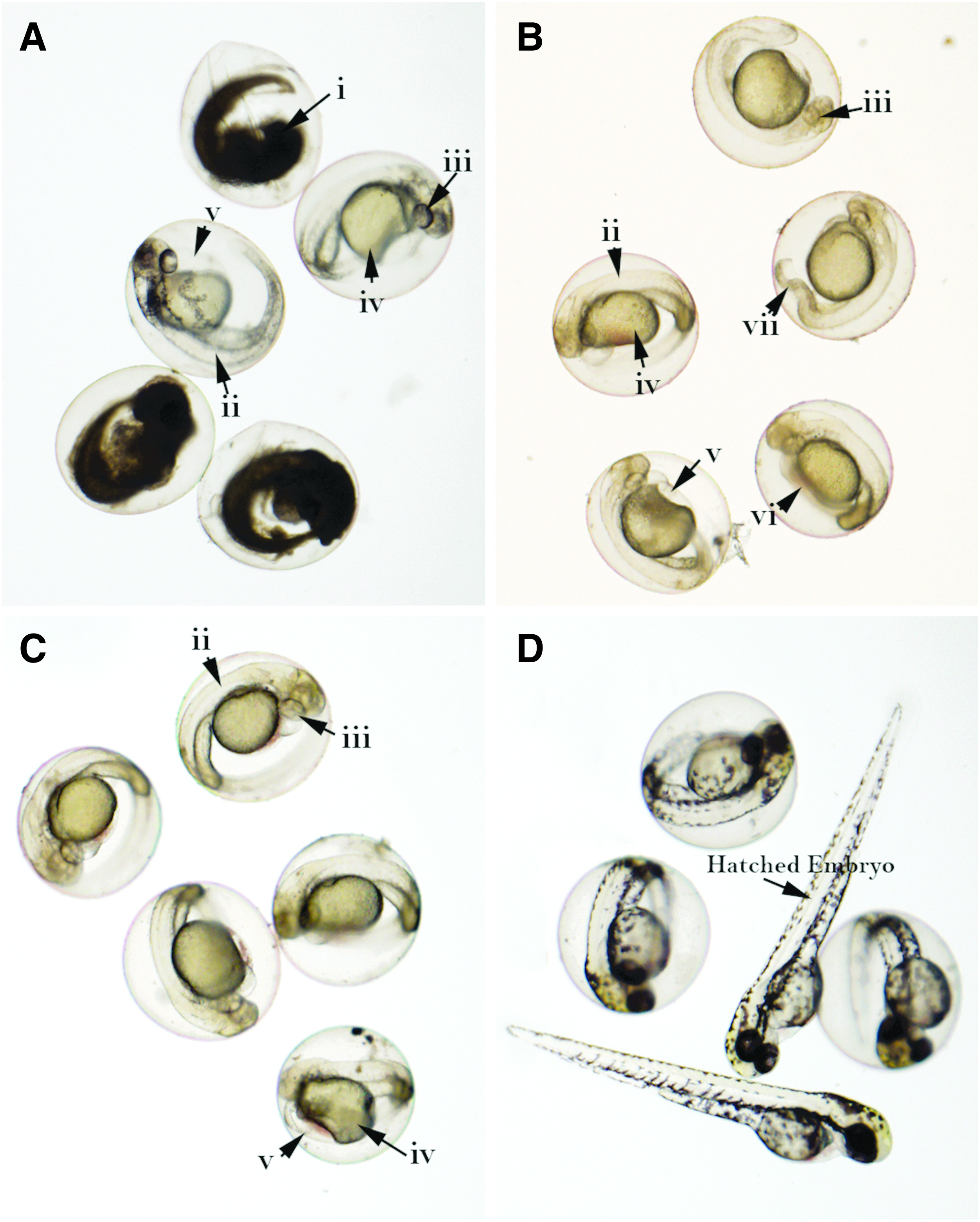
Day 2 embryos in Rx Visijet Clear
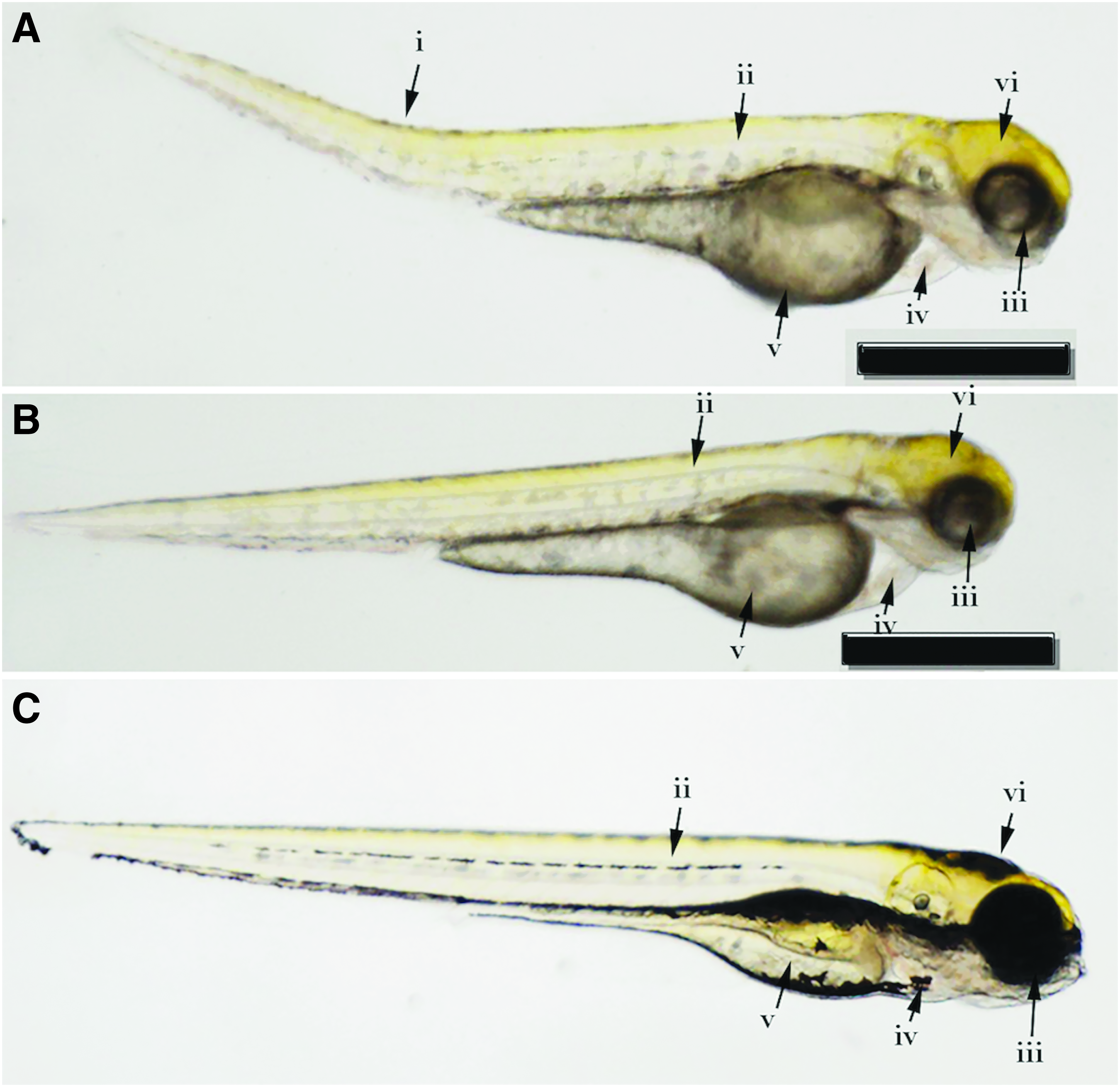
Day 4 representative larvae in MED620 assay
Additional test
Since toxicity is dose dependent, an additional test (Test 2) was performed to measure the effects of leachate concentration on the fertilized embryos. Assays in this test batch comprised 10 embryos, 2 AM-produced specimens, and 10 mL E3 medium in a soda lime silica petri dish. Tests were repeated for reliability. No lethal endpoint was observed in Visijet Clear assays; however, 96 h larvae in “ASB” Visijet Clear developed pericardial edema a day; this occurred a day after 72 h embryos in “Rx” Visijet Clear developed severe pericardial edema. In Visijet Crystal assays, ∼70% lethality was recorded in “ASB” Visijet Crystal on day 1 and test was discontinued. Although only 5% lethality was observed in “Rx” Visijet Crystal, the embryos were lethargic with pericardial edema, yolk deformations, and severe hypopigmentation on day 2, and test was concluded.
More than 50% lethality was observed in “ASB” MED assays on day 1. No lethal endpoint was recorded in “Rx” MED; however, behavior perturbations observed from day 2 to day 3 and the darkening of yolk sacs (Supplementary Figures S2, S3) in 96 h larvae indicated some degree of toxicity 8 in the assays. Figure 3 shows the survival rate of embryos and larvae exposed to AM materials within the 96-h period in Test 1 and Test 2. The survival rates are, however, not indicative of the overall biological performance of the materials.
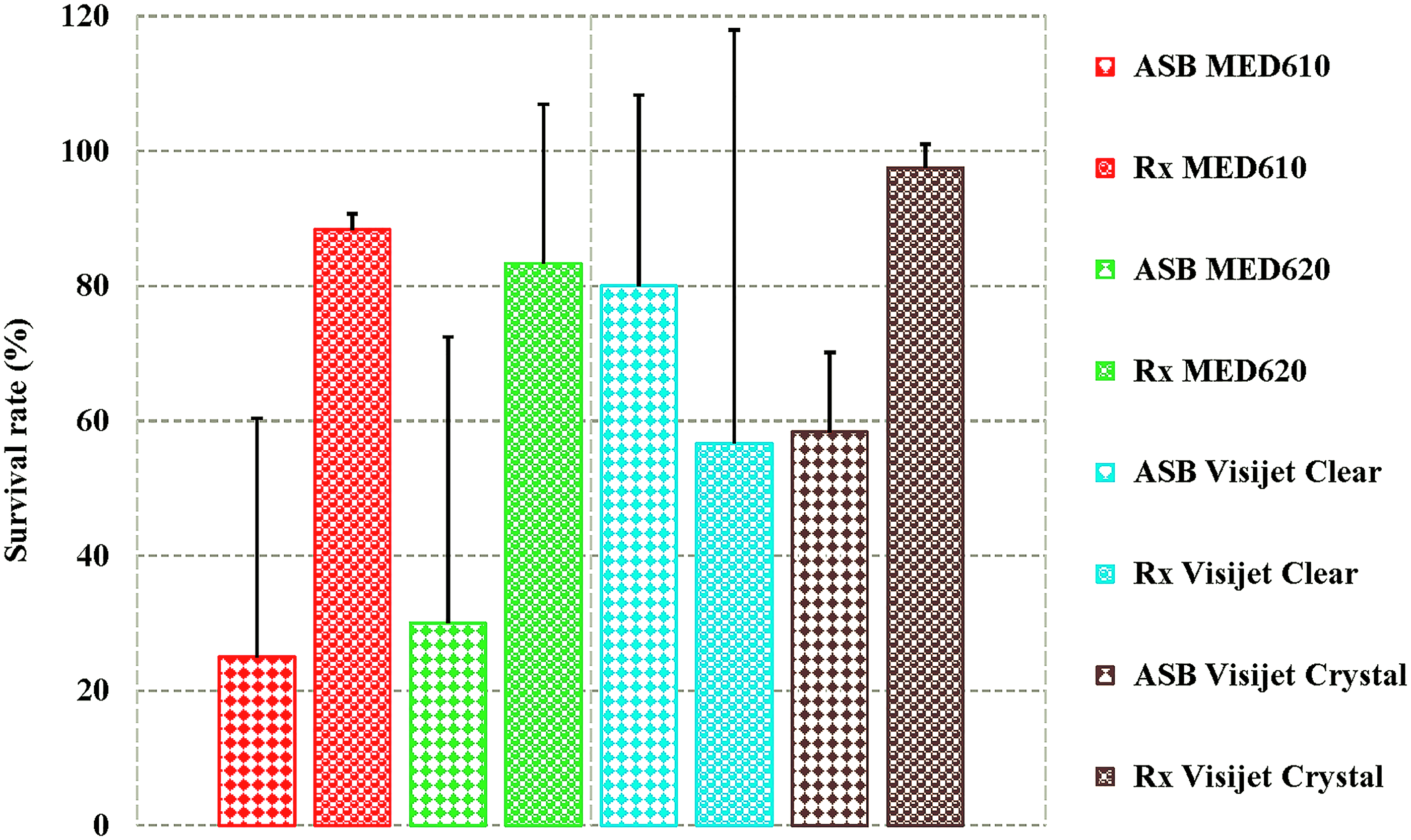
Survival rate of embryos and larvae exposed to AM materials within the 96-h period in Test 1 and Test 2. Error bars show standard deviation from the mean. AM, additive manufacturing. Color images available online at www.liebertpub.com/3dp
No developmental endpoint was observed in Telio CAD (Supplementary Figure S4) and control assays. As per OECD guidelines, the results are valid as the overall survival rates in the control groups were ≥90%. Larvae length (Fig. 4) measured at the end of the test on day 4 in Test 1 showed a significant difference (p ≤ 0.05, unpaired, two-tailed student's t-test) between “Rx” MED materials vs. control, and between “Rx” MED materials vs. Telio CAD. In Test 2, a significant difference (p ≤ 0.05) was found between “Rx” MED610 vs. control and between “ASB” Visijet Clear vs. control.
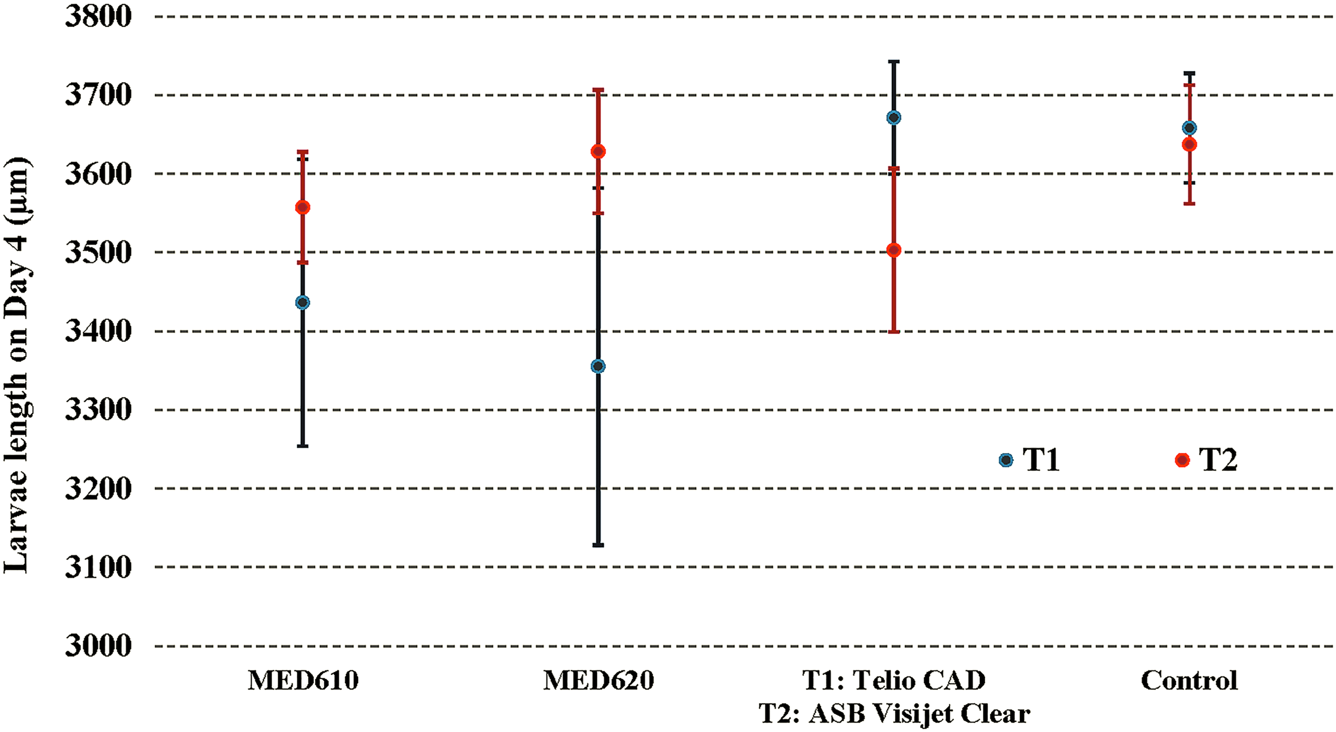
Growth retardation measured on day 4 in Test 1 (T1): MED610
Discussion
The safety data sheets for MED13,14 and Visijet Crystal materials 18 indicate they are acrylic formulations whereas Visijet Clear is a polycarbonate-like polymer. 17 The United States Pharmacopeia (USP) protocols are used to classify plastics in Classes I–VI, based on end use, type, and time of exposure of human tissue to plastics, of which Class VI requires the most stringent testing of all the six classes. 22 As per this classification, Visijet photopolymers are USP Class VI-capable materials, which, according to the manufacturer, are ideal for dental and orthopedic surgical guides, one-day crown preparation guides, and parts in other medical applications. 23 MED photopolymers, in contrast, are USP Class VI (and ISO 10993) certified materials with similar applications to Visijet polymers. Likewise, they are only approved for up to 24 h in the mouth.
Toxicological data show that “as-built” AM materials are generally unsafe in zebrafish assays; comparatively, Visijet materials performed better than MED materials. Ethanol treatment, however, proved effective in enhancing the biocompatibility of the MED materials but produced contrasting results in Visijet materials. The improved tolerance from ethanol treatment corroborates similar findings by Macdonald et al. 8 The biological performance of Visijet Clear improved in Test 2 and, surprisingly, was safer in “ASB” form than in “Rx” form. Although “Rx” Visijet Crystal recorded almost 100% survival rates in Test 1 and Test 2, tests were concluded on day 2 due to the severity of sublethal and teratogenic effects observed in embryos. Previous studies8,10 have also reported Visijet materials to be unsafe in zebrafish assays.
Although it is not unusual for zebrafish under 120 hpf to spend most of their time lying inactively on the bottom of the tanks until the inflation of their swim bladder, 24 those exposed to AM materials showed increased lethargy or behavioral perturbations, which often precede mortality. This sublethal endpoint according to Zhu et al. 10 indicates that photopolymer leachates directly affect the central nervous system or muscle contraction in zebrafish larvae.
Hypopigmentation in terms melanophore development and retinal pigment epithelium was a common sublethal effect observed in assays exposed to AM materials, with the most severe occurring in 48 h Visijet Crystal embryos. Chemicals such as anilines, phenols, and p-tert-butylphenol have also been linked to hypopigmentation in zebrafish embryos. 20 The photopolymer leachates also induced various degrees of teratogenic effects in the assays within the duration of test. The striking phenotypic similarities observed in Telio CAD and control assays confirm the absence of toxic residual monomer in the former. 25 Residual monomer refers to substances such as monomers, additives, and reaction products that are not firmly incorporated in the polymer network and may, therefore, leach and cause local and/or systemic side effects. 7 The acute systemic toxicity test is designed accordingly to determine the toxic potential or the irritant effects of toxic leachables that may be present in extracts of these biomaterials over a relatively short time. 26 In general, the zebrafish excels as a model system for developmental toxicity and offers advantages such as external fertilization, high fecundity, and ease of phenotype assessment over other vertebrates including mouse, in which aspects of organogenesis and disease pathology cannot be examined without interventions such as surgery or postmortem examination. 27
Researchers have attempted to improve the biocompatibility of photopolymers in 3DP. Inoue and Ikuta 28 were able to detoxify SLA parts with high temperature heat process in nitrogen atmosphere, but the procedure also impaired the transparency of the materials. Leonhardt et al. 29 suggested extraction of residual monomers with supercritical CO2 to improve biocompatibility. Oskui et al. 9 and Leonhardt et al. 29 reported improved biocompatibility outcomes for materials cleaned with isopropanol and postcured. Schmelzer et al., 11 in contrast, reported in their in vitro study that ethanol treatment was ineffective in improving the biocompatibility of MED610. It is worth emphasizing that although in vitro tests are faster, less expensive, more reproducible, and more scalable than other types of tests, they may suffer from a lack of relevance to the clinical use of materials, and this weakness is not trivial. 30 Nevertheless, the conflicting results further demonstrate the need for continual biological assessments of photopolymers, especially those for clinical applications, as often the medical profession is overly complacent in its acceptance of new materials without demanding proof of their safety and efficacy. 12
Conclusion
Within the limitations of the study, it is evident that the zebrafish biocompatibility assay is a reliable assessment tool for quantifying the toxicity of leachates in AM materials. Although ethanol proved an effective detoxicant for MED materials, conclusive evidence cannot be drawn regarding its use in a broader context due to the unique parameters (physical and chemical structure of the materials, printing process, and postprocessing methods) of the manufacturing process that eventually influence the clinical performance of the end-use devices. As a result, extreme caution must be exercised with the increased use of AM materials, particularly with 3D printers that may lack apposite settings to ensure the integrity of built parts. Furthermore, as the 3DP industry continues to grow exponentially with contemporaneous influx of new materials, it is imperative for practitioners and patients to understand both the advantages and limitations of the new technologies and materials.
Footnotes
Acknowledgments
The authors would like to thank Objective 3D (Australia) for kindly supplying the MED materials and 3D Systems (Australia) for supplying the Visijet materials for this study. G.J.L. was supported by the NHMRC (1044754, 1069284). The Australian Regenerative Medicine Institute is supported by funds from the State Government of Victoria and the Australian Federal Government.
Author Disclosure Statement
No competing financial interests exist.
References
Supplementary Material
Please find the following supplemental material available below.
For Open Access articles published under a Creative Commons License, all supplemental material carries the same license as the article it is associated with.
For non-Open Access articles published, all supplemental material carries a non-exclusive license, and permission requests for re-use of supplemental material or any part of supplemental material shall be sent directly to the copyright owner as specified in the copyright notice associated with the article.