
Abstract
Abstract
Biodegradable elastic poly(
Introduction
A 3D
Synthetic polymers may be easily processed into the desired shapes with satisfactory mechanical strength, and their degradation rates can be controlled to match the rate of regeneration of new tissue.7,8 Recently, improved tissue regeneration was achieved by repeated physical stress from an external source, such as systolic stress, compressive stress, and shear stress.9,10 Therefore, highly elastic scaffolds are required to transfer repeated physical stress from the external environment to the cells on the scaffold and thus facilitate mechanoactive tissue engineering.
In previous studies, synthetic biodegradable polymers such as poly(lactic acid) (PLA), poly(glycolic acid) (PGA), poly(lactide-co-glycolide) (PLGA), poly(ɛ-caprolactone) (PCL), poly(glycolide-co-caprolactone) (PGCL), and poly(
3D printing has been developed as an advanced technology to overcome the limitations of these conventional methods and may ultimately lead to the production of matrix scaffolds capable of more effectively promoting the regeneration of functional tissue.20,21 3D printing technology has emerged as a promising tool to fabricate scaffolds with high precision and accuracy, creating intricately detailed biomimetic 3D structures. The techniques currently being used to achieve 3D printing of scaffolds, which involve a layer-by-layer process, include, but are not limited to, direct 3D printing, fused deposition modeling, stereolithography, and selective laser sintering. These techniques have been used to produce scaffolds ranging from millimeter to nanometer size.22–25
The commercial orthopedic implant MEDPOR®, a nondegradable bead-type scaffold, is widely used as bone substitute and in prosthetics. According to a recent study, aciniform scaffolding with omnidirectional pore structure using micro-sized beads as a bead-type scaffold is a more favorable environment from the cell growth and functional perspective.26–29
We suggest that as feature of amorphous PLCL by molar ratio 50:50, surface stickiness and elastic properties can be applied to 3D self-assembled porous microbead-type scaffolds by linkage between beads under vacuum.
Therefore, the objectives of this study were (1) to design novel 3D self-assembled porous microbead-type PLCL scaffolds by rapid cooling of PLCL/dioxane into liquid nitrogen; (2) to evaluate the pore structure and dye release potential; and (3) to evaluate cell affinity on scaffolds by a fibroblast (NIH3T3) culture.
Materials and Methods
PLCL (molar ratio: 50:50; number-average molecular weight: 2.0 × 10⁵) was polymerized and analyzed using the methods described previously. 30 As indicated in Figure 1, PLCL solutions [1%, 5%, and 10% (w/v)] were obtained by dissolving PLCL in dioxane. Here, 3 μl aliquots of the PLCL solutions were dropped into liquid nitrogen and kept at −196°C for 10 min to allow phase separation. The frozen beads were added to molds (10 mm × 10 mm × 0.5 mm) and immediately lyophilized for 48 h. The 0.5-mm-thick 3D self-assembled porous microbead-type scaffolds thus obtained were used to investigate mechanical properties and carry out cell affinity tests.
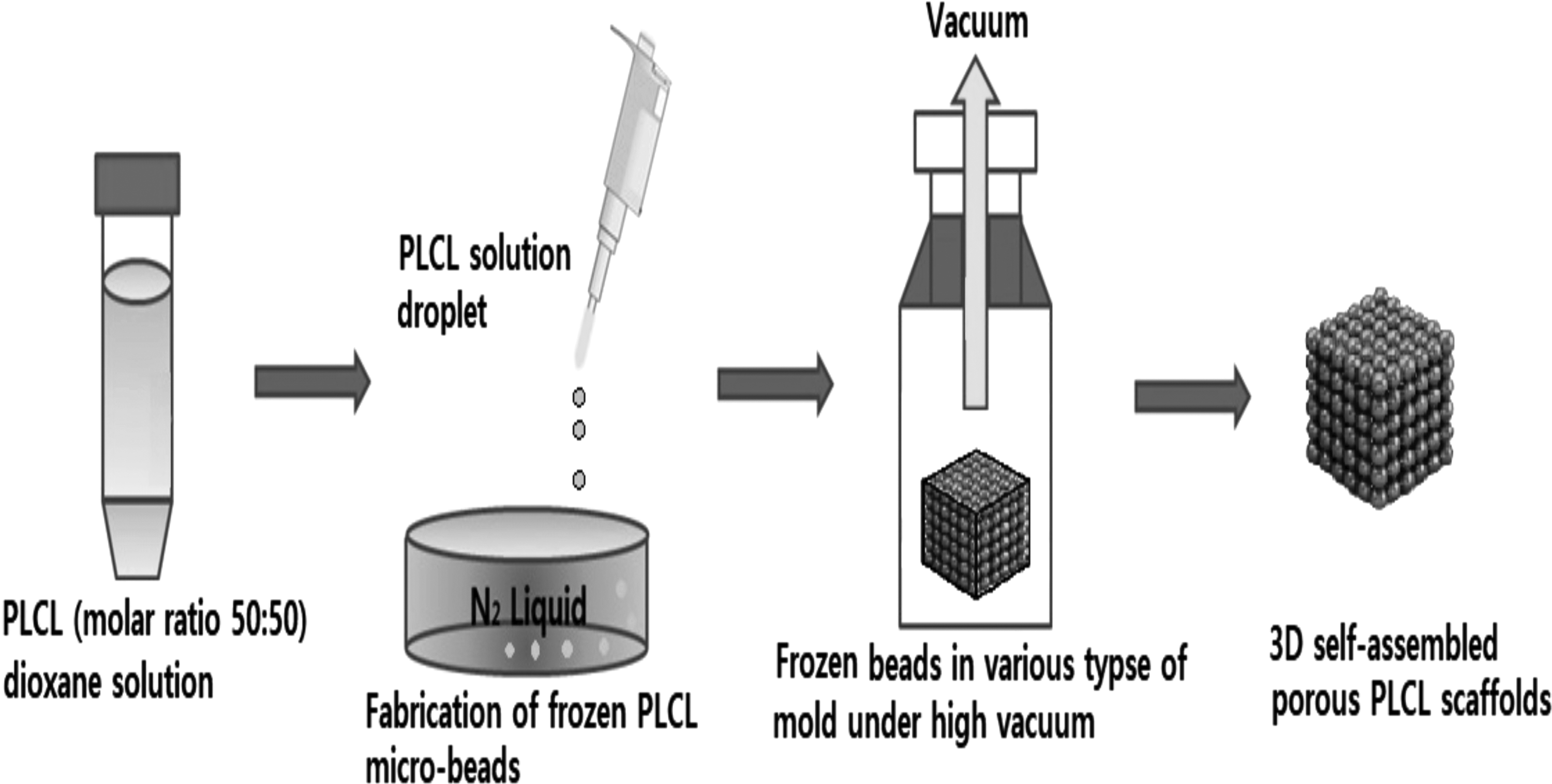
Schematic diagram of the 3D self-assembled porous microbead-type PLCL scaffolds using rapid cooling and lyophilization. PLCL, poly(
The surface and pore morphology of the beads and the matrix of porous bead-type PLCL scaffolds were observed using a field emission scanning electron microscope (FE-SEM: S-4700; Hitachi, Tokyo, Japan).
The porosity of the resulting scaffolds was evaluated, as previously described. 31 Briefly, each scaffold was submerged in a known volume (V1) of pure ethanol, and a series of vacuum-release cycles were performed to force the liquid into the pores of the scaffold. After completion of these cycles, the total volume of the liquid and liquid-impregnated scaffold was recorded as V2. On removal of the liquid-impregnated scaffold, the remaining liquid volume was recorded as V3. Finally, the overall scaffold porosity was given as [(V1 − V3)/(V2 − V3)] × 100%.
The mechanical properties, tensile strength and elasticity, of the scaffolds were evaluated using an INSTRON universal testing machine (Instron model 4467; Canton, MA). The tensile strength was measured using a gauge length of 10 mm and a 5N maximum load cell with a crosshead speed of 1 mm/min. Hydrated samples were prepared by immersing the 10 mm × 10 mm × 0.5 mm dry samples in phosphate-buffered saline (PBS) buffer at room temperature for 3 h. Recovery tests were carried out using a 5N load cell with a crosshead speed of 10 mm/min (strain = 5%, 50%, 100%, 150%, 200%, 250%). The recovery was calculated as Recovery (%) = 100 – [(L2 − L0)/(L1 − L0) × 100], where L0 indicates the original length, L1 indicates the extended length, and L2 indicates the final length after releasing the stress.
Drug release from the scaffolds was simulated using a dye, acid blue-25. A solution of acid blue-25 (1 mg/mL) in a distilled water/ethanol mixture (50:50, v/v) was prepared. PLCL scaffolds (10 mm × 10 mm × 0.5 mm) were submerged in the dye solution for 12 h under vacuum. Each scaffold was then suspended in 10 mL PBS and incubated at 37°C with rotation at 30 rpm for 0, 1, 2, 3, 5, 7, 9, and 12 h. After incubation, the releasate was concentrated by lyophilization, collected, and placed in a 96-well plate. The amount of released dye was determined by measuring the absorbance at 600 nm using a microplate reader.
Collagen-coated scaffolds and uncoated scaffolds were prepared to carry out cell affinity tests. For the collagen-coated porous bead-type scaffolds, 0.3 wt.% collagen solution (1000 ⌊l) was dispersed on the scaffold and dried at room temperature for 2 weeks.
The collagen-coated scaffolds and normal scaffolds (10 mm × 10 mm × 0.5 mm) were prewetted with a medium (Dulbecco's modified Eagle's medium with 2 mM
Results and Discussion
Characterization results of synthetic PLCL are shown in Table 1. As an amorphous phase polymer, low glass transition temperature (Tg) (∼0°C) and satisfactory mechanical properties were observed. Therefore, we supposed that surface stickiness is due to good mobility between polymer molecular chains at room temperature as a result of low glass transition temperature.
Characterization of Poly(l-Lactide-co-ɛ-Caprolactone) (50:50)
LA/CL, lactide/ɛ-caprolactone.
As an elastic polymer in amorphous phase, PLCL (molar ratio 50:50) adheres between beads. Therefore, scaffolds with various morphologies can be obtained through PLCL adhesion between beads under lyophilization, using molds of different shapes. Sheet-type elastic scaffolds comprising strongly attached beads are shown in Figure 2.

Photographs of porous bead-type elastic poly(
The morphology of single porous beads in the scaffolds from PLCL solutions was measured by FE-SEM observation. Bead size is somewhat variable, with relatively large diameters of ∼500–1000 μm. In addition, bead size is determined by drop size distribution, and drop size is affected by surface tension according to PLCL concentration. Therefore, somewhat smaller sized beads were observed for 5% PLCL compared with 1% and 10% PLCL. As indicated in Figure 3a and b, high interconnectivity and uniform pore morphology were obtained, and the pore size was decreased with increasing PLCL concentration. Scaffolds obtained from 1% PLCL showed ∼77% higher porosity and good interconnectivity (Table 2). In the case of 10% PLCL, a fully closed pore structure with low porosity was observed (Fig. 3c and Table 2). However, the porosity of the scaffolds obtained from 10% PLCL was ∼32%. We suggest that the porosity of the beads is affected only by the phase separation effect depending on the polymer concentration and cooling temperature.
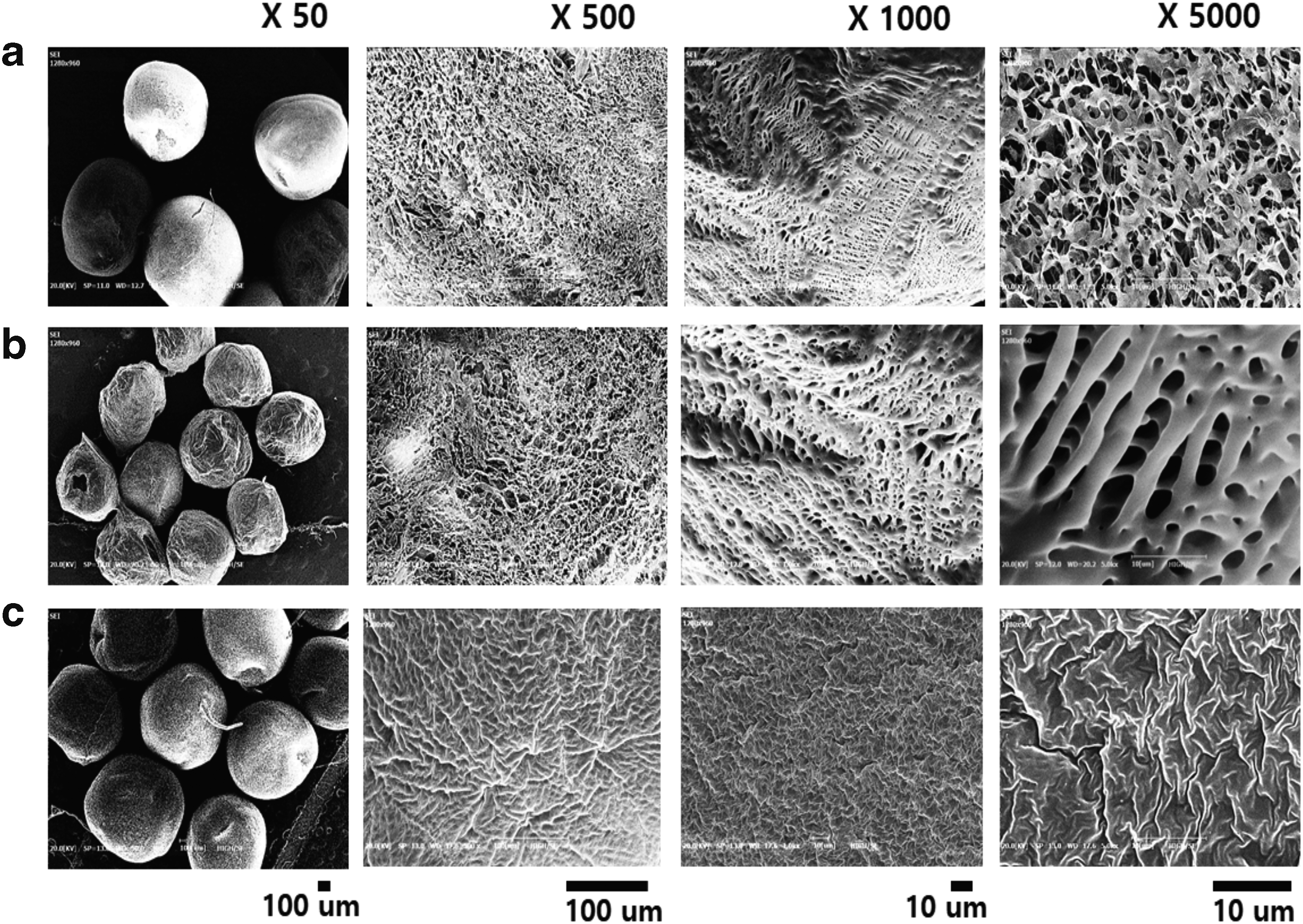
SEM images of beads on scaffolds with PLCL concentrations of
Porosity of Porous Bead-Type Elastic Poly(l-Lactide-co-ɛ-Caprolactone) Scaffolds (n = 5)
The tensile strength of the PLCL scaffolds increased with increasing PLCL concentration (Table 3) with 10% PLCL scaffolds displaying a 1.5-fold increased strength compared with the 1% PLCL scaffolds. The improvement in the mechanical properties is attributable to the increased PLCL concentration. Mechanical properties of porous polymer scaffolds are usually determined by polymer concentration via density and interconnected porous structure. In this study, we presume that a major cause of improved mechanical properties is increased PLCL concentration. On the contrary, all scaffolds are observed to have similar porous structure and bead size, unlike polymer concentration.
Tensile Strength of Porous Bead-Type Elastic Poly(l-Lactide-co-ɛ-Caprolactone) Scaffolds (n = 5)
Proper balance between porous structure via phase separation and mechanical strength is important. Therefore, the 5% scaffold with open pore structure and mechanical strength similar to that of the 10% scaffold was selected by comparison with the 1% PLCL scaffold, which had low mechanical strength and high porous structure, and the 10% PLCL scaffold, which had high mechanical strength and closed porous structure.
Thereafter, the elastic recovery of the porous bead-type PLCL scaffolds was measured. No difference in elastic recovery was observed as PLCL concentration was increased from 5% to 10% (Fig. 4), while the 1% PLCL scaffold broke at 200% strain. This result is possibly due to the low elongation at break caused by the poor adhesion strength between the beads at low PLCL concentrations.
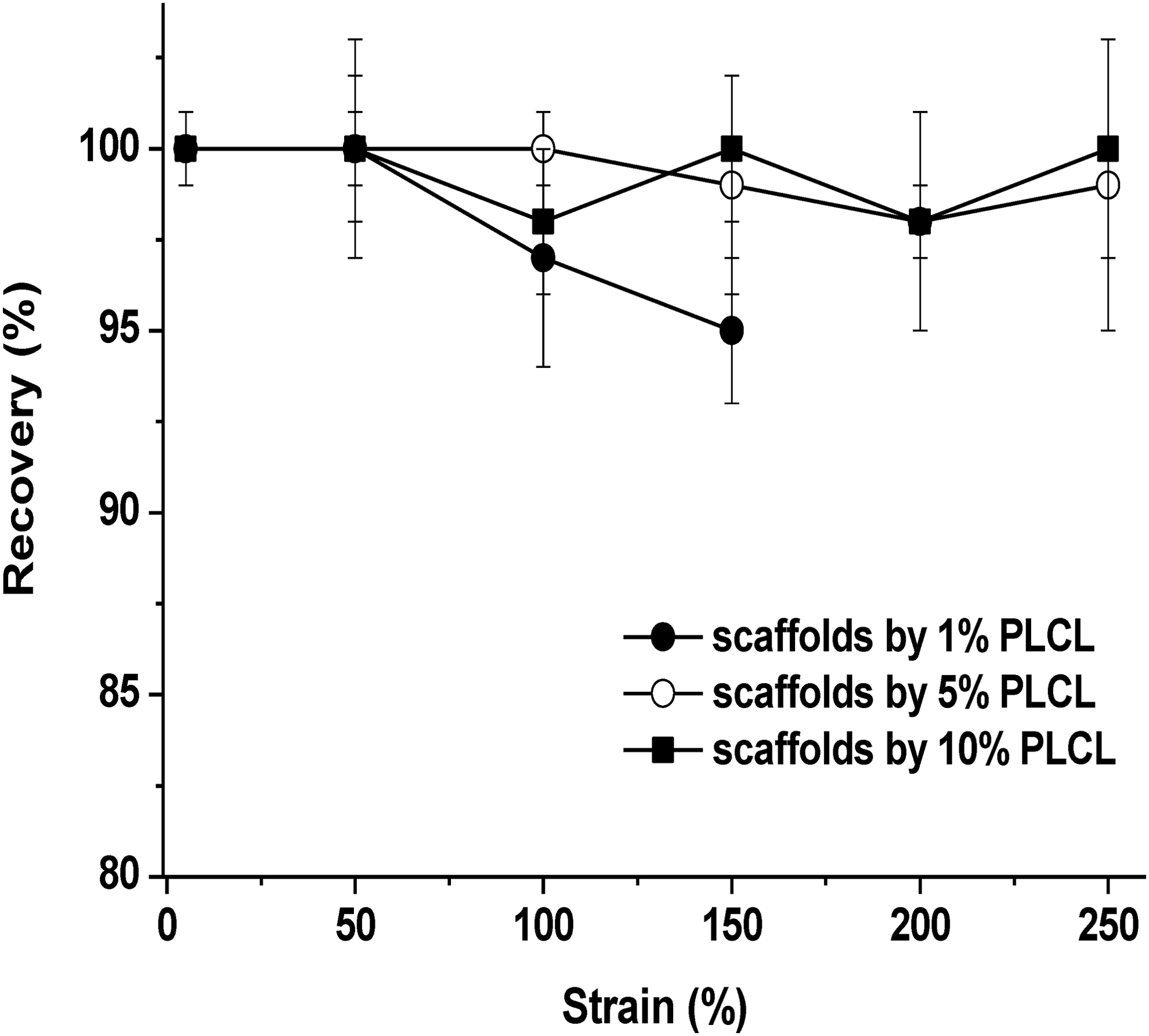
Correlations between elastic recovery and PLCL concentration of the porous bead-type elastic poly(
The amount of dye released from the scaffold is presented in Figure 5. The dye release pattern was strongly dependent on the 3D porous structure of the scaffold. The 1% PLCL scaffold showed an almost 1.5-fold higher initial release rate and sustained release behavior than the 10% PLCL scaffold. The 5% PLCL scaffold showed a similar trend as that of the 1% PLCL scaffold. We speculate that the low dye release efficiency of the scaffold prepared with 10% PLCL is attributable to its closed pore structure. However, the high release rate and sustained release behavior of the 1% PLCL and 5% PLCL scaffolds were due to better interconnectivity and the open pore structure. In addition, the higher initial release rate could be related to the faster diffusion over the larger surface area of the 3D porous structure.
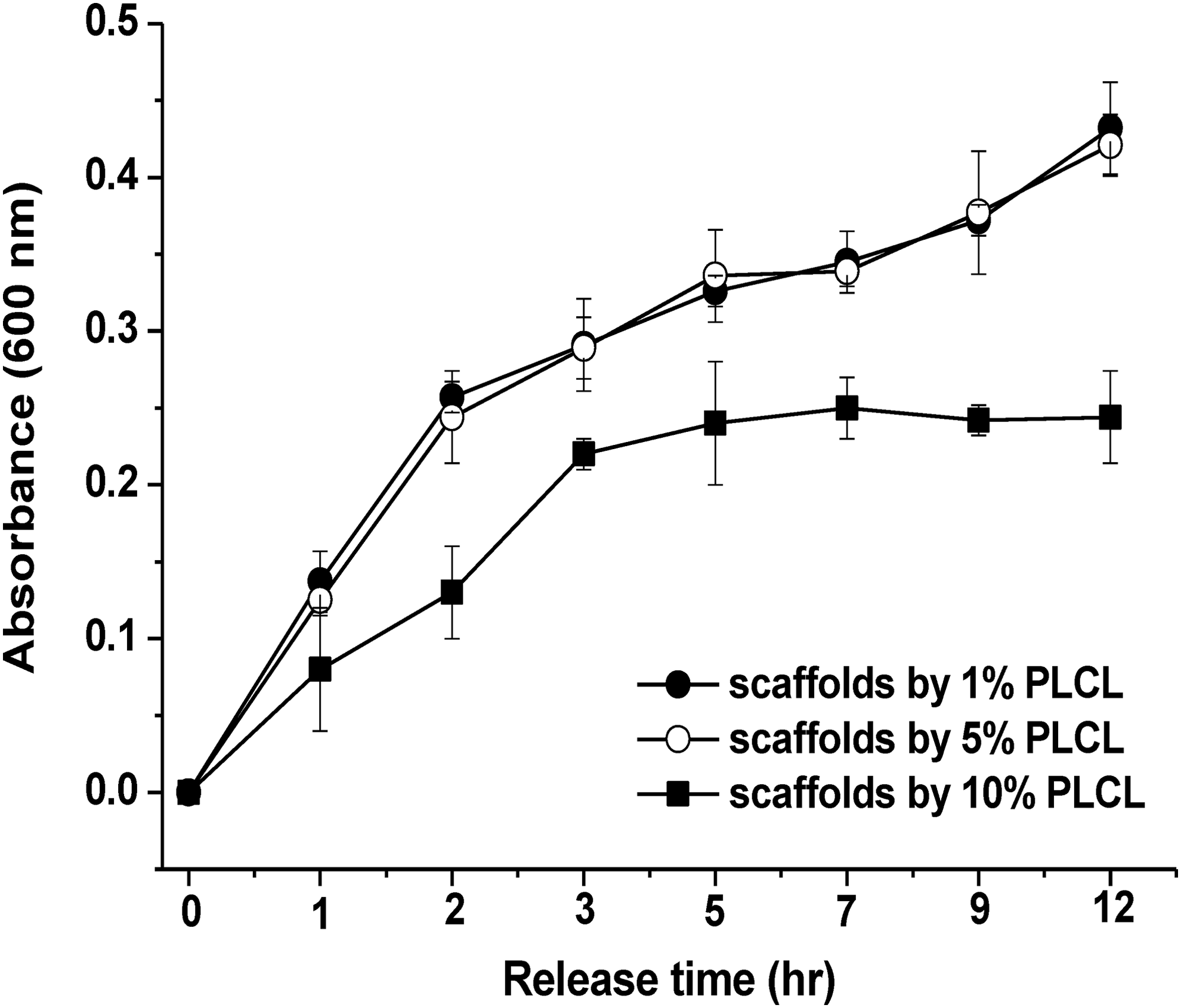
Acid blue-25 release test from porous bead-type elastic poly(
For the cell affinity evaluation, NIH3T3 cells were cultured on collagen-coated scaffolds and uncoated scaffolds. A very thin coating layer on scaffolds by a low-concentration collagen solution of 0.3% did not affect mechanical and morphological properties. Therefore, only collagen-coated scaffolds were prepared to carry out cell affinity tests. In addition, a homogeneous coating surface was observed by water contact angle on collagen-coated PLCL thin film (no show). The PLCL concentration had little effect on the initial cell adhesion of the scaffolds after a 4-h cell culture; however, after a 48-h cell culture, increased cell proliferation on the collagen-coated 1% and 5% PLCL scaffolds was observed (Fig. 6). Furthermore, as the cell culture time increased, a greater number of changes in cell adhesion and the growth properties were observed for the collagen-coated 1% and 5% PLCL scaffolds than that for the normal scaffolds. However, no significant difference was observed between the 1% PLCL and 5% PLCL scaffolds. The results of the tensile strength, recovery, dye release, and cell tests suggest that 5% PLCL scaffolds should serve as excellent substrates for cell adhesion and growth in tissue engineering. These scaffolds may therefore be applied to tissue engineering, for example, in vessel and cartilage regeneration, because of the retention of the elastic properties of 50:50 PLCL as well as the improved bioaffinity proffered by the collagen coating and the porous bead structure.

Correlations between cell adhesion and proliferation and PLCL concentration of the collagen-coated/uncoated PLCL scaffolds.
Conclusion
3D self-assembled porous microbead-type scaffolds were obtained by a simple, rapid cooling method using biodegradable elastic poly(
Footnotes
Acknowledgment
The present research was conducted by Baton-Zone service (3D Printing based customized self-healing implants) and the research fund of Dankook University in 2017.
Author Disclosure Statement
No competing financial interests exist.