
Abstract
Vascularization is essential for the regeneration of three-dimensional (3D) bioprinting organs. As a general method to produce microfluidic channels in 3D printing constructs, coaxial extrusion has attracted great attention. However, the biocompatible bioinks are very limited for coaxial extrusion to fabricate microchannels with regular structure and enough mechanical properties. Herein, a hybrid bioink composed of alginate (Alg) and silk fibroin (SF) was proposed for 3D bioprinting of microchannel networks based on coaxial extrusion. The rheological properties of the bioink demonstrated that the hybrid Alg/SF bioink exhibited improved viscosity and shear thinning behavior compared with either pure Alg or SF bioink and had similar storage and loss modulus in a wide range of shear frequency, indicating a sound printability. Using a coaxial extrusion system with calcium ions and Pluronic F127 flowing through the core nozzle as cross-linkers, the Alg/SF bioink could be extruded and deposited to form a 3D scaffold with interconnected microchannels. The regular structure and smooth pore wall of microchannels inside the scaffold were demonstrated by optical coherence tomography. Micropores left by the rinse of F127 were observed by scanning electron microscope, constituting a hierarchical structure together with the microchannels and printed macropores. Fourier transform infrared spectroscopy analysis proved the complete rinse of F127 and the formation of β-sheet SF structure. Thus, Alg/SF could form a double cross-linked network, which was much stronger than the pure Alg network. Moreover, cells in the Alg/SF scaffold showed higher viability and proliferation rate than in the Alg scaffold. Therefore, Alg/SF is a promising bioink for coaxial extrusion-based 3D bioprinting, with the printed microchannel network beneficial for complex tissue and organ regeneration.
Introduction
Three-dimensional (3D)
However, the bioinks for coaxial 3D bioprinting of biocompatible vascular-like structures with certain mechanical strength are very limited. A useful material for bioprinting should be easily manipulated by the bioprinter technology to be dispensed in complex 3D structures and maintain cellular viability and function, thereby providing structural and mechanical support to the structure. 1 Due to the good biocompatibility, degradability, and fast ion-cross-linking property, Alg is often used in coaxial printing to construct hollow fiber structures.5,6 Nonetheless, the insufficient mechanical strength of pure Alg hydrogel hinders its applications. The ionically cross-linked Alg loses mechanical properties over time with the diffusion of cross-linking ions, and it exhibits poor protein absorption and cell adhesion due to its highly hydrophilic nature. 7 Armstrong et al. 8 found that the pores on the cross-linked Alg surface were too small to transfer nutrients and metabolic waste, thus affecting cell migration and growth. Therefore, Alg is often used with other materials to improve its mechanical and biological properties, such as gelatin methacryloyl and chitosan.3,9
Silk fibroin (SF) is a natural material derived from silkworms, which has excellent biocompatibility and biodegradability, good breathability and moisture permeability, and nonirritating and good mechanical properties.10,11 SF-based bioinks have been extensively studied for their full aqueous phase at room temperature, cell compatible cross-linking modes, and strong mechanical properties.11,12 Normally, the sol–gel transition of aqueous SF is induced by temperature and pH. However, the gelation of pure SF itself is time-consuming. F127 is one of the most widely used nonionic polyoxyethylene–polyoxypropylene–polyoxyethylene (PEO-PPO-PEO) triblock copolymers, 13 with a nominal molecular weight of 12.5 kDa and a PEO/PPO ratio of 2:1 by weight, which can accelerate the conformational change of SF from random coil to β-sheet structure, due to the interaction between SF and the poloxamer with respect to hydrogen and hydrophobic bonding. 14 Collectively, it is possible to modify Alg bioink with SF in the presence of F127, to make it more suitable for coaxial extrusion-based bioprinting and enhance hydrogel properties.
In this study, we propose a novel bioink (Alg and SF) for coaxial 3D bioprinting. It is expected that the addition of SF to Alg can improve the mechanical properties of the printed structure and be beneficial for cell growth. The properties of the bioink and printed scaffolds were studied through rheological tests, Fourier transform infrared spectroscopy (FTIR) analysis, mechanical property, and cell compatibility evaluation. Optical coherence tomography (OCT) and scanning electron microscope (SEM) were also implemented to analyze the structure of the printed vascular-like microchannel scaffold.
Materials and Methods
Materials
Alg (W201502; Sigma), SF (20181120; Simet Biotechnology), F127 (SLBT0533; Sigma), and calcium chloride (10043-52-4; Sinopharm Group Chemical Reagent Co., Ltd) were prepared for printing. Dulbecco's modified Eagle's medium (DMEM; 8119035) and fetal bovine serum (FBS; 10091148) from Gibco were used for cell culture.
Preparation of bioinks and cross-linkers
Alg (5% w/v), F127 (13% w/v), and CaCl2 (5% w/v) solution were prepared in deionized water and sterilized by pasteurization. SF was dissolved in a 9.3 M lithium bromide solution at 60°C, followed by a dialysis (cutoff molecular weight of 3500) for 48 h to remove the salt. Then, the SF solution was lyophilized and stored at −20°C for further use. The sterilized Alg bioink was prepared by 5% Alg, with 5% CaCl2 as the cross-linker, whereas the Alg/SF bioink was prepared by 5% SF and 5% Alg, with 13% F127 and 5% CaCl2 as the cross-linker.
Experimental setup and 3D printing of scaffold with microchannels
The coaxial nozzle is formed by a shell nozzle (outer diameter 1.47 mm, inner diameter 1.07 mm) and a core nozzle (outer diameter 0.63 mm, inner diameter 0.34 mm), as shown in Figure 1a. The bioink and cross-linker were loaded into two syringes, which were driven by two microinjection pumps and connected to the shell and core nozzle, respectively. Then, the coaxial nozzle system was loaded on a 3D bioprinter (Regenovo, China). The bioink extrusion rate was set as 7 μL/s, the cross-linker solution was extruded at the rate of 5 μL/s, and the speed of the print head was set as 15 mm/s.

Coaxial nozzle with bioink (SF and Alg) and cross-linkers (Ca2+ and F127)
Rheological and compression tests
The rheological tests were carried out by an MCR301 rheometer (Annegro, Austria), using a 50 mm diameter plate. The viscosity and shear modulus of pure Alg and hybrid Alg/SF bioinks were characterized at 25°C. The shear modulus of cross-linked Alg and Alg/SF hydrogels were also characterized by testing the storage modulus G′ and the loss modulus G″ at 37°C. The frequency and strain were set as 1 Hz and 1%, respectively. The complex shear modulus, G* = G′+iG″, was investigated and the magnitude of the complex shear modulus is given by
The Alg and Alg/SF scaffolds with the size of about 20 × 20 × 3 mm were tested on a universal testing machine (AI-7000-S; GOTECH) at room temperature. The compressive force was applied parallel to the longitudinal axis of the samples with the rate of 1.0 mm/min. The compressive modulus was determined as the slope of the stress–strain curve within 0–10% strain. At least three samples were prepared for each group.
Microstructure analyzed by OCT and SEM
The OCT images of the scaffold were obtained by the Telesto-2 THORLABS system (TorLabs, Newton, NJ) with the measuring parameters including the center wavelength of 1310 nm, the imaging depth of 2.6 mm in air and 3.5mm in water, and the lateral resolution of 13 μm. The images of 9.5 × 9.5 × 3.58 mm3 (1024 × 1024 × 1024 dpi) at different positions of the scaffold sample were obtained by 1024 acquisitions without contacting the sample, and the scanning frequency was 48 kHz.
The Alg and Alg/SF scaffolds were lyophilized and fractured, followed by spraying with carbon. The microstructure of the scaffolds was observed on SEM (SU8020; Hitachi, Japan), and the pore size was analyzed by the ImageJ software.
FTIR analysis
One milligram of lyophilized uncross-linked Alg and SF, cross-linked Alg/SF/F127, and cross-linked Alg/SF (after rinse of F127) were separately blended with potassium bromide. Then, they were pressed into tablets and characterized by a Fourier transform infrared spectrometer (Nicolet 6700; Thermo) to analyze the compositions.
Cell activity and proliferation assay
The pre-hydrogel solution of Alg or Alg/SF/F127 was mixed with C3A cells (liver cancer cells, from ATCC, cultured in DMEM with 10% FBS) to make a bioink of 106 cells/mL. The bioink was then printed coaxially to make scaffolds with microchannels. To assess cell viability and distribution, the scaffolds cultured for 3 days were dyed with Calcein-AM (1 μmol/mL; Sigma) and propidium iodide (1 μmol/mL; Sigma) at 37°C for 30 min and then observed under an inverted fluorescence microscope (Ti-U, Nikon, Japan). Four samples were prepared for each group. The ImageJ software was used to count cells and calculate the survival rate.
alamarBlue was used to evaluate cell proliferation in the scaffolds. After culturing for 1, 5, 7, 9, and 14 days, 10% alamarBlue was added to the culture medium and kept with the scaffolds for 2 h. Then, 100 μL of the supernatant was pipetted to a 96-well plate. Optical density values were evaluated on a microplate reader at the wavelength of 590 nm, and all the values were normalized to day 1.
Statistical analysis
All experiments were independently repeated at least three times. Statistical analysis was performed by unpaired Students' t-test using SPSS 19.0 statistical software, and p < 0.05 was considered significant.
Results
3D printing of scaffolds with microchannels
The core flow of cross-linkers and shell flow of bioinks make the microchannel formation, whereas 3D motion of the coaxial nozzle deposits scaffolds with microchannels. Because the cross-linking reaction took place from the inside out, it can make sure the stable formation of the inner wall of microchannels and the firm adhesion of the outer walls of crossed microchannels between layers. When the printing of one layer finished, the nozzle was directly elevated to start another layer printing. The extrusion of bioinks and cross-linkers was continuous in the whole printing process to ensure the formation of interconnected and perfusable microchannels in the whole scaffold. For Alg/SF bioink, the formation of Alg network and SF network makes an interpenetrated double network, as shown in Figure 1b. The process of perfusion of Alg/SF scaffold with culture medium is shown in Figure 1c and Supplementary Video S1.
Rheological and mechanical properties
Rheological properties are important to indicate the printability of the bioinks. 16 In Figure 2a, the viscosity of uncross-linked Alg/SF decreases linearly with shear rate (from 0.1 to 100), indicating a sound shear thinning behavior, which is essential for hydrogel printing; while the viscosity of uncross-linked Alg decreases obviously only at the high-frequency section (from 10 to 100), and the viscosity of uncross-linked SF kept stable at a very low value, which is not suitable for printing. In Figure 2b, the uncross-linked Alg and Alg/SF had similar viscoelasticity especially at the high-frequency section, where the elastic shear modulus (G′) was lower than the viscous shear modulus (G″), indicating their liquidity. After printing and cross-linking, both Alg and Alg/SF solutions were converted to a gel state (G′ > > G″). Notably, the G* value of cross-linked Alg/SF was about 172.0 kPa, much higher than that of the cross-linked Alg (about 17.7 kPa), indicating the formation of double network interpenetrated with SF network of β-sheet structure and Alg network chelated by calcium ions. In Figure 2c, the compressive modulus of Alg/SF scaffolds was 16.0 ± 2.5 kPa, significantly higher than that of Alg scaffold (11.5 ± 1.6 kPa), demonstrating the formation of double cross-linked network of Alg/SF scaffold and the β-sheet SF can distinctly strengthen the Alg scaffold.

Viscosity of uncross-linked Alg, SF, and hybrid Alg/SF at 25°C
OCT characterization of 3D printing microchannels
OCT images in Figure 3a, b, e, and f show the internal cross section of the printed scaffolds with microchannels. Figure 3a, e and b, f, respectively, highlights the vertical and horizontal microchannels of the scaffolds, and regular and penetrating microchannels are clearly shown with the diameter between 500 and 600 μm. Compared with the microchannels in Alg scaffolds (Fig. 3a, b), those in Alg/SF scaffolds (Fig. 3e, f) are more smooth and regular, which are more convenient and efficient for the perfusion of nutrient solution. Notably, the microchannels in the OCT images have wavy walls, which were formed in the laying process of 3D printing. The wavy wall can enlarge the surface area of the microchannel and may be beneficial for the diffusion of oxygen and nutrient.

Cross section of the Alg scaffold
Figure 3c, d, g, and h shows the microstructure of Alg and Alg/SF scaffolds with microchannels. Compared with the smooth surface of pure Alg scaffold (Fig. 3c, d), the wall of Alg/SF scaffold (Fig. 3g, h) has a lot of micropores with the diameter of 5–20 μm, which was induced by the rinse of sacrificial F127.
FTIR characterization of Alg/SF before and after cross-linking
FTIR analysis was used to characterize the compositions of the hybrid Alg and SF polymers before and after cross-linking in this study. As shown in Figure 5, the uncross-linked Alg/SF keeps most of the characteristic peaks of Alg and SF, such as 1411 cm−1 carboxyl absorption peak of M monomer and 1604 cm−1 carboxyl absorption peak of G monomer of Alg, and 1650 cm−1 amide I peak of α-helical structure of SF protein. 17 During the printing process, SF was cross-linked by F127 to form a network, in which SF conformation was transformed from α-helix to β-sheet, which was demonstrated by the formation of 1628 cm−1 amide I peak of β-sheet in the cross-linked Alg/SF and Alg/SF/F127 curves. Alg was cross-linked by calcium ion to form another network. After perfusion and rinse with calcium chloride solution at room temperature, the strength and integrity of Alg network in the scaffold was improved. Compared with the cross-linked Alg/SF/F127 curve in Figure 4, disappearance of 1109 cm−1 ether bond peak in the cross-linked Alg/SF curve demonstrates that F127 was dissolved and rinsed by water, leaving a porous structure as shown in Figure 3h.
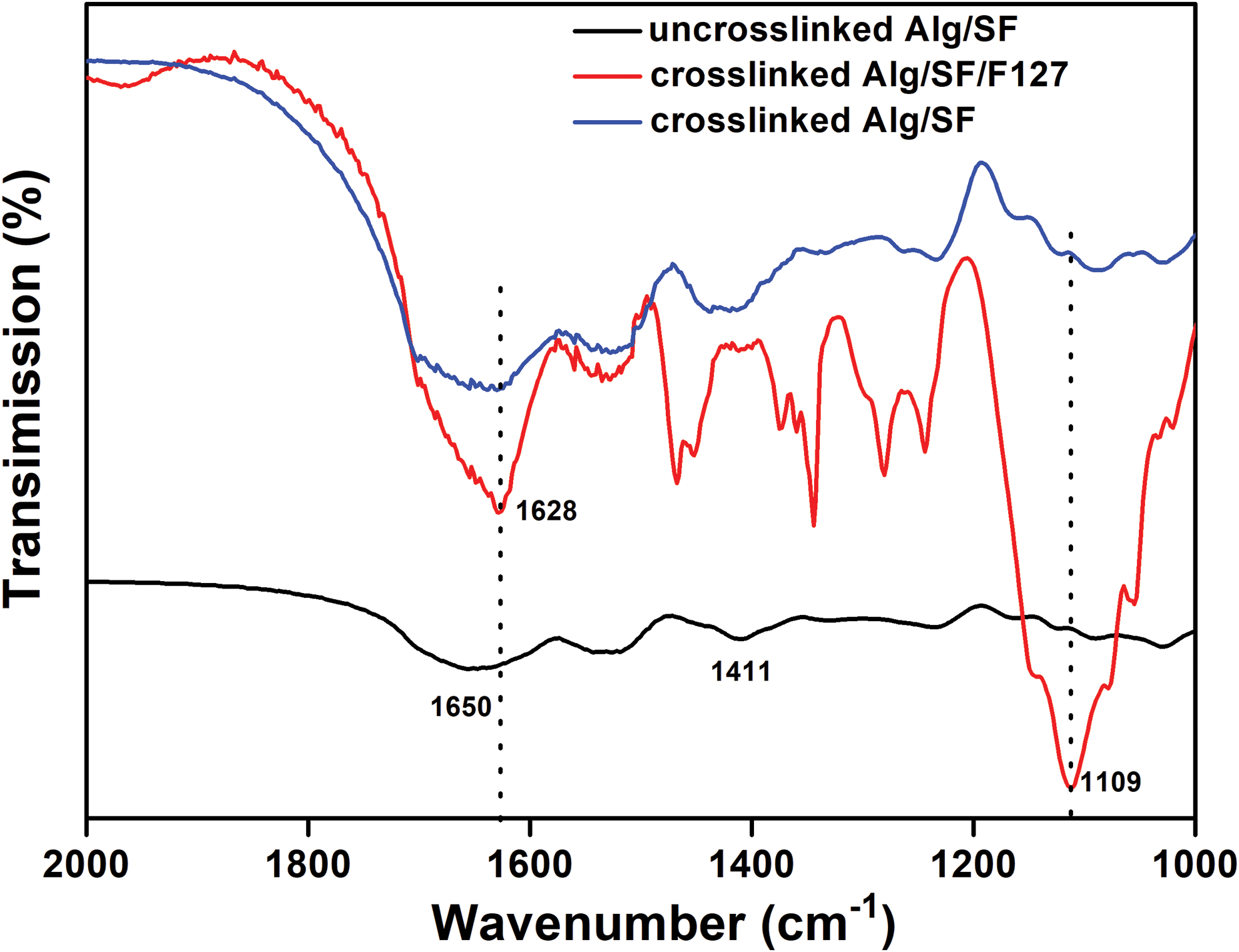
FTIR characterization of uncross-linked Alg/SF, cross-linked Alg/SF/F127, and cross-linked Alg/SF (after rinse of F127). FTIR, Fourier transform infrared spectroscopy. Color images are available online.
Fluorescent images of live and dead cells in the Alg
Cell viability and proliferation in the scaffold with microchannels
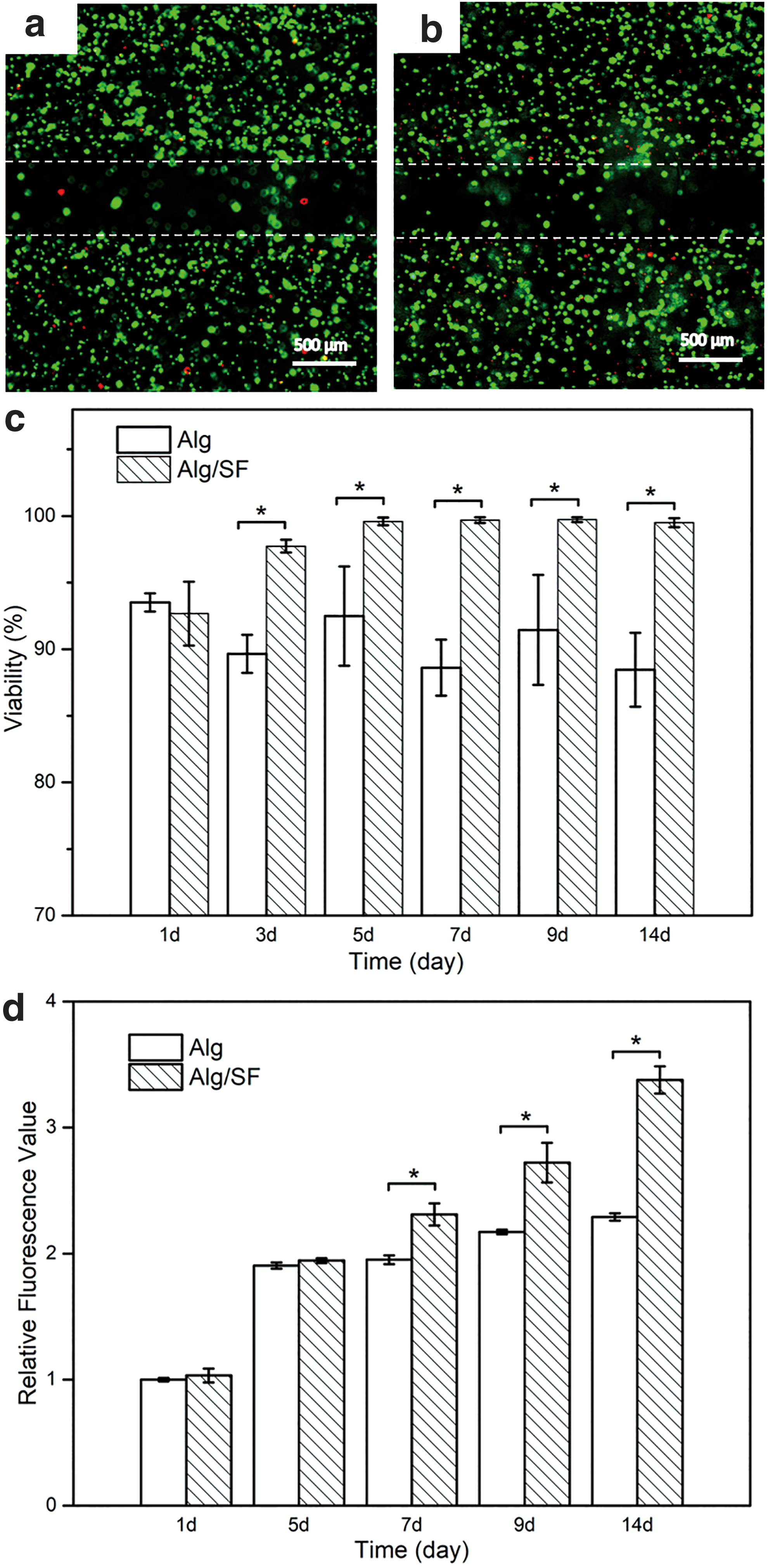
The fluorescent images of live and dead cells in the Alg and Alg/SF scaffolds with microchannels are shown in Figure 5a and b. The microchannels were obvious in the two scaffolds (indicated by dash line), and there were more dead cells (red points) in the Alg scaffold than in the Alg/SF scaffold. This can also be illustrated by the percentage of cell viability in Figure 5c. The cell viability in the Alg was about 93.5%, similar to that in Alg/SF scaffold (about 92.7%) at day 1 after printing. However, the cell survival rate of the Alg scaffold decreased with time to 88.4% at day 14, while that of the Alg/SF scaffold increased to about 99.5%. This may be due to the amicable interface of SF to cells and the fast proliferation of cells in the Alg/SF scaffold.
Cell proliferation rate is shown in Figure 5d. Both the cells in the Alg and Alg/SF scaffolds proliferated with time in 14 days. In the first 5 days, the proliferation rate was not different between Alg and Alg/SF group. However, the cells grew significantly faster in the Alg/SF scaffold than in the Alg scaffold from the seventh day.
Discussion
3D bioprinting of vascular-like structures based on coaxial extrusion is a promising method to produce bulk tissues and organs. However, it makes higher requirements for the bioinks, which should be biocompatible and come out of the nozzle smoothly with minimal pressure during printing and undergo rapid gelation by the fluid in the core nozzle. For example, the blend of gelatin and SF can be printed and cross-linked in situ without any cytotoxic effect, 18 but it is not suitable for coaxial extrusion and printing structures with hollow fibers. Poly (lactic-co-glycolic acid) or polyacrylonitrile can be dissolved in dimethyl sulfoxide and coaxially extruded in the water bath to form conduits19,20; however, the use of toxic dissolvent and too rapid solidification rate limit its further application in bioprinting. Actually, most of the bioinks used in 3D bioprinting based on coaxial extrusion are Alg or the composite of Alg and other materials.6,9,21–23 Since Alg bioink has major limitations, such as quick loss of mechanical properties during in vitro culture, lack of bioactive binding sites and resistance to protein adsorption, and slow and uncontrollable degradation,18,22,24 we developed an Alg/SF bioink with calcium ion and F127 as cross-linkers for coaxial extrusion-based bioprinting.
In the process of Alg/SF printing, F127 served as cross-linkers in the core nozzle with calcium ions. The cross-linkers and bioinks converged at the nozzle tip, where F127 and the calcium ions diffused into the bioinks instantaneously to make the cross-linking happen. This is different from the process reported before, in which both F127 and Alg were in the shell nozzle as bioinks. 8 In this study, F127 served as a cross-linker of SF as well as a sacrificial template, being completely rinsed after cross-linking, which led the formation of anisotropic micropores as reported. 8
The micropores, microchannels, and 3D printed macropores together constitute a hierarchical microchannel network and can be observed clearly by OCT and SEM, as shown in Figure 3. Specially, OCT is able to provide noncontact and undamaged investigation with high-resolution images of the inner structure of opaque 3D scaffolds, which is impossible to be realized by traditional optical imaging technology.25,26 The hierarchical microchannel network can enhance nutrient transport and may provide space for cells to grow, which can be demonstrated by the faster proliferation of cells in Alg/SF scaffolds (Fig. 5).
The perfusable interconnected microchannels and permeable walls with micropores can efficiently facilitate convective and diffusive mass transfers between the scaffolds and local tissues, which is very important for tissue engineering applications.27,28 It should be noted that the reduced shear force of Alg/SF bioink could also increase cell viability in the scaffold. Beyond the higher viability and proliferation rate shown in the results of Figure 5, the Alg/SF composite exhibited better compatibility than pure SF in other research. 29 Moreover, the Alg/SF scaffold has not only enhanced biological properties but also much stable structure than the pure Alg scaffold, which is essential for in vitro culture and postprocessing, especially for the in vivo implantation. 30
Conclusions
In this study, a new hybrid bioink composed of Alg and SF was developed for coaxial 3D bioprinting. The bioink had excellent viscosity and viscoelasticity for printing, and the printed scaffold had transversely and longitudinally interconnected microchannels, making it perfusable. The formation of double cross-linking network provided Alg/SF scaffold with smooth and regular microchannels and high compressive modulus. Due to the sacrificial F127, microporous structure appeared on the wall of Alg/SF microchannels, and thus, a hierarchical microchannel network was formed. The results of cell proliferation showed that the Alg/SF scaffold with microchannels was more conducive to cell growth. Therefore, the coaxial-based bioprinting of Alg/SF has a great potential for preparing large-scale vascularized tissue constructs.
Footnotes
References
Supplementary Material
Please find the following supplemental material available below.
For Open Access articles published under a Creative Commons License, all supplemental material carries the same license as the article it is associated with.
For non-Open Access articles published, all supplemental material carries a non-exclusive license, and permission requests for re-use of supplemental material or any part of supplemental material shall be sent directly to the copyright owner as specified in the copyright notice associated with the article.