
Abstract
A three-dimensional (3D) printing is a robotically controlled state-of-the-art technology that is promising for all branches of engineering with a meritorious emphasis to biomedical engineering. The purpose of 3D printing (3DP) is to create exact superstructures without any framework in a brief period with high reproducibility to create intricate and complex patient-tailored structures for organ regeneration, drug delivery, imaging processes, designing personalized dose-specific tablets, developing 3D models of organs to plan surgery and to understand the pathology of disease, manufacturing cost-effective surgical tools, and fabricating implants and organ substitute devices for prolonging the lives of patients, etc. The formulation of bioinks and programmed G codes help to obtain precise 3D structures, which determines the stability and functioning of the 3D-printed structures. Three-dimensional printing for medical applications is ambitious and challenging but made possible with the culmination of research expertise from various fields. Exploring and expanding 3DP for biomedical and clinical applications can be life-saving solutions. The 3D printers are cost-effective and eco-friendly, as they do not release any toxic pollutants or waste materials that pollute the environment. The sampling requirements and processing parameters are amenable, which further eases the production. This review highlights the role of 3D printers in the health care sector, focusing on their roles in tablet development, imaging techniques, disease model development, and tissue regeneration.
Introduction
Three-dimensional (3D) printing has been utilized in the manufacturing industry as an additive manufacturing technique for developing various products. The prime benefit of 3D printing (3DP) is the potential to deliver objects of any dimension and complex shapes modeled via computer-aided design (CAD). 1 The generated G codes from CAD software print the ink into products for utility. This technology has extensive possibilities in biomedical research, especially in tissue engineering and the pharmaceutical industry.
Research in tissue engineering, which has the potential of being the future of regenerative medicine, is considered the most complex yet impactful and useful way to regenerate tissue for in vivo transplantation that can prevent further deterioration of the patient's medical condition, thereby helping in its rectification. 2 Three-dimensional printers have changed the existing scenario of regenerative medicine as they can accurately print the tissue or organ by using the desired bioink. Three-dimensional printing can be utilized to design personalized dose-specific tablets 2 and 3D models of organs utilizing scanned images of patients that help plan surgery and understand disease pathology, manufacture cost-effective surgical tools, and fabricate implants and organ substituent devices for prolonging the lives of patients. 3
These tailored optimizations help in reducing the recovery time of patients. This technique has enhanced productivity as the production time usually ranges from a few minutes to hours, providing a superior solution to traditional manufacturing, which takes several days to weeks to get delivered. 4 In addition, 3D-printed products are ahead of other manufacturing processes regarding accuracy, resolution, reliability, and repeatability. 2 This review looks at recent improvements in 3DP technologies, focusing on how they can be used in different biomedical fields.
Three-Dimensional Printing Techniques
Several types of bioprinters are commercially available for addressing the health care system. In our previous manuscript, we detailed the various types of printing systems available for regenerative medicine. 5 The 3D printers exclusively utilized for tablet bioprinting are presented. Oral dosage forms are the most popular dosage forms in the world of pharmaceutical products. Three-dimensional printing technologies are widely used in the pharmaceutical industry to manufacture tablets. Therefore, research involving 3DP has been at the top of the priority list for researchers.
These techniques can be easily applied for manufacturing as simple as immediate-release tablets, that is, paracetamol, to complex polypills consisting of six drug combinations in a single pill, as shown in Figure 1. Three-dimensional printing can make efficient, more specific, and patient-centered dosage forms, such as multi-drug tablets like polypills and layered drugs; sustained release multidose tablets; high-infill drugs; and personalized medicines. This helps patients stay on therapy better.

Based on an extensive literature survey, it was found that the most sought-after type of bioprinting is extrusion-based printing, followed by laser-assisted and inkjet printing. 6 Table 1 describes the working principle of 3D bioprinters. Figure 2 represents the 3D printers used in tablet manufacturing. The working principles of commonly used bioprinters with their applications are discussed.
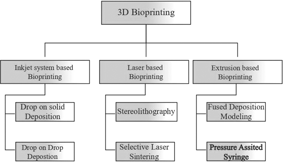
Types of 3D printers. 3D, three-dimensional.
Working Mechanism of Three-Dimensional Printers
3D, three-dimensional; CAD, computer-aided design; CAM, computer-aided manufacturing; SLS, selective laser sintering; UV, ultraviolet.
Inkjet printing
Among various bioprinting models, inkjet is the first bioprinting technology. This process of printing is performed in two steps where droplets (ink) are directed to the desired location of the substrate, followed by interaction between the droplets and substrates. Drop on solid deposition and drop on drop deposition are the types of inkjet bioprinting where drop on solid is repeated to create a layer-by-layer 3D structure. The thickness and space between the layers are optimized for improved adhesion 7 whereas in a drop-on-drop deposition, the printer head releases the droplets onto each other, thus producing a solid layer.
This makes a high-resolution 3D structure by producing droplets of around 100 m in diameter. The printable fluid is chosen with optimum viscosity and volatility to avoid nozzle clogging and fluid leakage, such that it is suitable for jetting and fast solidification. 8
Inkjet system-based printing for tablets
There is a widespread usage of inkjet printers for the construction of cell-free organ models. The applications include the structuring of skin tissue by using cells such as keratinocytes for the epidermis, fibroblasts to form the dermis, and collagen for the dermal matrix. 9 Acrylated peptides, acrylate poly (ethylene glycol), and human mesenchymal stem cells (hMSCs) are used to print the bone constructs. Graham et al used droplet-based bioprinting to generate renal tissue using human embryonic kidney cells (HEK cells) and MSCs. 10
The manufacturing of tablets using inkjet bioprinters differs from the other standard approaches. Khaled et al investigated the differences between conventional methods and bioprinting techniques for tablet fabrication. According to the findings, 3D inkjet printing leads to more porous and friable tablets. 11 The reason for higher porosity is the incomplete contact with the printed binder solution. 12
As a result, these features are helpful for the drug's quick disintegration with a minimal amount of water. Further, bioprinters use several types of layers to handle the difficulty of combining a complicated medication dose into a single tablet and controlling drug release. Thermal actuators are used by the thermal inkjet (TIJ) printer to create droplets. Recent investigations have employed TIJ printers to bioprint oral medicines with precise medication doses. 13
Laser-based printing
This technique has several features, such as being nozzle-free and a non-contact printing process that deposits the biomaterial on a substrate in a precise manner. High speed and precision are prime benefits of laser-based printing (LAB) to replicate the anisotropy and intricacy of the tissues using biological factors.
Stereolithography (SLA) and selective laser sintering (SLS) are further classifications of LAB where SLA utilizes ultraviolet (UV) light for the printing of tissue scaffolds that are printed precisely with controllable configuration and porous structure due to its high resolution. 14 This technique helps form tablets with smooth surfaces while providing modified or delayed release depending on the resin used. In addition, this technique does not require coating, thus reducing the volume of the drug and coating cost. On the other hand, laser is used as a power source to make solid 3D structures. The process of sintering is to compress the drug powder, thereby forming a tablet. 15
Extrusion printing/nozzle-based printing
Extrusion-based bioprinting is among the most widely applied approaches due to its flexibility and affordability. The basic principle of this technique is to produce continuous filaments via extrusion force. It can print biomaterials with an extensive variety of viscosities and concentrations of cells. Semi-solid extrusion (SSE) 3DP is commonly used in the tablet manufacturing industry. Three-dimensional printed tablets can be made chewable, with variable release profiles such as instant or controlled release, and unique properties such as high medication loadings that can be adapted to each patient's needs. 15
Further, SSE 3DP may be used to make immediate-release tablets with large drug loads, as evidenced by paracetamol tablets with drug contents of up to 80% (w/w). These tablets were able to release at least 90% of the medication after 10 min, and their physical and mechanical qualities met United States Pharmacopeia criteria. 16 Another study observed that the large drug loads could be achieved by using SSE in a study that generated levetiracetam tablets with a drug load of 96% (w/w).
The tablets were made in a variety of shapes (cylinder, torus, and oval), with nearly all of them achieving a drug release rate of more than 85% within 15 min. However, the torus-shaped tablets with 50% infill had the quickest drug release (97.45% in 2 min). Other studies have employed levetiracetam to make quick-release pills, but by altering the tablet volume, it was feasible to get varied drug doses. 17
Fused deposition modeling
Fused deposition modeling (FDM), also known as fused filament fabrication (FFF), is the most used 3DP technique adapted in the pharmaceutical, bioengineering, and food industry. A molten thermoplastic polymer filament is printed as a solidified plate when extruded through a high-temperature nozzle. Before extrusion, the drug and thermoplastic polymer are mixed by melting or incubating in specific solvents at desired temperature. This is generally used to modify the drug release profile, even for complex drugs. 18
Currently, the most common 3DP method utilized in pharmaceutical preparation is FDM. Three-dimensional printing may be used to create molds for pills or to directly manufacture pills utilizing medicine powders as raw materials. To print a 3D object, the printer nozzle lays the ink containing the binder and the powder of the active pharmaceutical ingredients (APIs) layer upon layer. For example, Goyanes et al incubated PVA (polyvinyl alcohol) filaments in an aminosalicylate solvent solution before using PVA filaments as raw material to print tablets with varying filling percentages using FDM 3DP technology.
As a result, 3DP has immense promise in individualized treatment because it can produce tablets in a variety of forms, sizes, and API percentages. Three-dimensional printing provides the ability to modify the outward shape and interior structure of tablets, allowing for variable release profiles. 19
Pressure-assisted syringe
Pressure-assisted micro-syringes are another type of nozzle-based printing that involves using viscous and semi-liquid materials extruded through a micro-syringe. This technique works like an inkjet printer, that is, the semi-liquid material is released by compressed air. This technology can produce microstructures of less than 5–10 μm. 20
Extrusion printing application
Extrusion bioprinting can print tissues by consuming various ranges of bioink such as cell-carrying hydrogels, micro-carriers, and cell aggregates. The liver model is constructed by alginate encapsulated HepG2 cells, whereas collagen bioink with Caco-2 cells is used to reconstruct intestinal villi. An attempt was made to construct the nervous system using this bioprinting system, consuming cervical ganglia and hippocampal neurons. 11 Cartilage constructs are formed with the combination of alginate and nanocellulose, which is an extremely printable bioink.
Three-Dimensional Fabrication of Biomimetic Tissues and Organs
Three-dimensional printing is precise and flexible creation of multiscale, multi-material, and multi-functional 3D biomimetic materials and structures. With the growing development of 3DP technologies, the challenges faced during the production of biomedical devices with advanced biomimetic materials and architectures for numerous purposes were addressed. Integration of 3DP with biomimicry encourages developments in the creation of functional materials and structures for future engineering systems, leading to breakthroughs in a variety of biomedical applications.
Scaffolds with 3D porous structures are used to obtain adequate mechanical characteristics for tissue regeneration. A milieu for cell adhesion and nutrient exchange is created by porous materials with adjustable pore size and distribution. 19 With the progress of tissue engineering, 3D scaffold-based implants play a significant role in replacing and repairing tissues and organs. Researchers attempted to establish a biological environment comparable to that of real tissue by using biomimetic scaffolds and inventing a printing technique. Recent advances in biomimetic scaffold manufacturing of soft and hard tissues are described. 21
Three-dimensional bioprinting of soft tissues
Skin tissue
Skin burns account for more than 11 million cases every year and account for the most common type of trauma worldwide. For small to moderately sized burns, primary closures are performed, which involves removing devitalized tissue followed by wound closure. However, for severe burns, such as those deep in the dermis, the common clinical intervention is skin grafting, which involves covering the wound bed with skin grafts.
Limited donor skin, biocompatibility, and immunological issues are the significant limitations of allografts. Three-dimensional bioprinting facilitates the printing of complex tissue architecture and in situ bioprinting, that is, printing pre-cultured cells with growth-promoting scaffolds on the burnt site, accelerates cell deposition at precise positions, limiting the need for multiple surgeries. 22
The robotically controlled bioprinters map the damaged skin via laser scans and with bioinks constituting collagen with primary adult human dermal fibroblasts and epidermal keratinocytes, deposit layer-by-layer on the defect area. Extrusion printers enable the deposition of bioinks on the dermis, including hair follicles, sweat, and sebaceous glands, which helps the accelerated maturation of skin. The use of 3D bioprinting for skin reconstruction after burns is promising as there is faster healing, reduced scarring, and better cosmetic outcomes. 23
Many tissue fabrication techniques have been used in the synthesis of skin tissues, culminating in the development of tissue replacements such as autologous split-thickness skin grafts, allografts, acellular dermal substitutes, and commercial goods such as Dermagraft and Apligraft. 24 Pourchet et al created a two-layered dermis and epidermis 3D cell printed full-thickness skin by utilizing new bioprinting technology. Gelatin and fibrinogen were combined to make the bioink.
Further, they implanted human dermal cells in the mentioned combination and printed a dermis construct. The skin replacements with a thickness of 5 mm were then created and seeded with human epidermal keratinocytes over the dermis construct. Natural human skin histological-like properties of the 3D cell were seen after 26 days of incubation. 25
Zhao et al created gelatin methacryloyl (GelMA) hydrogels in various concentrations for a monolayer skin modernizing method. The GelMA hydrogels have good physical characteristics, and they systematically vary the concentration of GelMA to govern keratinocyte adhesion, proliferation, and differentiation. To obtain a keratinocyte suspension, a hydrogel scaffold was employed, which was then used to produce the reconstruction of the categorized and functional epitomes.
This study found that by varying the concentration of GelMA pre-polymer solution, the physical and biological characteristics of the resulting hydrogels could be properly adjusted to suit the criteria for epidermal development. 24 The GelMA hydrogels aided in the creation of a stratified epidermis with a specific barrier function, such as electrical resistance and water loss prevention. Visually and physiologically, replacing natural human skin with 3D-printed skin is not difficult, with the 3D-printed skin being strengthened by histology and immunofluorescence qualities.
This approach has a wide variety of applications in skin design, toxicity research, and wound healing since it covers the entire thickness of the transdermal and localized wound formation. According to their findings, proteins and printing cells in nanodroplets had minimal effect on cell survival and function findings. 24
Brain tissue
The central nervous system has poor self-regeneration ability, and hence tissue engineering alternatives are considered to stimulate tissue healing, regeneration, and the recovery of neural functions. Recent work shows advancement in neural engineering by using fibrinogen-based bioink using an extrusion-based bioprinter, which has significantly shown increased survival and growth of the neurons.
Another study discovered neuronal markers on a fibrin/alginate printed scaffold with pluripotent stem cell-derived neural using Lab-on-a-Printer technology. 19 It was observed that the cells which expresses specific markers for mature neurons in a mimetically formed spinal cord measuring 1 cm (about 0.39†) in diameter with seven layers of fibrinogen loaded human-induced pluripotent stem cell (iPSC) derived neural progenitor cells. 19
The in vitro 3D glioblastoma tumor microenvironment has greatly aided high-throughput drug screening platforms. Stem cells in a fibrinogen/gelatin/alginate solution demonstrate high cell survival and proliferation rates. It was observed that biomaterials and cellular composition in bioink conserved the phenotype of the cell and promoted the screening of chemotherapeutical drugs that exhibited increased drug resistance. 26
Vascular tissue
The engineering of the blood vessels is considered quite challenging due to their small diameter, hierarchical organization, and hollow arrangement. Endothelial cell manipulation has been shown in experiments to effectively increase angiogenesis and vascular maturation during bioprinting. Sriphutikat et al aligned human umbilical vein endothelial cells embedded in fibrin-GelMA solution and witnessed the formation of capillary-like structures and the in vitro generation of networks of new blood vessels. 27
Extrusion-based bioprinters were used to print vascularized tissues-on-a-chip scaffold that contains gelatin, fibrinogen, and human neonatal fibroblasts inside microfluidics equipment. A hollow structure is constructed on a chip by using Pluronic F127, which allows the migration of endothelial cells outside and the building of interconnected channels that ultimately form a vascularized tissue. A mixture of platelet-rich plasma and alginate is used as a bioink to form a patient-specific bioink to print a fibrin-alginate network that is rich in growth factors to induce vascularization. 19
Liver regeneration
The liver comprises parenchymal liver cells that make up 80% of the hepatic mass (hepatocytes) and 20% of non-parenchymal cells, including kupffer cells, sinusoidal cells, pit cells, and hepatic stellate cells. 28 Despite the liver's having high regenerative ability, chronic liver damage does not restore the normal function, homeostasis, and microarchitecture of hepatic tissue. Therefore, liver engineering has the potential to bridge the gap between organ availability and finding an alternative orthotropic liver transplantation strategy. 29
Three-dimensional printing aims at developing bioartificial livers using bioinks to recreate 3D microarchitecture and extracellular matrix (ECM) protein composition. The hepatic ECM is a complex network of macromolecules that is not only involved in the regulation of cellular functions but also provides a natural extracellular scaffold for the cells. Biomaterials used in liver regeneration processes can be natural polymers that include polysaccharides (hyaluronic acid, chitosan), proteins (collagen, blends, gelatin, and fibrin), decellularized matrixes, or synthetic matrices (aliphatic polymers) etc. as represented in Table 2.
Biomaterials for Liver Regeneration
ECM, extracellular matrix; HUVEC, human umbilical vein endothelial cell; MSC, mesenchymal stem cell.
Xiang et al and Zheng et al created decellularized splenic scaffolds with well-preserved 3D ultrastructures that included the native vascular network as well as ECM components, in addition to decellularized liver tissue. 30 They discovered that a decellularized splenic scaffold exhibited similar ECM components, architecture, and mechanical characteristics, resulting in comparable performance to a decellularized liver scaffold.
When liver donation is not possible, the results show that a decellularized splenic scaffold can be used in place of a decellularized liver scaffold. Skardal et al revealed that extracts from acellular liver ECM or complete fresh liver tissue may be mixed with other natural polymers such as type I collagen, hydroxyapatite (Hap), or heparin-conjugated HA to generate hydrogels that increase primary human hepatocyte cell survival and function. 31
The benefits of ECM-based supplements are most likely attributable to the ECMs being a source of several unique biological components. Heparin-conjugated hydrogel has a stronger potential to support hepatocytes because heparin may firmly bind many growth factors, increasing their stability and bioactivity. Nakamura and Ijima used a similar method to create a scaffold of liver extracellular matrix (LEM) for hepatocyte cultivation. They attached HGF to the LEM scaffold to boost hepatocyte liver-specific activities. 32
Tubular organs
Three-dimensional printing plays a preeminent role in the treatment of tubular organs and tissues. Many tubular organs and tissues, including the trachea, esophagus, veins, arteries, and bladder, require therapeutic intervention for treatment to overcome diseases and malfunctions. Autologous transplantation is the most appropriate treatment for many organs apart from the esophagus and trachea due to non-existent autologous tissues. Fabrications of tubular organs and tissues have many limitations that are overcome by the 3DP technique.
It can form multilayered structures, providing a suitable microenvironment. A double-syringe Liquid-frozen deposition manufacturing platform was used for the development of a tissue-engineered trachea. In the 3DP constructs, MSC was seeded, which was implanted in mice, and they were able to remain tubular for 6 weeks. In addition, the scaffold showed excellent elasticity and sufficient mechanical properties to survive without collapsing and replacing a part of the rabbit trachea. This tissue-engineered fabrication of the trachea shows much potential in the future of customized tracheal tissue engineering. 33
Zhang et al fabricated a cell-laden urethra using PCL and PLCL polymers. The scaffold was fabricated so that it could mimic the original properties of the native urethra of rabbits. Two types of cells were seeded evenly: urothelial cells in the inner layer and smooth muscle cells in the outer layer of the scaffold. As a result, the scaffold proliferated and expressed cell biomarkers and maintained the microenvironment.
34
Shink'oka et al performed clinical trials of tissue-engineered vascular grafts fabricated using polymers such as poly
Tissue engineered vascular grafts (TEVG) was seeded with autologous vascular cells for the first few surgeries. Many limitations were observed for this method, including that it was time-consuming, the proliferation of cells took ∼60 days (about 2 months), and it was an invasive technique that required harvesting healthy cells from the patient. The limitations were overcome by seeding the in vitro scaffold with autologous bone marrow-derived mononuclear cells. The TEVG was found to be working fine even after 2 years of implantation without any indications of thrombosis, stenosis, or calcification. Atala et al. engineered bladder tissues to treat patients with end-stage bladder disease using scaffolds made of collagen and polyglycolic acid. 36
The scaffolds were bladder-shaped and seeded with urothelial and muscle cells. After 7 weeks, the bladder construct was implanted. It was observed that there was an increase in the volume of omentum-wrapped bladders along with a return to normal bowel functioning. The engineered bladder has the potential for the treatment of patients with cystoplasty. 36
Three-dimensional bioprinting of hard tissues
Cartilages
Cartilage tissue constructs are formed by combining rigid and soft biomaterials to match the microenvironment by supporting the mechanical and cell survival ability, thus promoting the preservation of cell viability. de Melo et al reported a printed ear-shaped structure with alternative layers of PCL with auricular chondrocytes and fibrinogen/gelatin/HA/glycerol/Pluronic F12, wherein PCL supplies strength and long-term stability, whereas fibrinogen-based bioink protects the cells from physical stress. 19
For cell survival, fabrication of cartilage tissue-like with spatially organized mechanical properties is highly stressful for cells. The cartilage tissue-like construct was stiff at the macroscopic level to simulate cartilage stiffness but maintained softness at the cellular level to preserve cell viability. To construct this structure, hMSC spheroid-laden fibrinogen bioink was bioprinted using an extrusion-based bioprinter in a support bath composed of poly-ethylene glycol (PEG)/alginate and supplemented with thrombin.
Results showed the softness (2.5 kPa) and highly diffusive capacity of fibrin, which contributed to high cell viability and chondrogenesis over a 21-day culture within the stiff (3 MPa) microenvironment. In another approach, both mechanical strength and cell survival were assured by bioprinting alternating layers of PCL and fibrinogen/collagen/chondrocyte bioink.
PCL was printed using an electrospinning technique, whereas the bioink was deposited over the PCL layer using an inkjet printhead. The construct presented a Young's modulus comparable to that of native cartilage (2 MPa) while it also favored cell spreading and chondrogenesis both in vitro and in vivo. 37
Bones
3DP has been extensively investigated in the field of bone tissue engineering. Leukers et al investigated HA scaffolds for bone tissue engineering utilizing 3DP. 38 They developed a unique test in which mouse calvaria 3T3-E1 (MC3T3-E1) cells were cultivated on scaffolds and maintained under static and dynamic conditions, followed by a histological analysis to measure the proliferation of these cells. In essence, the dynamic culture process produced a more powerful population than the static culture process.
By developing a scaffold arrangement with 45 inclined levels, the cells were formed into the framework, producing a practically indirect contact with the HA granules. Since the cells made the structure more integrated and prevented them from sliding down, this design hastened the seeding technique and boosted cell attachment. Cells are deposited in layers on the surface of the HA granules in a static culture, whereas cells grow within the cavities of the HA granules in a dynamic seeding process.
Brunello et al. explored the use of 3DP of powder for bone tissue engineering. 39 Powder-based 3DP is promoted as a unique bone remodeling approach since the outer frame, inside structure, permeability, and 3D-printed physical qualities of bone substitutes may all be altered and hence employed for specific reasons. 39 3DP of stem cells and polymer/bioactive glass compound scaffolds was accomplished for the engineering of bone tissue.
They employed 3DP methods to make a PCL/bioactive borate glass composite as well as human adipose stem cells in their study, using a two-syringe approach to make a scaffold with a biopolymer/bio-glass composite. 40 This composite's substance dissolved in an organic solvent as a scaffold, whereas concurrently printed cells remained suspended in the Matrigel. They discovered that the amount of borate glass in the scaffold might affect its printability, temporal bioactivity, degradability, and cell survival. The extrusion bioprinting approach has significant advantages in that it can create a scaffolding structure that supports cells while also providing form and mechanical integrity.
The extrusion bio-printing approach has significant advantages in that it can create a scaffolding structure that supports cells while also providing form and mechanical integrity. Extrusion bio-printers often have many syringes, one of which is specialized for printing scaffolding structures. The alternatives employed for this matter were polymer molten deposition and FDM with a polymer wire feed. Also, the pore size factor is significant since it could affect bone formation after implantation. 41
Three-dimensional bioprinted structures are of potential use in obtaining medical images for treating the disease condition of bone fractures. Kang et al reported that fibrinogen-based bioink stimulated the formation of vascularized bone tissue in vivo. 40 To ensure the mechanical strength and cell viability, 3D-printed PCL and PLGA are placed in cell-laden fibrinogen among the microchannels of the structure. Fibrin stimulates cell viability due to its softness, and the polymeric support of PCL and PLGA supplies compressive strength. 40
Strategies for 3D bioprinting fibrin-based soft and hard tissues
Some of the most common strategies for bioprinting fibrin-based 3D geometries and architectural mimicry of tissues include combining fibrinogen with viscous biomaterials with sol-gel properties, using a support bath to hold the printed structure, and in situ crosslinking. To stabilize gelatin for this purpose, its structure was altered to induce covalent crosslinking between the polymeric chains. GelMA is one such modified structure that is commonly employed in tissue engineering, particularly in 3D bioprinting. When exposed to UV radiation, GelMA is chemically crosslinked, allowing the development of structures with guaranteed bioink printability, mechanical strength, and good biocompatibility.
When coupled with fibrinogen, GelMA improves bioink printability. Further, due to the creation of an interpenetrated polymer network between GelMA and fibrin networks, the dual crosslinking capability of GelMA-fibrin results in a stronger and more durable construct. A third biomaterial, such as alginate, might be added to a gelatin/fibrin polymeric composite to boost its mechanical stability. By using dual crosslinking (enzymatic and ionic), alginate can prevent the early breakdown of a fibrin/gelatin mix. 42
Further, alginate-containing fibrinogen/gelatin has demonstrated efficacy in stimulating cell function when used as bioink in the construction of in vitro models useful for studying cellular mechanisms in 3D tissues such as hepatic mechanisms, metabolism, and tumor biology. Synthetic polymers, such as PEG, can also be added into fibrinogen to provide bioprintable and cell-friendly formulations, enabling variable printability, strong mechanical characteristics, and high cell viability for tissue engineering. 19
Three-dimensional printing for designing disease model
The prime objective of 3DP is to match the in vivo environment, and thus the biomaterials are cautiously selected to support tissue repair and regeneration. Three-dimensional printing is carried out to generate disease models that encode shortcomings as the in vitro disease models are insufficient to analyze the pathological condition. The 3D disease models validate the drugs and therapy treatments, drug-metabolizing, and dose responses. The models are effective in deciding the dose-response, minimize the use of animals for clinical trials, and closely resemble the in vivo model. They also help in planning surgery, wherein models are used to understand the disease pathology.
Liver models
Different 3D bioprinting approaches have been utilized to create liver tissue constructs. Faulkner-Jones et al reported the use of an inkjet-based bioprinter to encapsulate human iPSC and embryonic stem cell-derived hepatocyte-like cells (HLCs) in alginate hydrogels to create 3D ring structures. 7 Alginate was chosen based on its excellent biocompatibility, low immunogenicity, low toxicity, and hydrophilic nature. The cell-laden alginate droplets were exposed to calcium chloride solution followed by barium chloride before incubating in a culture medium.
The viability and albumin-secreting function of HLCs were well maintained following this valve-based bioprinting, which indicates the functioning of the 3D model. Extrusion-based bioprinting was used to generate a 3D hepatic structure by the scientists. 7 A five-layer alginate scaffold holding mouse-induced HLCs, each measuring 25 by 25 mm (about 0.98″), was constructed. During in vitro culture, the expression of albumin, ASGR1, and HNF4a gradually increased.
The construct was also transplanted in vivo, with increased proliferation and higher albumin expression observed. This work demonstrated the use of a 3D bioprinted liver scaffold as a practical choice for liver therapy. Kizawa et al also demonstrated a scaffold-free 3D bioprinting technology to build liver tissue that could stably maintain bile acid secretion as well as drug, glucose, and lipid metabolism for weeks. 43
This was achieved by connecting spheroids of human primary hepatocytes using the 3D printer. Their work provided insight into the long-term culture of 3D bioprinted liver constructs in vitro. In particular, the group studied the expression and activity of CYP3A4 enzymes and showed that both were maintained for around 2 months. To mimic the complex microarchitecture of the liver, Ma et al reported the use of DLP-based bioprinting technology to build biomimetic liver tissue at microscale resolution. 44
Witowski et al fabricated liver model that was under $150, full sized, and that maintains the similarity with the shape, structures of colorectal liver metastasis. 45 Computed tomography (CT) of patients was used to analyze the structures and printed using a desktop 3D printer. The structures were fabricated and filled with silicone. The basic idea of fabricating such model is to reduce operative time and improve short-term outcomes. 46
Heart model
Congenital heart surgery affects the baby because of pre-existing heart conditions after birth, which can be life-threatening and require immediate surgery in many cases. The surgery is done by an incision on the wall of the atrium, arterial trunk, or ventricle. The surgery is quite challenging and is predominantly performed in children, wherein the size of the thorax and heart are relatively small.
Three-dimensional printing recreates the anatomy of the patient's heart with the help of a database obtained from imaging the patient's heart, showing the pathology of the disease, and can be a better model for demonstrating and performing trial surgery Figure 3. 47 This model can be used for hands-on training for surgeons before the surgery of the patients, improving the surgical skills and success of the operation. Biglino et al used CT-angiography images of patients' congenital heart and made replicas via 3DP using resins as a study model. 48

Three-dimensional printed heart model (reproduced from Ref. 84 open access pharmaceutics).
The models were then provided to surgeons and surveyed to understand the disease in the printed model. After statistical comparison, it was estimated that 3D models were highly beneficial for surgeons to understand the disease physiology and polish their skills to perform surgery. 49
Renal model
The feasibility and accuracy of 3D-printed models in renal surgery were reviewed in detail by Sun and Liu and pointed out an effective utilization of 3D-printed renal models. The 3D-printed model can be used as a practical tool to understand invasive renal nephrectomy and explain to the patients their disease status. It can also clarify any doubts raised by surgeons and make them prepare for complicated surgeries. 50
Kyung et al fabricated a 3D kidney model for the patients who were supposed to undergo partial nephrectomy. 51 The main objectives behind developing this model are to have a better understanding of the individual tumors and to predict the surgical outcomes. Inhouse software (Asan Medical Center, Seoul, South Korea) helped in generating a virtual 3D model that showcased the anatomy of renal parenchyma, renal mass, renal vasculature, and collecting system.
The model was fabricated using an Objet 500 CONNEX3 3D printer (Stratasys), and fusion of fluid binding substances was used to print the model. With the help of CT imaging, 17 models were fabricated for 17 patients, and all the patients successfully underwent PN with complete removal of renal mass. The kidney model clearly presented the kidney parenchyma, mass, vessels, and collecting duct systems. In 14 cases, the collecting ducts opening was rightly predicted and the usefulness of the 3D printed kidney model was found to be satisfactory. It helps in improving the patient's understanding of the disease, surgical planning, and simplification of the process, thus helping in overall surgical outcomes. 52
Ligament and bone model
Three-dimensional printers demonstrate the efficacy and success of developing economically customized implants. Liu et al conducted an experimental study demonstrating a polylactic acid (PLA) screw developed for the anterior cruciate ligament (ACL) and implanted in a rabbit model. The screw model was designed using Rhinoceros software (ver. 4.0) according to the requirements of the ACL reconstruction model. The screws were made of biocompatible PLA and were orthogonal and porous with HA to improve osteoconductivity. 53
Screws also had MSCs for accelerating bone ligament healing via cell therapy. Post-implantation results showed that the screws were stable without cracking or rupturing and promoted tendon graft healing within the bone tunnel, which was confirmed by an elevated level of bone growth and favorable bone-graft interface formation. The study highlights the effectiveness of low-cost 3DP in personalized medical implant manufacturing. 54
Sterilization of 3D-printed structures in the form of surgical tools or implants can be a significant limitation. The use of thermoplastic biodegradable polymers in the 3D-printed forms cannot be autoclaved and hence may require radiation and ethylene oxide such as polyetheretherketone. 55
The conversion of radiography data into 3D-printed structures is referred to as 3Dgraphy. In an interesting report by Bagaria and Chaudhary, the 3D-printed models for nearly 50 different bone-related surgeries were developed and provided to surgeons before the operation. The effectiveness of these models was reported by all the surgeons who practiced and found better mental exercise and a realistic approach to the radiographic data that was transformed into 3D models.
Timeless Innovation Labs (Mumbai, India) developed an FDM-based Wanhao Duplicator Printer with a 0.1 mm (100 mm) resolution. Thermoplastic resin acrylonitrile butadiene styrene (ABS) was used as printing material, sterilized via ethylene oxide, and delivered to the surgeon before the surgery. 56
Takahashi et al fabricated a 3D temporal bone model using a powder bed of plastic and an inkjet printer. The basic objective of this model was to overcome the problem of reproducing air-containing spaces and middle ear structures. The air spaces were created using drainage holes that removed excess materials, and the middle ear part of the ossicle ligament was changed to a bony structure.
The assessment was done using macroscopic and endoscopic inspection of the model, comparing the CT of the model with the original, and some surgeons performed surgery to check the tactile sensation of the model. It was observed that almost all the structures were reproduced successfully and the model was sufficient for surgical training. 57
Table 3 demonstrates the commercial bioinks available along with the bioprinters that print the materials used for biomedical applications.
List of Commercial Bioinks
Alg, alginate; BMP, bone morphogenic protein 2; CHO, Chinese hamster ovary; Co11−, collagen; dECM, decellularized extracellular; FGF, fibroblast growth factor 2; GAG, glycosaminoglycans; GelMA, gelatin methacryloyl; GFOGER, hexapeptide sequence found in collagen; HA, hyaluronic acid; HAD, human acellular dermis; HAoEC, human aortic endothelial cells; Hap, hydroxyapatite; HDF, human dermal fibroblast; HepG2, human liver cancer cell line; HS, heparan sulfate; LAP, lithium phenyl, 2,4,6 trimethyl benzoyl phosphinate, photoinitiator; MEF, house embryonic fibroblast cells; MMP, matrix metalloproteinase; PCL, poly(ɛ, caprolactone); PLA, polylactic acid; PLGA, polylactic-co-glycolic acid ; RGD, arginine, glycine, aspartate motifs; SIS, small intestine submucosa; SMC, smooth muscle cells; TGF, transforming growth factor; TPP, tripolyphosphate; VEGF, vascular endothelial growth factor.
Three-Dimensional Printing in Medical Device and Implant Manufacturer
Device manufacturing by employing 3DP technologies gives precision and accuracy and has the ability to design complex structures. Table 4 lists the commercial 3D-printed medical devices available.
Devices Commercial Three-Dimensional Printed Medical
ABS, acrylonitrile butadiene styrene; BVS, bioresorbable vascular scaffold; CYP3A45, cytochrome P450 3A4; GGT, gammaglutamyltransferase; RPTEC, renal proximal tubule epithelial cells.
Surgical devices
MakerBot Replicator 2 (MakerBot, Brooklyn, NY) bioprinter with MakerBot MakerWare software generates Gcode to develop a PLA-based surgical retractor. The instrument measured 17 cm × 1.5 cm × 4 mm and was printed with 75% infill. Retractors are ubiquitously used instruments in all types of surgery that are used to hold the underlying tissues and organs. The developed retractor was sterilized with glutaraldehyde at room temperature, was found to maintain the aseptic condition, and retained the strength of PLA. 58 The study shed light on effective and repeatedly sterilizable retractors that can be created at a low cost using 3DP technology.
Surgical graspers
Surgical graspers employed during surgery have evolved with the evolution of surgical methods. Open surgery has evolved into minimally invasive surgery. Minimal invasive surgeries give an added advantage of smaller incision size by preventing the direct exposure of the area and decreasing the risks of infection. Devices employed for minimally invasive surgery have evolved into intelligent devices with excellent maneuverability and better reachability, and they have been devised with several degrees of freedom.
The instruments must be flexible to turn and move through curves of variable diameters to accomplish the minimally invasive surgery (MIS). Conventional devices available for MIS are stainless steel with a jaw at their end, manually actuated, 59 as shown in Figure 4A. These conventional devices have low maneuverability and are not able to follow tortuous and complex curves. To improve steerability and flexibility, structures with complex shapes need to be integrated.
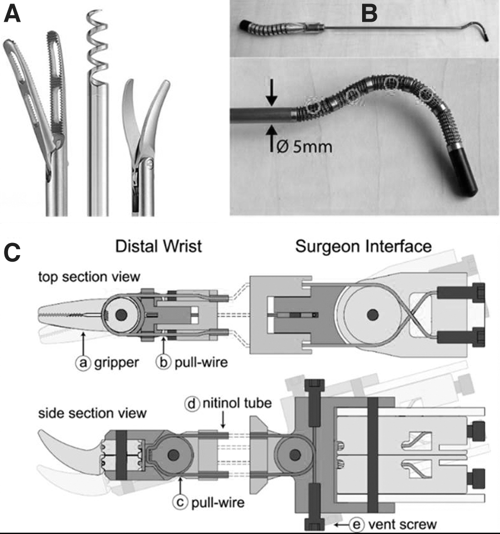
Gerboni et al proposed a manually actuated device, the Helixflex, a bioinspired, maneuverable instrument for skull base surgery, shown in Figure 4B. The Helixflex had a four degree of freedom steerable tip with excellent stiffness. 60 Riojas et al have reported a similar surgical device with an elbow and arm shown in Figure 4C. 61 Although these handheld devices have provided flexibility and maneuverability, they are still complex devices with complex shapes and are not suitable for sterilization. 3DP technology can solve the given challenges.
Culmone et al have fabricated a Helicoflex of R5 epoxy photopolymer resin for MIS with 10 degrees of freedom using three 3D-printed parts and can perform fluid motion on paths with curvature. 62 Henselmans et al have 3D printed a manual device, Memo Flex II, for MIS with 32 degrees of freedom using R5 photopolymer. 63
Surgical tool
Surgical tools include scissors, needle drivers, forceps, and scalpels. Conventional surgical tools are fabricated using stainless steel, which provides durability due to high stiffness and smoothness; high heat resistance for sterilization; and easy washing. However, these stainless-steel tools are heavy, incompatible with imaging modalities, and require a longer manufacturing time. Moreover, heavy tools carried by the medical staff during washing and surgeries cause extra physical effort and become of utmost significance when transported in bulk to the hospitals during a pandemic. 64
Also, the presence of ferromagnetic material during magnetic resonance imaging (MRI) and CT would cause substantial damage to image formation due to magnetization.65,66 However, 3DP technology provides a solution by reducing the production time and easing the fabrication of surgery-specific lightweight devices. Several resins have been employed for the fabrication of surgical tools, including Polyamide,67,68 ABS, Polypropylene reinforced with glass fiber fillers, nylon reinforced with glass fiber fillers, and polyphthalamide reinforced with carbon-fiber fillers.8,69
George et al have 3D-printed surgical tools using DuraForm EX plastic powder and are further employed for inguinal hernia repair. The major limitation of the given 3D printed needle was the slippage of the needle during suture driving. 69 Researchers in polymer matrices have introduced carbon-fiber filler to fabricate lightweight 3D-printed surgical tools and increase their strength. Polyphenylene sulfide (PPS) resin reinforced by short carbon-fiber fillers (A630T-30V) was employed by Mekata et al to fabricate 120 mm long ads on tweezers, DeBakey tweezers, long forceps, long needle holder, cooper scissors, and Metzenbaum scissors. 70
Due to PPS resin reinforced with short carbon-fiber filler, the weight reduction took place by one-third compared with stainless-steel surgical tools and had higher mechanical performance than other resin-based surgical tools. 70
A needle driver holds the suturing needle to close the skin during suturing and other surgical procedures. Fontanelli et al have fabricated a 3D-printed robotic needle driver laparoscopy tool that mimics the motion of a human hand with additional degrees of freedom and have demonstrated the reduction in time of suturing. 71
Surgical models
Preoperative planning and practice are of immense importance to avoiding medical errors during clinical practice. Imaging tools such as MRI and CT scans have been used as primary tools for planning. However, additional aid for preparation would assist in complex surgical procedures involving atypical anatomical abnormalities. They could help in improving spatial sense and anatomical precision. Three-dimensional printing technology has provided the ability to create patient-specific organ models, which will provide an effective way to perform spatiotemporal mapping and preoperative preparation.
Simulation surgeries on 3D-printed models would allow surgeons to adopt better strategies and conduct feasibility tests. 72 Blaszczyk et al have reported the fabrication of a 3D-printed arterial Circle of Willis model using PLA for the clipping of intracranial aneurysms and demonstrated the steps required to produce patient-specific 3D models from medical image data, which is of significant importance for preoperative planning. 73 Researchers have reported that surgeons trained with physical models or prototypes have better skills than those who did not have that training. 74
Typically, surgical prototypes are 3D printed using Nylon 12 printed by SLS, filaments by FFF, silicone extrusion by direct ink writing, resin polymers through photopolymerization by SLA, and liquid polymers through material jetting followed by UV curing. In mono-material prototypes, it is not easy to distinguish various anatomical features of the prototypes. Due to this, multi-colored prototypes are more suitable and preferred for preoperative preparations. 75
In addition to cardiovascular and orthopedic models, 3D prototypes have also been designed to mimic various tissues. Different tissues have been 3D printed after considering the following factors, including elasticity, density, and shore. Materials employed for printing soft tissues include poly (vinyl) alcohol, phytagel, polyvinyl chloride, agar-agar, gelatin, hydrogels, or silicones. Due to their biocompatibility, cell adhesion, and proliferation, hydrogels have been gaining popularity. However, it is still challenging to tailor their mechanical properties. 76
Life support systems
The scarcity of life support systems such as oxygen concentrators, incubators for prematurely born babies, and ventilators has been observed during pandemics. Especially during the COVID-19 pandemic, a considerable fraction of the population got hospitalized and needed life support systems and other devices shown in Figure 5. 77 However, the availability and production of life support systems were so lethargic that many countries faced a shortage of life support systems. To overcome this shortage, an initiative to set up new plants for the manufacturing of life support systems was taken by the government.
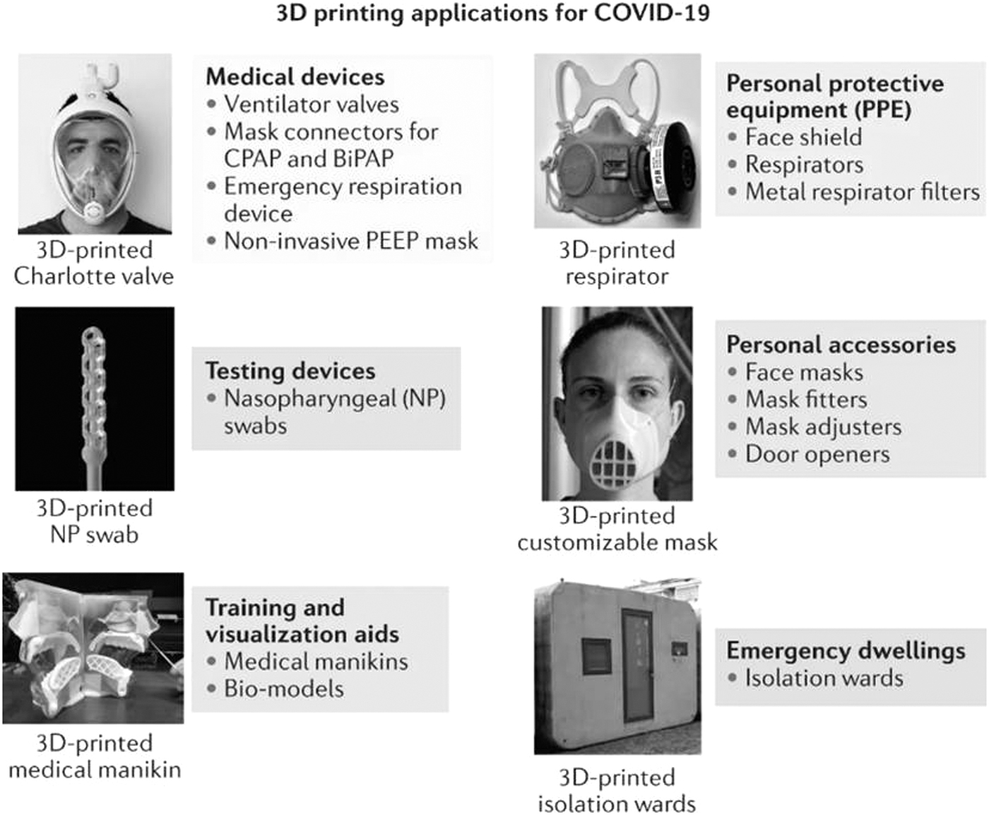
Manufacturing for life support systems and other device manufacturing supported by 3D printing (reproduced from Ref. 49 open access journal of clinical medicine).
As a result, the setting up of new plants has increased production. However, the time required during the fabrication remains the same. Moreover, countries required an effective solution to overcome this shortage. Due to this, the manufacturer turned to 3DP technologies to fabricate the life support systems, owing to the lesser time required for manufacturing.
Three-Dimensional Printing in Pharmaceutical Application
Three-dimensional printing technology is the most innovative and powerful tool for precisely manufacturing precisely developed dosage forms, tissue engineering, disease modeling, and pharmaceutical and medical applications. Recent advances in 3D pharmaceutical applications include multipurpose drug delivery systems with enhanced release characteristics; modifiable and tailored dosage formulations and transplants considering the anatomy of the patient; as well as cell- and tissue-based materials for regenerative medicine.
Tailored approaches to drug delivery and personalized medicines made with the help of 3DP aim at meeting the desired manufacturing of individualized pharmacogenomics, physiological, and anatomical demands. The irrefutable benefits of 3DP are highlighted in this part. 78
Three-dimensional printed tablets for communicable/infectious diseases
Lately, a lot of infectious diseases have created havoc around the globe, be it Coronavirus, Ebola, Zika, or Swine flu. Three-dimensional printing technologies can be widely used for combating such viral diseases and against disease-causing pathogens such as bacteria, parasites, and fungi. Haitian cholera and Escherichia coli are examples of such bacteria causing such outbreaks affecting the gastrointestinal tracts of the infected patients.
Tuberculosis is yet another disease caused by a bacterium that is common in countries all over the world. A carefully constructed drug that is specific and targeted for the pathogen can only lead to better results in the treatment of such infections, and it may reduce unnecessary exposure of the drug to other body cells. 79
Three-dimensional printing can provide an effective and precise delivery system for targeting specific sites by constructing a tablet (one layer at a time) batch of the desired fabrication. Although the FDA approved the first 3D printed drug in 2015, nearly all 3D printed drugs are still awaiting approval. Utilizing this technology can lead to the replacement of large batch production of the drug with personalized and limited production.
This can help avoid extra costs and material loss. In such emergency times, it can make a significant difference. Also, by altering the release kinetics of the drug, the frequency of the drug administration can be reduced, which can be advantageous to the health care workers involved in treating such infections in patients as there will be less exposure to the pathogen for them. 80
Communicable diseases show a variety of symptoms that, if controlled at once, can lead to fast healing as well as provide comfort to the patients. When applied to specific drug classes, 3DP can produce remarkably effective results. One of these examples includes analgesics, where immediate release of analgesics can be easily achieved by enhancing the dissolution rate of the drug. Three-dimensional printing has been used to develop matrices providing a high surface area to volume ratio, hence leading to the quick onset of action of the drug, for example, paracetamol (acetaminophen). 81
Yet another possible application of 3DP and CAD includes the treatment of tuberculosis that is caused by Mycobacterium tuberculosis. 82 Short-term combination therapy of drugs is recommended for the treatment, which should be closely monitored. Compartmentalized tablets can be designed with tailored release kinetics of multiple drugs. The drugs as well as the doses can be easily personalized as per the requirements of the patient, hence being able to provide patient-specific treatment.
This can be achieved by altering the thickness of the material to reach the required degradation rate, leading to the release of the drug at specific time intervals. This controlled release reduces the systemic exposure of the drugs as well as their frequency of administration. The drugs prescribed in this multidrug therapy include isoniazid (INZ) and rifampicin (RFC). A bilayer tablet providing optimum dose delivery can be prepared using FDM.
For this purpose, INZ is formulated in a hydroxypropyl cellulose (HPC) matrix that releases the drug in the stomach, and RFC is formulated in a hypromellose acetate succinate matrix that allows the drug to be released in the upper intestine. These were made into drug-containing filaments by hot-melt extrusion (HME), which were further used to print the final tablet. 83 These 3D-printed tablets can provide better clinical efficacy and avoid the interaction of RFC with stomach acid that may lead to degradation.
The PEG-coated tablets can also be easily manufactured using FDM printing that optimizes the floating ability of the drugs that need to be gastro-retentive, increasing the bioavailability of the drug at the targeted site. This can be especially useful for antibiotic drug administration in gastrointestinal tract-related infections. The PEG can also be used to facilitate bioactivity and provide an adhesion site, improving the protein absorption and the cell adhesion of peptide sequences.
Various hydrogels used in 3DP can change the physical and biological properties of the drug. The hydrogels possess excellent mechanical properties without the presence of any cytotoxic agents. When exposed to a specific light, temperature, or ion concentration, it undergoes a sol-gel transition. 84
Three-dimensional printed tablets for non-communicable/non-infectious diseases
Since most severe non-communicable diseases are chronic, long-term medications are prescribed to the patients. This often involves complex dose regimens, combination drugs, and multiple dosing. The main target of drug development in such cases is patient compliance. Consistent treatment is the key to the management of such diseases. The frequency of dosing and the side effects are the major factors that affect the adherence of patients to the therapy.
Here, 3D-printed dosage forms involving personalized medicines and polypills can provide an effective means to increase the acceptability of such therapies. The need for swallowing multiple drugs at a time can be uncomfortable for patients, especially elderly patients. Hence, it would be wise to combine those medications in a way that they do not affect the individual effects of the drugs.
Three-dimensional printing techniques can resolve such problems by formulating polypills according to the dosage requirements of the patient. This is especially useful to patients who require medications for multiple associated diseases. Three-dimensional printing allows the formulation of the desired dosage form with predetermined release kinetics that can provide optimum pharmacokinetic responses.
Diabetes mellitus
Diabetes mellitus is one of the most prevalent chronic disorders all over the world. Beta-cell dysfunction, impaired production of hepatic glucose, and peripheral insulin resistance are the main causes of the disease, resulting in hyperglycemia. The major causes of high morbidity and mortality in diabetes are medication non-compliance with the patients due to the complexity of dose regimens, adverse effects, cost, and polytherapy.
In a recent study, metformin-loaded polymeric fibers were constructed with the help of PVA, and tablets were prepared using the FDM printing technique. The resulting tablets show better release profiles due to the high area to volume ratio of the tablet. These tablets can have variable infill percentages and easily swallowable dimensions, which is generally not the case with conventional drugs. Three-dimensional printed polypills may also provide an efficient solution to the problem.
Some recent studies have been performed to fabricate a bi-layered polypill containing two frequently prescribed drugs, metformin and glimepiride. 85 They are both often prescribed in different dose combinations. Manufacturing such customized polypills can lead to increased convenience for the patient as well as an increase in the adherence of the patient to the therapy.
For this purpose, Eudragit® RL (sustained-release layer) and PVA are used to incorporate Metformin and Glimepiride, respectively. Such bi-layer tablets provide flexibility, desired release kinetics, and personalized fabrication of the drugs, which can prove to be highly beneficial in the management of diseases such as diabetes mellitus. 86
Neurodegenerative diseases
In such diseases, progressive degeneration of the structure and working of the nervous system occurs, leading to neuropsychological changes. Effective and novel therapeutic strategies can be utilized to reduce the predictable decline in functional and cognitive performance in such cases.
3DP Immediate Release FDM technology can be used to prepare oral tablets of low-dose pramipexole (1 mg), Polyox™, and Eudragit. 87 Drug-loaded Eudragit EPO-Polyox filaments can be produced from FDM and later printed into tablets of the desired size and shape.
These tablets do not require any disintegrating agents and have good drug-release and drug-release properties. This study demonstrates that 3D FDM printing can easily be applied to manufacture personalized low-dose drugs such as risperidone, aripiprazole, and donepezil, which are used to treat neurological disorders such as Parkinson's. 88
Chronic lung disease
Chronic lung diseases are generally caused by active or passive tobacco smoking, chemical fumes, dust, or some occupational hazards. The most common chronic lung diseases include asthma, pulmonary fibrosis, chronic obstructive pulmonary disease, asbestosis, and pneumonitis. Certain drugs having narrow therapeutic windows can be formulated with the required pharmacokinetic properties for the treatment. For example, theophylline extended-release tablets can be produced using 3D FDM and HME technology.
HPMC hydrogels can be used as the required polymers for this purpose. This may help formulate more specific and feasible drug delivery of narrow therapeutic window drugs. 89 Similarly, the HPC matrix can be used to make gastro-retentive floating tablets using FDM and HME 3DP technologies. 90 HPC and ethylcellulose-based filaments can also be used to make different shapes of theophylline dosage forms that pulsate and float. 91
Cancer
For first-line treatment of cancer, currently around 20 oral medications are approved. Here too, patients who do not properly adhere to the therapy have more chances of relapse and morbidity. The reasons for non-adherence are mostly the multiple and frequent administration and adverse reactions to the drugs. Attempts have been made to formulate 5-fluorouracil biodegradable patches for the treatment of pancreatic cancer. Extrusion-based 3DP is utilized to provide high loading efficiency. This reduces the exposure of the drug to other organs and hence reduces the side effects. 92
Drop-on-Powder 3D-printed tablets loaded with 5-fluorouracil can also be used to produce oral tablets. The powder carrier can be hydrates of calcium sulfate, vinyl polymers, and carbohydrates with 2-pyrrolidone as the inkjet ink, which was later coated with Soloplus®. This can be used to formulate personalized dosage forms with high accuracy and dosage. 93 Some initial research for magnetic printed pills on 5-fluorouracil, which gets triggered by the help of applied magnetic fields, is going on, which could turn out to be a game-changing concept. 94
The two-cell culture helps in estimating and analyzing the in vitro response, which provides clues for in vivo assays. The 3D assays using 3D cultures and 3D models directly mimic the microenvironment and properties that respond to the materials used for model development. In the case of the 3D model, the cells interact with the surrounding material and secrete their own ECM, creating comfortable conditions for further cell interaction, proliferation, and expression of biochemicals. This establishes cell behavior for a prolonged period and is beneficial for clinical applications as shown in Figure 6.

Three-dimensional printed tumor model demonstrating vascularization.
The 3D-based models are successful in understanding the pharmacokinetics of drugs. Three-dimensional printing has greater precision and helps in establishing spatial relationships between the elements, including the growth factors, ECM materials, and cells to develop as desired tissue/organ. 95 GelMA was 3D printed into a mesh-shaped scaffold with 5637 and T24 bladder cell lines. Once the 3D-printed model was established, it was validated with the rapamycin mammalian target (mTOR) pathway and Bacillus Calmette-Guerin (BCG).
The mTOR pathway is the most mutated signaling pathway in many cancers, and BCG is the drug of choice for bladder cancer treatment. The results demonstrated the efficacy of the 3D model similar to the patient cancer model. The developed 3D bladder cancer model showed higher drug resistance and cell-cell interaction similar to the in vivo microenvironment. 95
Mental health disorders
They are disorders related to mood, behavior, and thinking. The main aim of the therapies prescribed is to reduce the signs and symptoms and prevent relapse, which is possible if the patients adhere to the therapy.
Not long ago, the FDA approved Spritam®, the first 3D printed drug for levetiracetam, for the treatment of partial-onset seizures, primary generalized tonic-clonic seizures, and myoclonic seizures. It is a fast-disintegrating tablet, so it does not require swallowing. This increases the acceptability of the tablet among geriatric patients. SLS technology is used for printing Spritam. 96
Also, the aqueous solubility of drugs like haloperidol can be enhanced using FDM 3DP technology. Glutaric acid combines with polymers such as Kollidon® VA64 or with a mixture of Affinisol™. 97 In the same way, aripiprazole orodispersible tablets can be made with similar polymers to make up for the fact that the drug does not dissolve in water.
Cardiovascular disorders
These diseases are related to complications in the blood vessels and the heart. Adherence to the therapy is again a key factor that affects mortality. Fixed-dose combination drugs are preferred in such cases, which provide better efficacy and compliance with patients. Three-dimensional printing can prove to be extremely useful in formulating such combination tablets. These Polypills are formulated generally through FDM, having dose flexibility to provide scope for personalized medication.
The drug combination that is prescribed often is Enalapril maleate and hydrochlorothiazide. Attempts have been made to formulate a bi-layered tablet using the filaments of both the drugs produced via the HME method 98 ; attempts also have been made to formulate layered polypills having six drugs, including paracetamol, naproxen, caffeine, chloramphenicol, prednisolone, and aspirin. This is another example of the polypill that can provide better compatibility with patients prescribed with such combinations. 99
For some antihypertensive drugs, accelerated release is preferred, such as hydrochlorothiazide, which needs enhanced dissolution when administered orally to release the drug through channels. This can be very useful for treating edema as well.
Rheumatoid arthritis
Very recently, the FDA approved the IND of a Chinese company, Triastek, for the drug T19, utilized in the treatment of rheumatoid arthritis. The company is working on its own 3D technique, melt extrusion deposition, for the formulation of the drug. The formulation enhances the therapeutic effects of medicines while improving patient compliance by lowering their side effects. It works as a chronotherapeutic drug delivery system that releases the drug at specific times according to the requirements per the circadian cycle of the body. 89 Figure 7 represents the culmination of biomedical applications involved in 3DP. 100

Applications of 3D printing for medical purposes.
Four-Dimensional Printing and Biomedical Applications
All the materials present in the environment respond and acclimatize themselves to external stimuli by constructing highly sophisticated structures with the ability to radically shift their roles throughout space, time, and spatial dimensions. There are ongoing efforts to improve the resolution in 3DP, accuracy, and printing speed so that it can more produce natural structural motifs. Smart materials are the ones that can change form in response to an external stimulus. Four-dimensional printing alludes to the integration of geometry, smart materials, and additive manufacturing along with the fourth dimension, which is the time factor, where the material reacts to pH, heat, temperature, etc. 119
Stimuli-responsive materials can be made using smart material, such as those activated by perspiration or the release of a drug in a biodegradable hydrogel. 120 In one such experiment, it was observed that 4D-bioprinted materials provide precise control through self-folding, unwinding, inflation, and deswelling to transport and distribute medications, biomolecules, and cells. Theragripper (TG), which is a thermo-responsive, is made up of biodegradable poly(propylene fumarate) and biocompatible poly(N-isopropylacrylamide-co-acrylic acid) with regulated medication release via its porous layers.
The TGs become inactive at 32°C, which enables them to spontaneously grab tissue when they are introduced into the body at freezing temperatures. This characteristic immobilizes TGs at a particular spot and prolongs medication release, regulating drug concentration and decreasing adverse effects. 121 Patients suffering from inflammatory bowel disease and gastrointestinal cancer may benefit from this experiment. This is because they will not have to endure unpleasant drug delivery methods or take drugs for systemic chemotherapy. TG has a number of advantages; however, the endoscope cannot reach the small intestine in patients receiving this treatment. 122
Biocompatible and clinically approved PCL is a popular 4D bioprinting polymer. PCL triol, castor oil, and multi-isocyanate were the three components that were used in the production of biocompatible shape-memory polymers that had structure capabilities that could be customized. Compared with PCL, these polymers enable MSCs adhere together, proliferate, and transform more. Both materials had acceptable deformation properties and biocompatibility; however, none of the investigations indicated that physical stimulation of deformation affected cell behavior. 123
Through the use of SLA, Miao et al were able to print a 4D reprogrammable nerve conduit. The conduit is made of soybean oil epoxidized acrylate (SOEA) with 0.8% graphene. Utilizing laser-induced gradient internal stress allowed for the execution of the 4D dynamic process of the various reactions of SOEA. Utilizing laser-induced gradient internal stress allowed for the execution of the 4D dynamic process of the various reactions of SOEA. Graphene helps alter conduit curvature, conductivity, and nerve differentiation. 124
The following experiment is only one example of many that have been conducted to discover more about 4D bioprinting and its potential medicinal uses. One of the advantages of 4D printing is that it is superior than traditional production for product quality and speed. It permits dynamically reconfigurable buildings employing smart materials and offers beneficial, responsive systems that may change medicine, engineering, and other professions. Future exploration will focus on improving the dynamic character of gadgets. 125
Future Prospective and Conclusion
Extensive research on 3D bioprinting over the past decade demonstrates its broad uses and prospects in tissue engineering. However, additional study on bioink creation and 3D bioprinting techniques is necessary to solve problems such as vascularization, biomanufacturing concerns, and unsuitable characteristics. The development of multimaterial hydrogels, precise bioprinting processes, and the combination of different printing techniques can all contribute to the advancement of bioprinting applications in health care. Some of the most crucial areas that need to be improved include printing procedures.
This review provides an overview of the advances in 3D bioprinting techniques in the health care sector, as well as an introduction to the associated methodologies that researchers typically explore. One of the advanced and innovative 3D applications is tablet printing. The commercial tablets are manufactured, and patients were fitted to the manufacturer's dose advice instead of fitting the dose to the needs of the patient. This problem can be solved by 3D printing the tablets to match the needs of the patients' dosage.
Individualized therapy such as diabetes and thyroid requires 3D-printed dosage forms. By varying the number of dosage forms, the 3D printing will generate tablets for immediate and pulsative release, transdermal dosage forms, etc. The technology is beneficial as it is based on a personalized drug delivery system that can be of immense use for chemotherapy or treatment, with the added advantage of reducing adverse effects. Three-dimensional printing of cells and tissues is conducted for organ fabrication through this essential research; an attempt has been made to pass over the most relevant applications of 3D bioprinting techniques, such as tissue regeneration, cancer investigations, manufacturing of 3D-printed materials, etc.
The benefits and limitations of existing biomaterials and 3D bioprinting processes to compare them are also mentioned. Given the fast advancement of 3D bioprinting technology, further study is needed to retain it as a viable fabrication method in biomedical systems.
Footnotes
Acknowledgments
The authors wish to acknowledge the Department of Science and Technology- Science Engineering Research Board, Early Career Research Award (ECR/2018/000709), and Indian Council for Medical Research (ICMR Adhoc-5/3/8/53/2020—ITR) for funding the research.
Authors' Contributions
P.A.—contributed to writing original draft preparation, data curation; G.A.—contributed to writing one section in manuscript; A.P.—contributed to writing one section in manuscript; V.M.—contributed to designing tables and edited the manuscript; V.S.—provided conceptualization, designed and edited the manuscript; D.P.—designed and edited the manuscript; and K.S.V.—conceptualized, designed, and edited the manuscript.
Author Disclosure Statement
No competing financial interests exist.
Funding Information
This work was financially supported by the Department of Scientific and Industrial Research, Government of India [Ref No. E-1719/JS&FA/DSIR; DSIR/CRTDH/DPSRU(AH)2021].