
Abstract
Objective and methods:
The crucial symptom of atopic eczema is itch. Acupuncture has been shown to exhibit a significant effect on experimental itch; however, studies focusing on clinical itch in atopic eczema and corresponding mechanisms are lacking. The study design was a unicenter, single-blinded (observer), prospective, randomized clinical pilot trial with an additional experimental part. In 10 patients with atopic eczema, we investigated the effect of acupuncture treatment (n = 5) compared to no treatment (n = 5) on itch intensity and in vitro basophil CD63 expression upon allergen stimulation (house dust mite and timothy grass pollen) in a pilot trial.
Results:
Mean itch intensity in a visual analog scale was rated significantly lower in the acupuncture group (−25% ± 26% [day 15–day 0]; −24% ± 31% [day 33–day 0]) than in the control group (15% ± 6% [day 15–day 0]; 29% ± 9% [day 33–day 0]). From day 0 (before treatment) to day 15 (after 5 acupuncture treatments) as well as day 33 (after 10 acupuncture treatments), the acupuncture group showed less CD63 positive basophils than the control group regarding stimulation with house dust mite and grass pollen allergen at various concentrations (5 ng/mL, 1 ng/mL, 0.5 ng/mL, or 0.25 ng/mL).
Conclusions:
Our results show a reduction of itch intensity and of in vitro allergen-induced basophil activation in patients with atopic eczema after acupuncture treatment. Reducing basophil activation can be a further tool in investigating the mechanisms of action of acupuncture in immunoglobulin E-mediated allergy. Due to the limited number of patients included in our pilot trial, further studies are needed to strengthen the hypothesis.
Introduction
Materials and Methods
Trial design
The study design was a unicenter, single-blinded (observer), prospective, randomized clinical pilot trial with an additional experimental part. Acupuncturist and observer were different individuals.
Subjects
Ten (10) patients with atopic eczema (8 male, 2 female; mean age: 25.2 ± 4.5 years) with a history of atopic eczema >10 years and disease severity of SCORAD (Scoring Atopic Dermatitis score) greater than 20 as well as allergic rhinitis with sensitization to Phleum pratense and Dermatophagoides pteronyssinus were included in the study (Table 1). Patients with intake of drugs or topical treatment with potentially systemically active topical immunosuppressive agents were excluded or asked to stop medication 10 days prior to the study and during the whole study period in order to avoid possible side-effects or treatment-related changes. The study was approved by the local ethics committee of the Technische Universität München and conducted according to Declaration of Helsinki principles; participants had given their written informed consent prior to inclusion in the study.
SD, standard deviation; Acup, acupuncture; con, control; SCORAD, Scoring Atopic Dermatitis score; D., Dermatophagoides; Spec, Specific; IgE, immunoglobulin E; Bet v 1, major birch pollen allergen, Betula v 1.
Interventions
After block randomization, the following procedures were carried out before treatment (day 0), after 15 days (5 acupuncture treatments, respectively), and after 33 days (10 acupuncture treatments, respectively). Acupuncture treatments were carried out twice a week by an experienced acupuncturist. Individual acupuncture points were chosen, however, always including the following acupuncture points: Quchi (LI 11), Hegu (LI 4), ZuSanLi (St 36), and Xuehai (Sp 10). Stainless steel needles (0.25 × 40 mm) were inserted 2–3 cm for a period of 20 minutes. Patients in the control group received no acupuncture intervention, and showed up only for study examination visits.
Patients were advised to continue their topical therapy as before the study (potentially systemically active agents were not allowed; topical class I and II corticosteroids were allowed).
Outcome parameters
The severity of atopic eczema was evaluated by SCORAD (range 0–103). It is a clinical tool developed by the European Task Force on Atopic Dermatitis for assessing the extent, severity, and subjective symptoms of atopic dermatitis. 12 Itch was rated on a visual analog scale (VAS) between 0 and 100, and blood samples (36 mL) were drawn to measure basophil activation (at least 1 hour after a meal).
Basophil activation test (BAT)
The FLOW-CAST Basophil Activation Test® (Bühlmann Laboratories AG, Schönenbuch, Switzerland) was used for the quantitative determination of in vitro basophil activation. 13 Fifty microliters (50 μL) heparinized whole blood was first incubated with 20 μL stimulation buffer for 10 minutes at 37°C and then with 50 μL of allergen extract solution (D. pteronyssinus or P. pratense, Bühlmann Laboratories AG,® Schoenenbuch, Switzerland) diluted in buffer at a final concentration of 5 ng/mL, 1 ng/mL, 0.5 ng/mL, or 0.25 ng/mL, 50 μL stimulation buffer (negative control), or a highly specific monoclonal anti-FcɛRI antibody (positive control) for 40 minutes at 37°C. The degranulation process was stopped by 1-mL blocking buffer on ice. Twenty microliters (20 μL) of phycoerythrin-conjugated anti-IgE and fluorescein isothiocyanate conjugated anti-gp53 were added and incubated for 30 minutes in an ice bath. Erythrocytes were destroyed by adding 2 mL lysing solution (B-BAT-LYR; Becton-Dickinson) for 10 minutes at room temperature. Cells were washed twice with washing solution and resuspended in 200 μL washing solution. Flow cytometric analysis was performed within 2 hours using a FACScan (Becton-Dickinson Immunocytometry System, Heidelberg, Germany) and CellQuest TM software. According to the instructions of the manufacturer, the basophil population was gated by the presence of phycoerythrin-conjugated anti-IgE, and the expression of gp53 (CD63) was analyzed on this gated cell population. Acquisition was performed on 1000 cells for each sample, and results are given as the percentage of basophils expressing gp53.
Statistical analysis
If not mentioned otherwise, mean values ± 95% confidence intervals are given. After having checked all parameters as normally distributed by the Kolmogorov-Smirnov one-sample test, differences between treatment groups were evaluated by the unpaired samples t-test. P-values less than 0.05 were considered as statistically significant. All tests were performed two-tailed. The analyses were performed using SPSS version 13 (SPSS Inc., Chicago, IL).
Results
Ten (10) patients with atopic eczema (8 male, 2 female; mean age: 25.2 ± 4.5 years) were enrolled. None of the patients had been on systemic immunosuppressive drugs within 3 months prior to the study. None of the patients dropped out or experienced a serious adverse event. The corresponding clinical characteristics are shown in Table 1. There were no significant differences between groups except for specific IgE to timothy grass pollen, which was 43 ± 31 IU/mL in the acupuncture group compared to 77 ± 10 IU/mL in the control group (p = 0.04).
SCORAD
Compared to day 0 (before treatment), on day 15 as well as day 33 mean SCORAD was rated slightly lower in the acupuncture group (−6.1 ± 10.1 [day 15–day 0); −5.6 ± 17.6 (day 33–day 0]) than in the control group (4.4 ± 1.6 [day 15–day 0]; 3.5 ± 3.4 [day 33–day 0]) with no significant differences between groups.
Itch intensity (VAS)
Compared to day 0 (before treatment) on day 15 (p = 0.024) as well as day 33 (p = 0.022), mean itch intensity was rated significantly less in the acupuncture group (−25% ± 26% [day 15–day 0]; − 24% ± 31% [day 33–day 0]) than in the control group (15% ± 6% (day 15–day 0); 29% ± 9% [day 33–day 0]).
Basophil activation test
Compared to day 0 (before treatment) on day 15 (after 5 acupuncture treatments) as well as day 33 (after 10 acupuncture treatments), the acupuncture group showed a reduction in the percentage CD63 positive basophils compared to the control group for all performed tests and concentrations (positive control, D. pteronyssinus (D) (5 ng/mL, 1 ng/mL, 0.5 ng/mL or 0.25 ng/mL) and timothy grass pollen (T) (5 ng/mL, 1 ng/mL, 0.5 ng/mL, or 0.25 ng/mL).
Mean differences in the percentage of CD63 positive basophils between day 0 and day 15 were significantly higher in the acupuncture treatment group compared to the control group for positive control stimulation with anti-FcɛRI antibody (p = 0.028) as well as timothy grass pollen 5 ng/m (p = 0.002) and 0.5 ng/mL (p = 0.012). Results are shown in Table 2 and Figure 1.
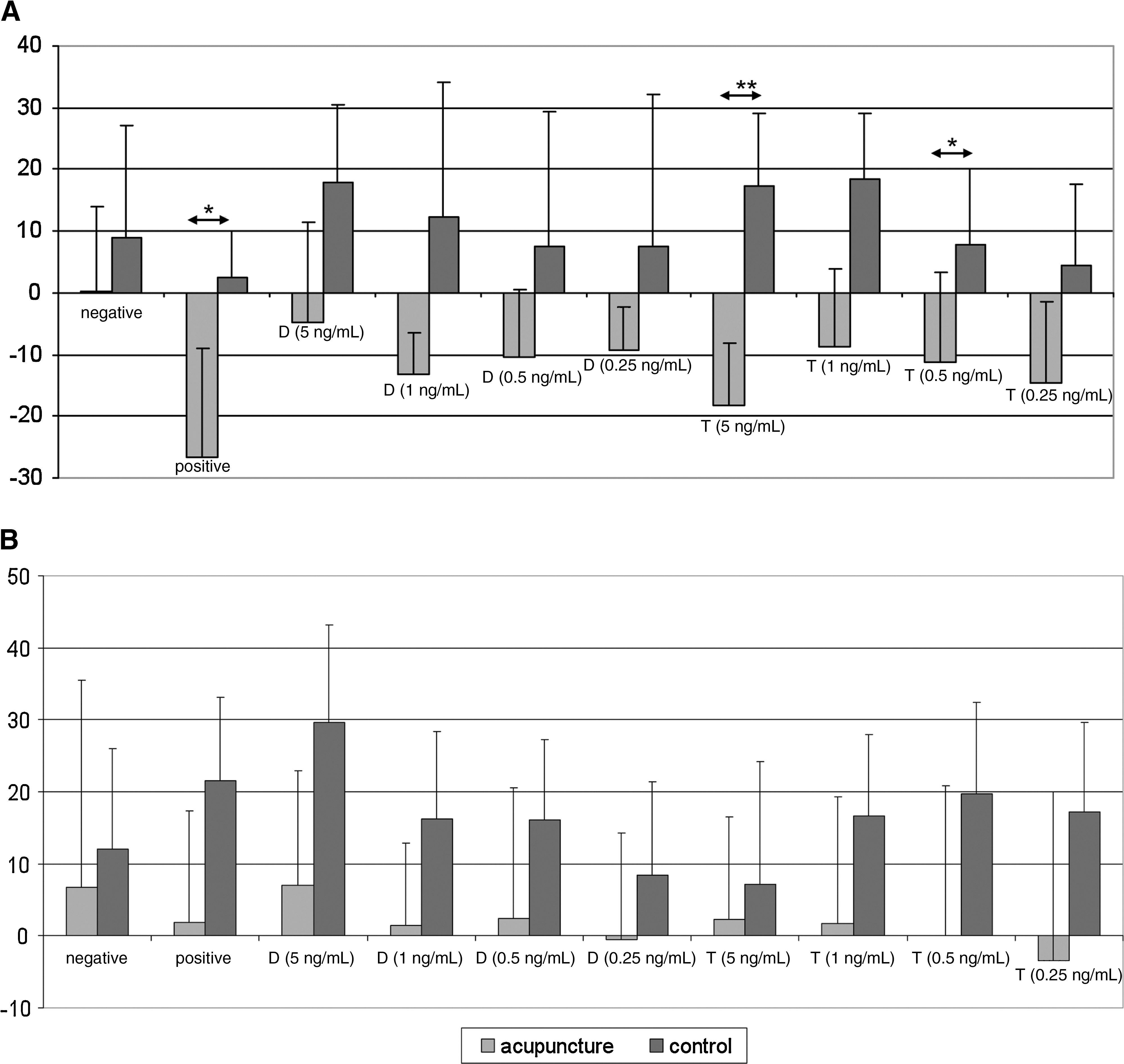
Mean differences in the percentage of CD63-positive basophils between days 0 and 15 (
D, Dermatophagoides pteronyssinus; T, timothy grass pollen; VAS, visual analog scale; SCORAD, Scoring Atopic Dermatitis score.
Discussion
In this pilot study, we observed significant reductions of basophil activation as well as itch intensity by acupuncture. While basophil activation was generally reduced in the acupuncture group, it reached statistically significant levels for timothy grass stimulation as well as the positive control stimulation. The general reduction of basophil activation by acupuncture—even in the positive control stimulation with anti-FcɛRI antibody—points to a reduction of spontaneous activation of basophils and an allergen-independent mechanism of acupuncture action. We have no explanation for the relatively stronger effect of acupuncture at day 15 compared to day 33 of treatment; further studies are needed for clarification.
Although there are several studies on the effect of acupuncture on experimental itch, 4,5,14 –16 so far no controlled study investigated the effect of acupuncture on basophil activation or on itch in atopic eczema. Only a few studies have so far investigated the effect of acupuncture or related techniques on atopic eczema. 17 –22
Experimental studies in human and animal models have shown a potential influence of acupuncture on various immunoregulatory substances such as various neurotransmitters and cytokines 23,24 ; as some of them are known to activate basophils or reduce itch, 25,26 this might be an explanation of the observed effect of acupuncture on basophil activation and itch in the current study. Further experiments have also shown local inhibition of activation and proliferation of mucous mast cells as well as secretion of substance P and vasoactive intestinal peptide in the colon of rats, 27 and suppression of IgE production and modulation of Th1/Th2 cell response by electroacupuncture in 2,4-dinitrophenylated keyhole limpet protein immunized mice. 28 In the field of pain, acupuncture has been shown to specifically affect brain regions 29 –31 that have also been found to play an important role in the central modulation of itch. 26,32 –34 Future studies will have to focus on the underlying mechanisms.
Limitations of this pilot study are the low number of included patients as well as the lack of a placebo control group controlling for point-specific effects. As patients with potentially systemically active topical immunosuppressive agents were excluded, modulation of itch and especially in vitro basophil activation by active treatment with drugs can be regarded as highly improbable.
In this study, patients were treated with an individualized selection of acupuncture points chosen by the acupuncturist evaluating the procedure of acupuncture. Another approach regarding future studies can be a (semi)standardized point scheme characterizing the effect of a special selection of acupuncture points. Future larger-scale studies are also needed to evaluate the effect of acupuncture on clinical and in vitro outcome parameters of atopic eczema and corresponding implications.
Reducing basophil activation can be an additional tool in investigating the mechanisms of action of acupuncture in IgE-mediated allergy. Due to the limited number of patients included in our pilot trial, further studies are needed to strengthen the hypothesis.
Footnotes
Acknowledgments
The study was financed by the ZAUM-Center for Allergy and Environment, Technische Universität München.
Disclosure Statement
No competing financial interests exist.