
Abstract
Background:
Recently the authors reported the experimental evidence of the developing concept of Electro Magnetic Information Transfer (EMIT) of specific molecular signals directly and continuously on target cell picking up the molecular signals from the source chemical effector. This was in agreement with the pioneering work of Jaques Benveniste suggesting that the electronic transmission of the 4-phorbol-12-myristate-13-acetate (PMA) signals could be transferred to target neutrophils by an oscillator when coupled to two electromagnetic coils demonstrating the same biologic activity and so mimicking the biologic function of the original chemical active molecule. The present work is the further development of recent research designed to verify the hypotheses that water could record and replay the EMIT from biologic active chemical molecules.
Methods:
Retinoic acid, a well-known chemical differentiating agent, was placed at room temperature in the input coil connected to an oscillator (VEGA select 719), while culture medium for human neuroblastoma cell (LAN-5) and NT2/D1 stem teratocarcinoma human cells was placed into the output coil and exposed to signals for 1 hour. At the end the oscillator was switched off and LAN-5 neuroblastoma and NT2/D1 stem teratocarcinoma cells were seeded, respectively, into the medium conditioned as reported into an incubator under controlled conditions. After 5 days of incubations, cells were examined by different strategies such as morphological and biochemical parameters.
Results:
It was demonstrated that the electromagnetic signals coming from the retinoic acid molecule could be recorded and stored by the aqueous system of the cell culture medium. Cells seeded in the electronically conditioned medium received physical information generating a statistically significant decrease in metabolic activity and changes in phenotypical structure with protrusion typical of differentiated neuronal cells.
Conclusions:
These experimental results provide some evidence that water could be tuned in a resonant manner by the EMIT procedure appropriately carried through a carrier frequency provided by the oscillator in a manner that seems related to the chemical structure of the source molecule as, in this case, retinoic acid.
Introduction
These data were in agreement with the previous work 2 of Thomas et al., in which they showed that normal human neutrophils reacted to 4-phorbol-12-myristate-13-acetate (PMA) transmitted via an electronic oscillator, by reducing cytochrome C as when they were directly exposed to PMA. The Thomas finding of the electromagnetic molecular signal arising from biologically active molecules was recently confirmed by Montagnier et al. 3 in a work were they discovered a novel property of DNA, namely, the capacity of some DNA sequences to emit electromagnetic waves when a resonance is generating with the ambient electromagnetic background (background noise is so presumably providing the conditions for generating a stochastic resonance effect).
The present work is the report of the further development of the authors' recent research aimed to verify the hypotheses that an aqueous system could record, store, and replay the EMIT from a biologically active molecule.
Materials and Methods
Cell cultures
LAN-5 cells and NT2/D1 stem teratocarcinoma human cells were grown in RPMI (Gibco Laboratories, Scotland) supplemented with 10% fetal calf serum (Gibco Laboratories, Scotland) and antibiotics (110 IU/mL of penicillin and 0.1 mg/mL of streptomycin) at 37±0.3°C, and 5% CO2 as carbon source and subcultured twice a week at a 1:5 ratio. For every experiment, control and exposed cells were taken from the same flask.
Transmission apparatus
For the transmission experiments to cells, the input coil coupled was operated at room temperature and was coupled via a homemade amplifier (Gain 0.25 dB from 1 to 100 Hz maximum output voltage 20V p-p, maximum output current 1A, maximum power 20 W rms) to wave generator VEGA select 719. The output (target) coil was placed in the cell incubator. The target coil was made of 85 turns of 2-mm copper wire, 17 cm long and 9.5 cm wide and fed at 100 mV from the wave generator. The source tube containing 5 μM RA was placed inside the input coil. The target coil in the incubator contained RPMI LAN-5 cells and NT2/D1 culture medium. Figure 1 is a diagram representing the assembly of the experimental apparatus for transmission to cells. The numbers in the following sentence refer to the numbers in the diagram in Figure 1. The signal from the retinoic acid (RA) solution in the coil (1) was fed (2a) into the electronic amplifier (2), then from the electronic amplifier (2b) the signal was transferred (3a) to a wave generator (3). In the wave generator, the electronic signal corresponding to RA was superimposed on a 7-Hz sinusoidal frequency carrier modulated at 3 kHz. From the wave generator then, the signal (3a) was fed into the cell incubator (4) where the target coil (5) was placed to irradiate the RPMI LAN-5 cells and NT2/D1 culture medium for the LAN-5 cells inside it. During the experimental procedure, the electrical parameters remained constant.

Experimental apparatus and assembly. 1—Source signal coil, 2—Electronic amplifier, 2A—Input signal in the electronic amplifier, 2B—Output signal from the electronic amplifier, 3—Wave generator, 3A—Input signal in wave generator, 3B—Output signal from wave generator, 4—Cell incubator, 5—Target coil.
Cellular metabolic activity and proliferation by WST assay
LAN-5 and NT2/D1 cells were seeded to cell culture medium electronically conditioned with RA EMIT by Vega select 719. For each experiment, LAN-5 and NT2/D1 cells were plated into 25-mL 4.2×5.2-cm-base Corning flasks (2.0×10⁵/mL cells in a total volume of 5 mL). The flasks were kept in culture for up to 5 days with or without RA-EMIT-conditioned medium. Cells were then counted and metabolism determined by the water-soluble tetrazolium salt (WST-1) method. The experiment was repeated three times.
The quantification of LAN-5 and NT2/D1 metabolic activity, as an index of cellular proliferation, was performed by a colorimetric assay based on oxidation of tetrazolium salts (Cell Proliferation Reagent water-soluble tetrazolium salt (WST)-1; Roche Diagnostics, Basel, Switzerland). Cells were cultured for up to 5 days in a normal humidified incubator in the presence of the RA-EMIT-conditioned medium (exposed), or in normal medium (control) and they were analyzed by means of the formazan dye every 24 hours. WST reagent diluted to 1:10 was added in the wells at 4 hours, and at 1, 2, 3, and 6 days after plating, and then incubated for 2 hours in humidified atmosphere (37°C, 5% CO2). Quantification of the formazan dye produced was performed by absorbance measurement at 450 nm with a scanning multiwell spectrophotometer (Biotrack II; Amersham Biosciences, Little Chalfont, UK).
Immunofluorescence
For immunofluorescence staining, the cells were grown in Labtek chamber slides.
The cells were then washed with phosphate-buffered saline (PBS) with Ca/Mg and fixed with absolute ethanol for 5 minutes, then incubated with the specific monoclonal antibodies, anti-200-KDa neurofilaments (Sigma) appropriately diluted for 1 hour at room temperature. Cells were then washed three times with PBS and incubated with fluoresceinated antimouse IgGF(ab')2 fragment (Sigma), (appropriately diluted) for 1 hour at room temperature.
Reverse transcription polymerase chain reaction
Total RNA was extracted from cells using TRIzol Reagent (Life Technologies, Merelbeke, Belgium) according to the manufacturer's instructions. Typically, 5–10 μg total RNA per 10 cm2 dish of cell culture was obtained. Reverse transcription polymerase chain reaction (RT-PCR) was used to evaluate relative mRNA levels of neurofilament protein (NF-200) in control and RA-EMIT conditioned medium exposed LAN-5 and NT2/D1cells. One microgram (1 μg) of total RNA was used to synthesize first-strand cDNA with random primers using 100 U of ImProm-II TM RT-PCR kit (Promega, Madison, WI) according to the manufacturer. The reaction was also carried out in the absence of reverse transcriptase (RT) to check for genomic DNA amplification. The NF-200 subunit-specific primers used for PCR were: 5′-aagtgaacacagatgctatgcg-3′ 5′-ctgtcactccttccgtcacc-3′. The 18S was used as internal controls, because these genes are uniformly expressed during development. The subunit-specific primers used for PCR were as follows: 5′-tttcggaactgaggccatgattaag-3′ 5′-agtttcagctttgcaaccatactcc-3′. An aliquot (2 μL) of RT reaction was PCR-amplified in a final volume of 50 μL, by using 20 pmol of each primer, 200 μmol/L of each dNTP, and 0.5 U of Taq DNA polymerase (T.Aquaticus, Amersham-Pharmacia). PCR was carried out in a Bio-Rad I Cycle instrument. The thermocycling conditions for each pair of primers were as follows: denaturation at 95°C for 3 minutes followed by 30 cycles of denaturation at 95°C for 30 seconds, annealing for 45 seconds at 62°C, elongation at 72°C for 1 minute and a final polymerization step at 72°C for 5 minutes for NF-200 and 20 cycles for 18S. The amount of template and the number of amplification cycles were preliminarily optimized for each PCR reaction to avoid conditions of saturation. Aliquots (5 μL) of the reaction products were run on 1% agarose gels containing ethidium bromide (0.5 μg/mL) to mark and visualize the PCR products. Gels were then photographed under UV light with a Versadoc (Bio-Rad) instrument. These experiments were replicated three different times.
Statistical analysis
Statistics were performed with Student's t-test with p<0.05 as the minimum level of significance.
Results
Electronically transmitted RA EMIT-conditioned medium effect on LAN-5 and NT2/D1 cell metabolism
The cell growth rate was analyzed by the WST-1 both in LAN-5 and NT2/D1 cells as control (not grown in electronically transmitted RA-EMIT-conditioned medium) or grown in electronically transmitted RA-EMIT-conditioned medium. An inhibition in the cell metabolism in the cells grown in electronically conditioned RA-EMIT medium was statistically (p<0.01) significant after 5 days' exposure (Fig. 2).
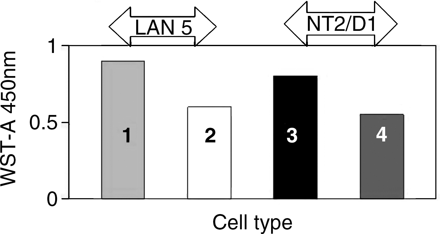
Electronically transmitted retinoic acid Electro Magnetic Information Transfer (RA-EMIT) conditioned medium effect on human neuroblastoma cell (LAN-5) and NT2/D1 cell metabolism. The cell growth rate was analyzed by the water-soluble tetrazolium salt (WST-1) both in LAN-5 (1) and NT2/D1 (3) cells as control (not grown by RA-EMIT medium) or grown by RA-EMIT-conditioned medium (2, 4). A statistically significant inhibition in cell metabolism (p<0.01) was detected after 5 days' culture.
Electronically transmitted RA EMIT-conditioned medium effect on LAN-5 and NT2/D1 cell morphology
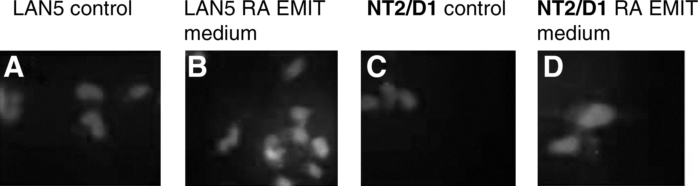
By phase contrast and scanning electron microscopy LAN-5 and NT2/D1control cells appeared small, polygonal, without neuritelike structures. The exposure to electronically transmitted RA-EMIT-conditioned medium induced morphological changes toward a more neuronal phenotype: The cells were stretched out and rich in neuritelike structures with blebs, mimicking the same effect induced by RA treatment (Fig. 3).

Electronically transmitted retinoic acid (RA)-conditioned medium effect on human neuroblastoma cell (LAN-5) and NT-2/D1 cell morphology by contrast microscopy. EMIT, Electro Magnetic Information Transfer. Contrast microscopy of LAN-5 cells in absence
Electronically transmitted RA EMIT-conditioned medium effect on LAN-5 and NT2/D1 on neurofilament expression
Figure 4 shows the indirect immunofluorescent analysis of control and exposed LAN-5 and NT2/D1 cells with anti-200 neurofilaments. While control cells were slightly or not positive for NF 200 (CTR), the neurofilament protein became more fluorescent after exposure to the RA EMIT-conditioned medium (EXP). The same results were achieved by RT-PCR analysis for mRNA expression coding for NF-200 (Fig. 5).
Electronically transmitted retinoic acid (RA)–conditioned medium effect on human neuroblastoma cell (LAN-5) and NT-2/D1 cell by NF-200 indirect immunofluorescence (red fluorescence; shown in black and white) of LAN-5 cells and NT2/D1 in absence

NF-200 mRNA reverse transcriptase polymerase chain reaction (RT-PCR) analysis on electronically transmitted retinoic acid (RA) effect on human neuroblastoma cell (LAN-5 cell) and NT2/D1 cells. Left bars
Conclusions
Since the time of Galvani over the past centuries, much evidence has been accumulated indicating that living systems have evolved so as to make practical use of electromagnetic fields. It is currently well established that all the physiologic activities that contribute to the functional organization and maintenance of stability of living systems are associated with electromagnetic activities. 4 Living organisms might be considered aggregates of electromagnetic fields that are embedded within or correlated with atomic and molecular structures. 5 –8 Informative Medicine as an evolution of electromagnetic medicine is still in its beginning. 9 Nevertheless, the evidence reported here (that the EMIT mediated through the aqueous system procedure could tune eukaryotic cell cultures toward cell differentiation and maturation, 10 involving physiologic processes and epigenetic expression leading to specific phenotypical features) allow one to foresee some possible future application of the EMIT mediated through an aqueous system procedure for the treatment of some human degenerative diseases.
The mechanism of the interaction and signal transduction 11 –14 between the physical agent and the biologic target still remains to be understood, so that experiments are in progress to define the biophysical mechanisms involved in providing an explanation regarding how water molecules present in an aqueous system could record, store, and replay the specific electromagnetic information pattern coming from a specific molecular activity.
These further experimental findings provide some essential preliminary basic support to the possibility of transferring Informative Medicine paradigms to clinical practice to bedside applications.
Footnotes
Acknowledgments
This work has been partially supported by a grant from NAMED [Natural Medicine].
Disclosure Statement
The authors declare that they have no competing interests.