
Abstract
Objectives:
The objective of this study was to investigate the change of mean blood flux in Hegu acupoint after contralateral Hegu acupoint was stimulated.
Methods:
Healthy volunteers (N=140) were placed in a temperature-controlled room as a resting state for 60 minutes. Then the measurements of skin blood flow were carried out in pre- and postacupuncture stimulation every 30 minutes over a total of 180 minutes using a Moor full-field laser perfusion imager. The change ratio of mean blood flux in Hegu acupoint was used to evaluate the contralateral stimulation effect in different groups.
Results:
After acupuncture of the right Hegu acupoint (AR), the change ratio of mean blood flux in the left Hegu acupoint was increased significantly compared with the control group 60 minutes after acupuncture stimulation (p<0.01). Then the blood flow in the AR group was sustained at a higher level compared with that of the control group (p<0.05). However, the change ratio of mean blood flux in the right Hegu acupoint increased significantly in the 30 minutes after contralateral acupuncture stimulation (p<0.01). From that point on, the change ratio was sustained at a higher level in the AR group than in the control group until 150 minutes after stimulation (p<0.05).
Conclusions:
On stimulation of either the left or right side of Hegu acupoint, the change ratio of mean blood flux was increased in the contralateral Hegu acupoint in a period from 30, 60, to 150 minutes after stimulation, respectively.
Introduction
Recently, increasing attention has been focused on the relationship of acupuncture and circulation. 3 –5 In Traditional Chinese Medical (TCM) theory, the effects of acupuncture depend on the special sensation in a local acupoint after stimulation, which might be related to the blood perfusion changes in acupoints or meridians. 6 When an acupoint was stimulated adequately, the blood perfusion of this point continued to increase whereas the blood perfusion of the nonacupoint only changed slightly by the same acupuncture stimulation. 7 In patients with primary Raynaud phenomenon, auricular electroacupuncture can reduce the frequency and severity of attacks with little influence on skin perfusion. 8 These results indicated that the change pattern of blood perfusion resulted from acupuncture stimulation is still not clear. Previous studies have been shown that thermostimulation could result in an increase in blood perfusion in the contralateral 9 and ipsilateral 10 side of the dorsal hands, while there was no effect on the blood perfusion in the periumbilicus area. 10 According to the Neijing theory, if someone has disease in the left side of the body, the treatment point is usually selected in the right side, and vice versa. Recently, a systematic review analyzed the contralateral acupuncture and ipsilateral acupuncture effect on the poststroke patients with hemiplegia. 11 Although this system review and meta-analysis could not come to a definitive conclusion, it indicated the importance of distinction between contralateral acupuncture and ipsilateral acupuncture. In the present study, the aim is to investigate the change of mean blood flux in the Hegu acupoint after the contralateral Hegu acupoint was stimulated.
Methods
Subjects
One hundred and forty (140) healthy volunteers were recruited in this study (Fig. 1). All subjects were students from the China Academy of Chinese Medical Sciences and Beijing University of TCM. All subjects had no history of diseases and had not taken any medicine in the past 6 months. Each subject provided informed consent and had an adequate understanding of the procedure and purpose of this study.
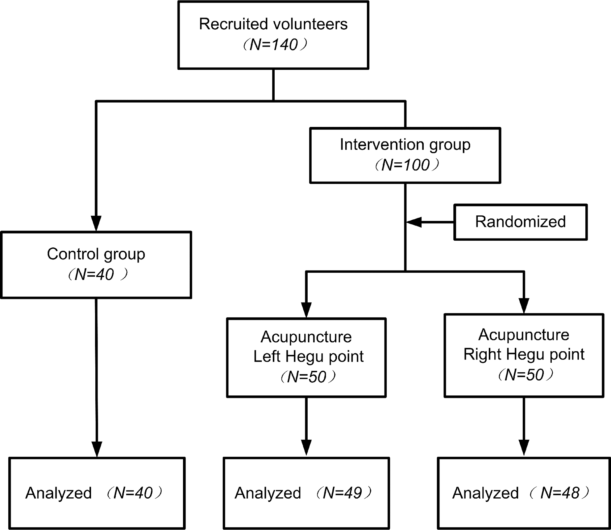
Flow diagram of participants in the study.
Procedures
Protocol for measurement of mean blood flux
Upon arrival at the laboratory, subjects were placed in a temperature-controlled room (24°C–26°C) as a resting state for 60 minutes. Measurements of skin blood perfusion were carried out using a Moor full-field laser perfusion imager (moor FLPI, Moor Instruments Ltd., UK). Before recording, both hands of the subject were immobilized with a cylindrical object to ensure positioning. The scan parameters were as follows: high resolution/250 frames; number of images=10; exposure time=8.3 ms; time interval=10 seconds. Measurement was carried out by Moor FLPI every 30 minutes over a total of 180 minutes. During the experiment, the laboratory room was kept in a dark light condition and the protocol for measurement was strictly abided by. The measurement process is illustrated in Figure 2.
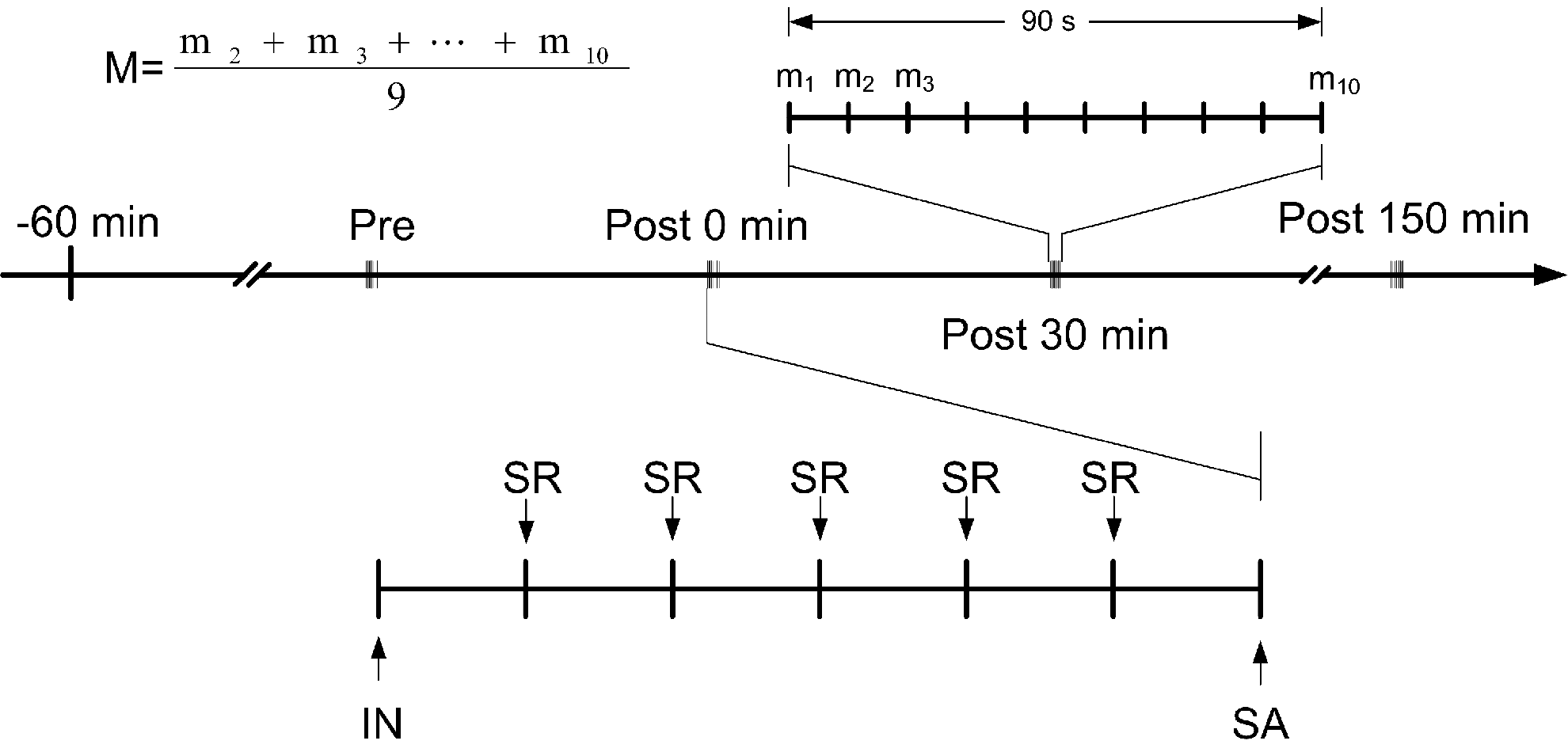
Procedure of acupuncture and measurement. Pre, pre-acupuncture; Post, postacupuncture; IN, insert needle; SA, stop acupuncture; SR, slowly rotate the needle every 5 minutes.
Acupuncture protocol
For acupuncture, a small acupuncture needle, 0.25×25 mm (100112, Zhen Huan), was gently inserted in a depth of 15 mm in the Hegu acupoint (LI4). The position of the Hegu acupoint was confirmed according to previous studies. 6,12 The needle was slowly rotated every 5 minutes for a total of 30 minutes during an acupuncture session 13 in order to maintain the soreness and numbness sensation of de qi. The acupuncture process is illustrated in Figure 2. In the acupuncture left Hegu point (AL) group, just the left Hegu acupoint was acupunctured, whereas in the group receiving acupuncture of the right Hegu acupoint (AR), the right Hegu acupoint was stimulated. In the control group, all subjects maintained stillness, without any intervention.
Image analysis protocol
After the blood flux images were acquired, the mean blood flux of the Hegu acupoint was acquired using Moor Full-Field Laser Perfusion Imager Review (V3.0, Moor Instruments Ltd., UK). In any time point (pre, post 0 minutes, 30 minutes, 60 minutes, 90 minutes, 120 minutes, 150 minutes), the mean value (M, as shown in Fig. 2) of blood flux in the Hegu acupoint was represented by averaging of the last nine frames, which were acquired in 90 seconds (Fig. 2). In every group, the change ratio of mean blood flux in Hegu acupoint was recorded as Ri,
Statistical analysis
The group differences were analyzed using SPSS software. The change ratio of mean blood flux (%) was expressed as mean±standard deviation. Statistical analysis was performed using independent-sample t-test. Values of p<5% were considered statistically significant.
Results
In this study, a total 137 subjects were included in the final statistics (Fig. 1). There were 2 subjects in AR group and 1 subject in the AL group did not finish the measurement. The reason was due to the hands movement during the measurement. Detailed information of subjects is summarized in Table 1. There were no significant differences in age and body mass index between the groups of control, AL, and AR.
SD, standard deviation; AL, acupuncture left Hegu point; AR, acupuncture right Hegu point.
After acupuncture in the right Hegu acupoint, the change ratio of mean blood flux (MBF) in left Hegu acupoint is shown in Table 2 and Figure 3. During the period of 0 minutes to 30 minutes postacupuncture, there were no significant differences between acupuncture right group and the control group (p>0.05). From 60 minutes to 150 minutes postacupuncture, the change ratio of MBF in the acupuncture right group increased significantly compared with that of the control group (p<0.05 or p<0.01).
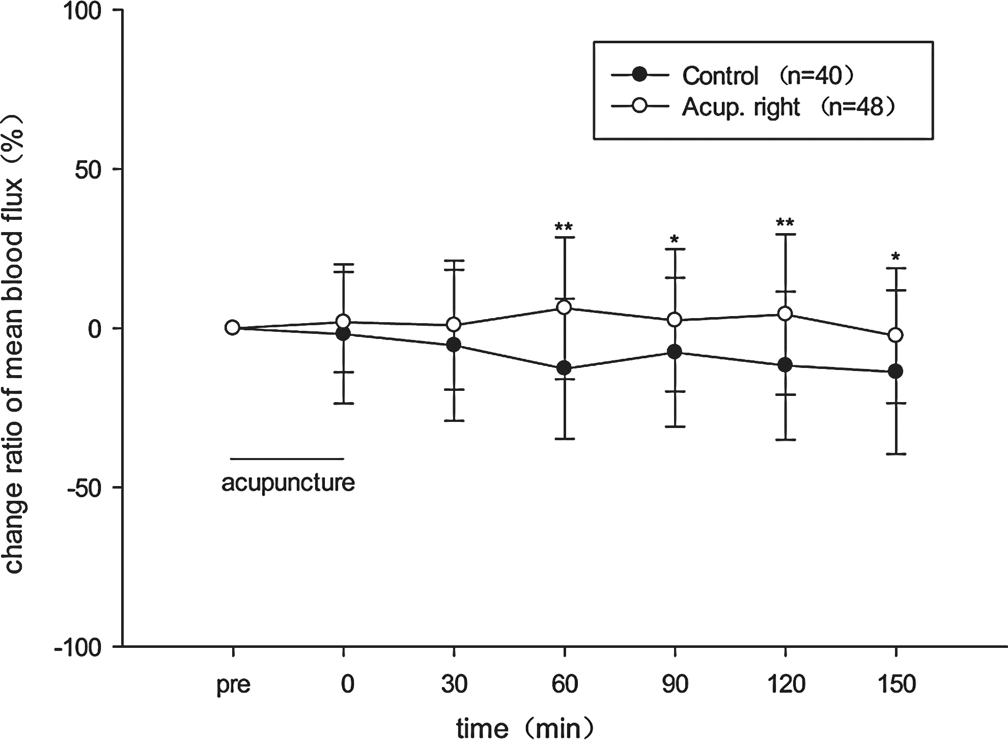
Change ratio of mean blood flux in left Hegu acupoint. Data are expressed as mean±standard deviation. *p<0.05, **p<0.01, control vs Acup. right.
p<0.05; ** p<0.01, versus control group.
SD, standard deviation; AR, acupuncture right Hegu acupoint.
The change ratio of MBF in the right Hegu acupoint after acupuncture in the left Hegu acupoint is shown in Table 3 and Figure 4. At the time of 0 minutes postacupuncture, there were no significant differences between the acupuncture left group and the control group (p>0.05). From 30 minutes to 150 minutes postacupuncture, the change ratio of MBF in acupuncture left group increased significantly compared with that of the control group (p<0.05 or p<0.01).
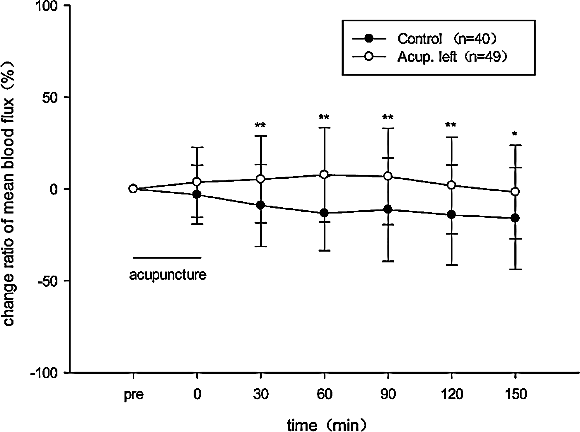
Change ratio of mean blood flux in right Hegu acupoint. Data are expressed as mean±standard deviation. *p<0.05, **p<0.01, control vs Acup. left.
p<0.05; ** p<0.01, versus control group.
SD, standard deviation; AL, acupuncture left Hegu acupoint.
Discussion
The principle of acupuncture for treatment lies in the acupoint stimulation, which regulates viscera functions through activating meridian function. So in the theory of TCM, if a Hegu acupoint is stimulated on one side, the function of Large Intestine meridian (LI) located on the other side might also be activated. As a result, the running of qi and blood that flow in both LI meridians was changed. The authors' previous study 14 has indicated that the CO2 volume of skin respiration in the same name acupoints had a high degree of correlation, which suggested that there had been functional linkage between the same name meridians. These results were supported by the studies showing that stimulation on different acupoints located on the same meridian induced certain similar functional magnetic resonance imaging (fMRI) activation patterns in the brain. 15
According to the previous study, the MBF was larger at the acupoints than in their surrounding tissues, which indicates that the MBF can be used as an index for discriminating differences in the microcirculatory conditions between acupoints and their surrounding tissues. 16 Thus in this study, the cross-interaction of Hegu acupoints through the index of the change ratio of MBF was investigated.
It has also been shown that acupuncture can not only increase general circulation 17 and circulation in specific organs, 18 but also change the skin microcirculation as well. 4,12,19,20 The skin blood perfusion regulation is controlled by the autonomic nervous system. 21 In this study, stimulation by acupuncture caused a significant increase in change ratio of blood flux in the contralateral Hegu acupoint, which might suggest that contralateral stimulation by acupuncture plays a significant role in the general autonomic nervous system. These results support the result that autonomic nervous system participates in the formation of the acupuncture effect. 7,22
It is reported that cutaneous vasodilation might be related to the activation of the adenosine receptor in the local microcirculation system. It is suggested that increase of local blood perfusion in an aucupoint after acupuncture might be related to the adenosine receptor activation. A previous study 13 indicated that insertion and manual rotation of acupuncture needles in Zusanli (ST36) acupoint resulted in an increase in the extracellular concentration of adenosine, whereas the level of adenosine in the contralateral acupoint was not changed significantly. Therefore, in the contralateral side, the blood increase in an acupoint after stimulation may be independent of the adenosine receptor activation.
It has been shown that acupuncture at different acupoints exhibited differential hemodynamic neural responses in some brain areas, 23 and these specific acupuncture-associated modulatory effects could be sustained during the stimulation phase, and even remained in the subsequent resting epoch. Signal changes in some brain regions during the postrest epoch of the acupuncture were even higher than that of the stimulation epoch. 24 In this research, the change ratio of MBF in a contralateral Hegu acupoint was sustained at a higher level than that in the control group for at least 150 minutes after stimulation, which provided the supplemental evidence for the results found in the fMRI. During the first 90 minutes after stimulation, the responses of change ratio of MBF have the different models in contralateral Hegu acupoints, which suggested that the acupuncture effect of bilateral Hegu acupoints might be asymmetry within a special period after contralateral stimulation.
According to other research, 25 under anesthesia condition, thermostimulation resulted in an increase of blood perfusion in the rat ipsilateral foot, and had no effect on the blood perfusion in the contralateral-side foot. These results indicated that this asymmetry phenomenon was strengthened by the anesthesia. In other words, synchronous changes of blood perfusion in the bilateral side might be related to the wakefulness status.
Footnotes
Acknowledgments
This research was supported by the National Natural Science Foundation of China (Project No. 81001553).
Disclosure Statement
No competing financial interests exist.