
Abstract
Aim:
This present work was aimed to investigate wound healing-related biologic activities of traditional herbal formulas used for wound treatment in southern Thailand.
Methods:
Water and ethanol extracts of the formulas (THR-SK004, THR-SK010, and THR-SK011) were tested for their antibacterial potency against methicillin-resistant Staphylococcus aureus (MRSA) and -susceptible S. aureus. Anti-inflammatory activities of the extracts were assessed by detection of the inhibition of lipopolysaccharide-induced nitric oxide production. Anti-oxidant activities and cytotoxicity of the extracts were also measured.
Results:
Among the tested formulas, ethanol extract of THR-SK010 consisting of four herbs: Curcuma longa L., Areca catechu L., Oryza sativa L., and Garcinia mangostana L., was found to possess promising antibacterial activities with MIC90 of 4 μg mL−1 against MRSA isolates. This ethanol extract offered the highest anti-inflammatory activity as well as DPPH and hydroxyl radical scavenging activities.
Conclusions:
Remarkable antibacterial, anti-inflammatory, and antioxidant activities as well as low toxicity on Vero cells of THR-SK010 ethanol extract provide scientific information to support the topical use of the formula for wound treatment. This information proposes the potential to develop a new generation of phytopharmaceuticals based on traditional knowledge.
Introduction
Research on wound healing agents is one of the developing areas in modern biomedical sciences. Medicinal plants are important sources of new chemical substrates that have therapeutic effects; therefore, extensive research has been carried out in the area of wound healing management with medicinal plants. 5 Medicinal plants, which form the backbone of traditional medicine, have been the focus of very intense pharmacological investigations in several countries, including Thailand. Moreover, several herbal formulas of China, 6 India, 7 Ghana, 8 and Nigeria 9 have been identified and their use was documented. In Thailand, particularly the southern part of Thailand that has more than 3000 traditional healers, 9 –11 many herbal formulas are in current use but these herbal formulas have never been investigated. Recent studies suggested that the services of traditional healers should be incorporated into contemporary health care provision of the Thai health care system. Thus, the need to justify and document ethnomedical practice is important.
In this article, the objectives were to investigate the relevance of biologic activities to wound healing, including antibacterial, antioxidant, and anti-inflammation activities of Thai traditional herbal formulas used for wound treatments. Moreover, the cytotoxicity of these herbal formulas was additionally tested as supporting information for further in vivo studies.
Materials and Methods
Plant materials and preparation of extracts
Plant parts as described in Table 1 were locally collected and reference voucher specimens were deposited at the Faculty of Traditional Thai Medicine, Prince of Songkla University, Hat Yai, Songkhla, Thailand. The formulas consist of equal amounts (100 g) of their medicinal plant components, which were dried and ground into fine powder. The powdered formulas (100 g) were submitted to solvent extractions by maceration with distilled water at room temperature for 3 days or with ethanol for 7 days (500 mL each solvent). After filtrations through a Whatman No. 1 filter paper, aqueous filtrates were freeze-dried, and ethanol filtrates were removed with a rotatory evaporator, kept at 55°C until they were completely dry. Yields (%; wt/wt) of each extract were calculated as the ratio of the weight of the extract to the weight of the crude herb powder, and are shown in Table 1. Samples were stored in a sterile screw-capped bottle at −20°C and dissolved in 10% dimethylsulfoxide (DMSO; Merck, Germany) before use.
Extraction yields of ethanol/water extracts.
Wound healing-related biologic activities
Anti-inflammatory activity
Inhibitory effect on nitric oxide (NO) production by murine macrophage-like RAW264.7 cells was evaluated using a method modified from a previous report.
12
Macrophage cells RAW264.7 (4×10⁵ per well) were seeded in a 24-well plate overnight. With 200 μg lipopolysaccharide (LPS) per milliliter of medium, the herbal formulas samples at various concentrations (7.8–250 μg/mL) were added and incubated at 37°C in a humidified atmosphere containing 5% CO2 for 48 hours. NO production was determined by measuring the accumulation of nitrite (
where A is
Antioxidant activity
DPPH-based radical-scavenging activity of the extracts was measured from the bleaching of the purple methanol solution of DPPH. An aliquot of 100 μL of the formula extracts at different concentrations (7.8–250 μg/mL) was mixed with 100 μL of DPPH solution. The mixture was then incubated at room temperature for 30 minutes. Absorbance of the solution was measured at 517 nm and DPPH radical-scavenging activity was calculated. 13
Measurements of hydroxyl free radical–scavenging activity of the formula extracts were taken using a previously described method followed by slight modifications. 14 The reaction mixture containing 100 μL of ferric sulphate (0.5 mmol) and 75 μL of 3% hydrogen peroxide was incubated in the dark room for 5 minutes. An aliquot (50 μL) of brilliant green reagent at concentration of 0.435 mmol was subsequently added into the mixture. After 10 minutes of incubation, the extracts (25 μL) were then mixed with the reaction mixture. The absorbance was measured at 624 nm after incubation for 10 minutes under dark conditions. Results were expressed as percentage of inhibition.
Scavenging of superoxide radical was determined by the method described by Samark et al. (2009), with slight modifications. Superoxide radicals were generated in 1 mL 20 mmol Tris–HCl buffer pH 8.0 containing 0.05 mmol nitroblue tetrazolium (NBT), 0.01 mmol phenazine methosulphate, and the test compounds were preincubated for 2 minutes. The reaction was initiated by the addition of 0.078 mmol NADH. Blue chromogen, which formed due to NBT reduction, was examined at 560 nm. Results were expressed as percentage of inhibition. 14
Gallic acid (Sigma-Aldrich Chemic GmbH, Spain) was used as a positive control and an extract diluent and 1% DMSO (Sigma-Aldrich, USA) was added as a negative control. DPPH based-, hydroxyl, and superoxide–radical-scavenging activities were calculated by using the following equation,
where A is optical density of the control solution and B is optical density of the test solution.
Anti-staphylococcal activity
Determination of minimum inhibitory concentration
Antibacterial activity tests were carried out against clinically isolated methicillin-resistant Staphylococcus aureus (MRSA) NPRC R001–R020 (n=20) and methicillin-susceptible S. aureus (MSSA) NPRC S001–S020 (n=20). S. aureus ATCC 29213 was added as a standard control strain. Cultures for experiments were prepared from 24-hour Mueller-Hinton broth (MHB, Difco, France) and the suspensions were adjusted to a McFarland standard No. 0.5 and subsequently diluted with fresh MHB to achieve a bacterial culture concentration corresponding to 106 CFU/mL.
Minimum inhibitory concentrations (MICs) of the formula extracts were tested by the CLSI broth microdilution method. 15 A 96-well sterile microtiter plate (Nunc, Denmark) was prepared by dispensing 100 μL of the inoculated broth plus an aliquot of 100 μL of twofold serial dilutions of the extract (0.48–1000 μg/mL). An aliquot of 100 μL of 1% DMSO was employed as a negative control. The plate was incubated for 24 hours at 37°C and the bacterial growth was measured by recording the absorbance at 620 nm, using a microplate reader (Sunrise, Tecan, Switzerland).
Time-kill assay
Time-kill assays were used to determine the time required for the effective formula extract, THR-SK010 ethanol extract (THR-SK010E), to eliminate the growth of MRSA NPRC R001, MSSA NPRC S003, and S. aureus ATCC 29213. Bacterial inoculum prepared as described above (1 mL) was mixed with 1 mL of MHB containing THR-SK010E at final concentrations of MIC, 2×MIC, 4×MIC, and 8×MIC. The tubes were incubated at 37°C and samples (100 μL) were taken at 0, 2, 4, 6, 8, 10, and 18 hours. Bacterial growth of each sample was measured by recording the absorbance at 620 nm and serial 10-fold dilutions were made and plated on Mueller-Hinton agar (MHA, Difco, France). The total viable count was determined after overnight incubation at 37°C.
Cytotoxicity assay
Cytotoxicity activities of the formula extracts against Vero cell were determined by green fluorescent protein (GFP)-based assay at the National Center for Genetic Engineering and Biotechnology, National Science and Technology Development Agency, Pathumthani, Thailand. Ellipticine used as a positive control exhibited cytotoxicity against Vero cell line with IC50 of 1.48 μmol.
Results
Wound-related biologic activities
Anti-inflammatory activities of ethanol and water extracts of the formulas were tested by measuring their effects on the pro-inflammatory mediators nitric oxide in LPS-challenged macrophages RAW 264.7 cells. As demonstrated in Table 2, NO inhibitory results revealed that THR-SK010E and THR-SK011 ethanol extract (THR-SK011E) exhibited NO production inhibition activity with IC50 values of 71.06 and 72.67 μg mL−1, respectively, while other extracts were apparently inactive (IC50>100 μg mL−1).
DPPH, 2,2-diphenyl-1-picrylhydrazyl; NA: not applicable.
Since it is well known that reactive oxygen species are harmful to the wound-healing process, this study was additionally undertaken to evaluate antioxidant activity of the herbal formulas. Scavenging abilities for DPPH, hydroxyl, and superoxide radicals of the extract are shown in Table 2. THR-SK010E displayed the highest DPPH and hydroxyl radical scavenging activities, with the IC50 values of 19.24 and 13.58 μg mL−1, respectively. THR-SK011E possessed the highest superoxide radical scavenging activity, with the IC50 value of 75.00 μg mL−1.
Antibacterial activities
Table 3 demonstrates antistaphylococcal activity of the extracts of the formulas. Remarkable antibacterial effects, expressed as MIC90 of the THR-SK010E against clinically isolated MRSA (n=20), colonization origin MSSA (n=20) isolates, and S. aureus ATCC 29123, were 4, 8, and 4 μg mL−1, respectively. None of the water extracts of the formulas displayed anti-staphylococcal activity (the MIC ranges from 250 to 1000 μg mL−1).
MIC90 of ethanol/water extracts on the tested isolates.
MICrange of ethanol/water extracts on the tested isolates.
MICs, minimum inhibitory concentrations; MRSA, methicillin-resistant Staphylococcus aureus; MSSA, methicillin-susceptible Staphylococcus aureus.
Figure 1 illustrates the results of the time-kill curves for representative MRSA and MSSA isolates (NPRC R001 and NPRC S003), and a reference strain (ATCC 29213; data not shown). Numbers of viable MRSA decreased 1.3–2.3 Log CFU mL−1 following the treatment with THR-SK010E at MIC, 2MIC, 4MIC, and 8MIC for 2 hours and declined 1.9–5.8 Log CFU mL−1 after treatment for 18 hours. The growth of S. aureus ATCC 29213 and MSSA isolate was inhibited for 18 hours and the bacterial viability decreased 3.8–7.7 and 2.0–6.3 Log CFU mL−1, respectively, after 18 hours.
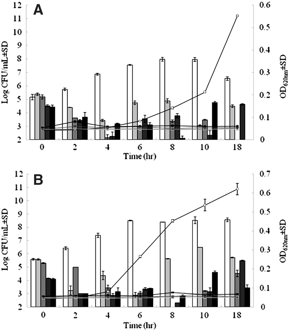
Time-kill assays for methicillin-resistant Staphylococcus aureus (MRSA) NPRC R001
Cytotoxicity
THR-SK004 and THR-SK010 ethanol extracts displayed moderate cytotoxicity, while the other extracts exhibited low cytotoxicity on the Vero cell line. Fifty percent (50%) inhibitory concentration (IC50) of THR-SK004E and THR-SK010E were 169.72 and 45.68 μg mL−1, respectively (Table 2). The IC50 value of the positive control ellipticine used in this study was found to be 1.48 μg mL−1. THR-SK010E against MRSA and MSSA isolates showed an index of 11.42 and 5.71, respectively.
Discussion
Wound healing is a complex process initiated in response to an injury, and which restores the function and integrity of damaged tissues. Three (3) overlapping phases consisting of inflammation, proliferation, and remodeling are well known and represent a normal wound-healing process. 5 Even though inflammation is a normal and necessary step in the wound-healing process, 16 uncontrolled inflammation of wounded tissues can severely damage healthy cells and impair wound healing. 17 Colonized or infected wounds tend to develop an abnormally prolonged inflammatory state that generates large amounts of nitric oxide. 18 Therefore, the hypothesis that traditional herbal formulas promote wound healing by modulating inflammation was tested by detecting nitric oxide production of LPS-stimulated macrophages after treatment with the formulas. The current study's results revealed that THR-SK010E and THR-SK011E exhibited a NO production inhibition activity. Previous reports have shown that Curcuma longa, 7,19 Areca catechu, 20 Oryza sativa, 21 and Garcinia mangostana, 22 which are herbal components of THR-SK010, possessed anti-inflammatory activity. Similarly, herbal components of THR-SK011 including Ceiba pentandra, 23 Aloe barbadensis, 24 Tinospora crispa, 25 Senna siamea, 26 and Chromolaena odorata 27 [29] reportedly had that activity. Thus, the herbal formula extracts as well as other medicinal plants that inhibit pro-inflammatory mediators such as NO would be beneficial in the treatment of inflammation and wounds. 17,28,29
Oxygen-derived radical as well as nonradical oxidants are essential parts of healing and can trigger the various beneficial pathways of wound healing. 30 However, at high concentrations, they can induce severe tissue damage and even lead to conversion of a normal growth tissue into a malignant tumour. Topical applications of compounds with free-radical-scavenging properties in clinical studies 31 as well as in an animal model 32 have been shown to improve wound healing significantly. The potent antioxidant activity of both THR-SK010 and THR-SK011 against free radicals that was demonstrated in this study may be due to the potent activity of active constituents of the herbal components such as curcuminoids from Curcuma longa, 33 xanthones from Garcinia mangostana, 34 aloesin from Aloe barbadensis, 35 and phenolic compounds from Chromolaena odorata. 36
Prevention of bacterial infections is another important aspect in the treatments of cutaneous wounds. Infected skin wounds, particularly by S. aureus, are frequently associated with delayed wound closure, prolonged inflammation, and severe scar formation. Considering the strictest criteria proposed by Cos et al. (2006), extracts with a selective activity and MIC values below 1–50 μg/mL are considered noteworthy. 37 In this regard, THR-SK010E can be categorized as a useful antimicrobial agent as it has developed MIC varying between 2 and 4 μg/mL. More importantly, the therapeutic indexes of the formula were more than 10, indicating that the extracts exhibited no cytotoxicity. 38 With the exception of Oryza sativa, antibacterial activities of Garcinia mangostana, Curcuma longa, and Areca catechu have been documented. Previous studies on Garcinia mangostana have revealed that benzene and ethanol extracts of the plant exhibited anti-MRSA activity with MICs of 80 and 39–50 μg/mL, respectively. 39,40 α-Mangostin, which was isolated from Garcinia mangostana, had the highest activity against MRSA (MIC=1.57–12.5 μg/mL). 39 It should be noted that the formula ethanol extract (MIC=2–4 μg/mL) had a potent anti-MRSA activity similar to that of α-mangostin and approximately 10 times greater than that of Garcinia mangostana ethanol extract. In addition, Curcuma longa ethyl acetate extract 41 as well as the active constituents of this plant, curcumin 42 and turmeric oil, 43 have been proved for anti-MRSA potencies, but their MIC values (MIC=100–1000 μg/mL) are largely higher than that of the formula extract. Although anti-MRSA activity of Areca catechu has never been reported, Karphrom et al. (2009) found that ethanol extract of the plant had moderate antistaphylococcal activity with the MIC value of 780 μg/mL. 44 Previous ethnobotanical surveys stated that many diseases, including skin diseases and wounds, were treated using a combination of more than one plant, and traditional medical practitioners believe that combining more than one plant increases effectiveness of medicines. 45,46 However, there are few pharmacological studies that support this information. 7
Conclusions
The main purposes of wound care are to reduce risk factors that inhibit wound healing; to enhance the healing process; and to lower the incidence of wound infections. Therefore, the remarkable antibacterial potency and wound healing–related biologic activities obtained from this study provide scientific information to support the utilization of THR-SK010 for wound treatments. Close investigations into the in vitro and in vivo wound healing activities by the formula are therefore warranted and currently being pursued in the current authors' laboratory.
Footnotes
Acknowledgments
This work was supported by Agricultural Research Development Agency (ARDA). The authors are thankful to Miss Stefania Vignotto for editing of the manuscript.
Disclosure Statement
No competing financial interests exist.