




Abstract
Objective:
To assess the immediate effects on vertical mouth opening, orofacial mechanosensitivity, and lumbar and suboccipital mobility after adding a myofascial induction technique to a multimodal protocol in subjects with temporomandibular disorders (TMD).
Design:
A randomized and double-blind controlled trial was carried out.
Settings/Location:
University-based physical therapy research clinic.
Subjects:
Sixty subjects (35±11.22 years) with TMD, and restricted mobility of the mandibular condyles and the first cervical vertebrae, were recruited and randomized to either a control group (CG) (n=30) or an experimental group (EG) (n=30).
Interventions:
The CG underwent a neuromuscular technique over the masseter muscles and passive hamstring muscle stretching. A suboccipital muscle inhibition technique was added to this protocol in the EG.
Outcome measures:
Primary measurements were made of vertical mouth opening and pressure pain threshold of the masseter muscles. Secondary outcome measures included pressure algometry of the trigeminal nerve, suboccipital range of motion, and lumbar spine mobility, assessed with the sit-and-reach (SAR) test and lumbar forward bending. All evaluations were collected at baseline and immediately after intervention.
Results:
In the intragroup comparison, the EG observed an increase in suboccipital flexion (p<0.001; F 1,29=14.47; R 2=0.33) and the SAR test (p=0.009; F 1,29=7.89; R 2=0.21). No significant differences were found in the between-group comparison for any variable (p>0.05).
Conclusion:
The inclusion of a myofascial induction maneuver in a protocol combining local (neuromuscular treatment) and distal techniques (hamstring stretching) in subjects with TMD has no impact on improving mouth opening, suboccipital and lumbar mobility, and orofacial sensitivity to mechanical pressure.
Introduction
T
The etiology of TMD includes biological, environmental, and social factors. 4 TMD have been defined as a psychosocial phenomenon 8 that is related not only to local disorders of the temporomandibular joint (TMJ), but also to a global dysfunction that influences the health status. 9,10 There is, however, a certain discrepancy with regard to the link between TMD and aspects such as body posture 11 or muscle performance. 12
A biomechanical, 13 neurological, 14 and fascial 15 relationship has been observed between the cervical spine and the TMJ. From a holistic viewpoint, the “fascial connection theory for TMJ and other parts of the body” 16 and the “tensegrity theory” 17 offer a conceptual approach that provides a theoretical background to understand how restriction of movement of one part of the body may involve dysfunction in distal parts. Both models propose plausible explanations to understand the possibility of transferring mechanical forces from the TMJ to the rest of the body within the myofascial system. 18 The so-called myofascial chains may also help to explain the relationship between the TMJ and the whole body. 19
Myofascial induction (MYO) techniques are noninvasive and harmless procedures that have been purported to positively affect healthy individuals 20 and subjects with TMD, 21 and to have an impact on general aspects of the organism. 22 Likewise, neuromuscular techniques on the masticatory muscles seem to improve mouth opening, self-perceived pain, and mechanosensitivity in subjects with myofascial trigger points in the masseter muscles. 23
Muscles should not be considered as isolated entities. 24 Recent evidence suggests that to release tension in a soft tissue, muscular stretching can be applied to adjacent and/or distal structures that may be related through myofascial force transmission to the targeted area. 25 This connection has been previously concluded between suboccipital and hamstring muscles (HSM). 26
Therefore, the purpose of the present study was to primarily assess the immediate effects on vertical mouth opening (VMO) and orofacial mechanosensitivity after adding a myofascial induction maneuver to a multimodal protocol combining local (neuromuscular treatment) and distal techniques (hamstring stretching) in subjects with TMD. Secondary outcome measures included suboccipital and lumbar mobility.
Methods
Study design and randomization process
A double-blinded, randomized, and controlled trial was carried out. Participants were selected and randomized to either a control group (CG) or an experimental group (EG). Randomization was made using a randomized number table designed by an online company. An outside coworker safeguarded the sequence for those participating in the study.
Sample size
Sample size estimation was made using the Granmo software, version 7.12 (Granmo; IMIM Hospital del Mar, Barcelona, Spain). For a unilateral contrast, taking into account an α-value of 0.05 and a statistical power of 90%, 25 subjects were needed in each group to detect a difference equal or superior to 14% in mouth opening, as the outcome measure that defines mandibular functionality. A common standard deviation of 16% and a loss to follow-up rate of 8% was assumed.
Blinding
The study subjects were informed of the general aspects of the trial. Participants and evaluators who collected data were unaware of the treatment allocation group and the aims of the study to ensure participant blinding and outcome assessor blinding. 27 The therapist who performed the intervention did not participate in the assessment.
Sample and study procedure
Based on a nonprobabilistic convenience sampling, 67 individuals were consecutively selected from the clinical consultancy of one of the researchers. Figure 1 lists the flow chart diagram of the participants during the process. The final sample included 60 subjects, 41 women and 19 men (35.16±11.62 years), who were distributed into the CG (n=30) and the EG (n=30). The inclusion criteria were (1) diagnosis of having myofascial pain in the TMJ, with or without limited opening and bilateral pain, for at least 6 months, according to the Research Diagnostic Criteria for Temporomandibular Disorders 28 ; (2) a positive response to the anamnesic index for TMD 29 ; (3) age between 18 and 50 years; (4) presence of local and referred pain after manual pressure of tense bands in the masseter muscles 30 ; (5) restricted mobility in the anterior–posterior condilar mobility test 31 ; and (6) restricted mobility of the first cervical vertebrae (C1) in the cervical flexion-rotation test. 32 –34 The exclusion criteria were (1) a previous cervical whiplash; (2) severe traumatisms, surgery, and/or fractures in the mandibular condyle, TMJ, craniofacial region, and/or any spinal level; (3) degenerative, systemic, rheumatic, or tumoral disorders; (4) being under psychiatric treatment; (5) having received manual therapy within eight weeks before data collection; (6) being under orthodontic treatment; and (7) consumption of analgesics or anti-inflammatory drugs within 48 hours before the study.

Flowchart diagram of the study subjects.
After participants accepted to be included in the trial, they signed an informed consent form. The clinical trial has been developed according to the ethics guidelines for research in human beings. It was approved by the Institutional Review Board and it was registered in the Australian and New Zealand Clinical Trial Registry with registry number ACTRN12614000412639.
Evaluators
Measurements were made at baseline and 5 minutes after intervention by 2 therapists with over 5 years' clinical experience who were trained in managing the assessment tools. The intervention in both groups was performed by another therapist with over 7 years' experience in manual therapy, including specific postgraduate training courses in myofascial induction and neuromuscular techniques.
Outcome measurements
Vertical mouth opening
VMO was assessed with the subject in the supine position using an electronic digital caliper (Powerfix, London, United Kingdom). 35 This procedure has proven to have high intratester reliability. 36 The mean of three consecutive measurements, with a resting period of 45 seconds, was taken as the reference value. The standard error of measurement (SEM) for repeated evaluations in subjects with TMD is between 2.1 and 2.9 mm. 37,38
Pressure algometry
A digital algometer, model FDX 25 (Warner Instruments, Greenwich, CT), with a 1 cm2 contact area was used to measure pressure pain threshold (PPT). PPT is defined as the minimum pressure needed to provoke pain and/or disconfort. 39 With the subject lying supine, evaluations were made in both sides on (1) the anatomical site described for the location of tense bands in the masseter muscles, close to the mandibular angle, 20,30 and (2) the three emerging branches of the trigeminal nerve: supraorbital (V1), infraorbital (V2), and mental (V3) nerves. 40 Three measurements were performed with a 30-second interval between them, taking the mean as the reference value. 20 Pressure algometry has shown a high interexaminer reliability, ICC=0.91 (95% confidence interval [95% CI] 0.82–0.97). 41 The SEM in healthy individuals seems to range from 0.52 to 0.64 kg/cm2. 41
Suboccipital range of motion
Suboccipitalrange of motion (ROM) was assessed with a digital inclinometer (DI), model ACU002 (Lafayette Instrument Company, Lafayette, IN). The DI is an electronic device that displays absolute angular inclination with a resolution of 0.1°. 42 The DI has two different sensors. With the subject standing still and with the head and the back resting on the wall, one of the sensors was placed along the sagittal suture, as a reference line perpendicular to the bridge of the nose, whereas the other was placed on the wall. 43 The subject was asked to gently look upward to measure suboccipital extension and then to look downward to evaluate suboccipital flexion, without moving the back or head from the wall (Fig. 2). Any compensatory movements were prevented by the assessor. Participants performed their maximal pain-free ROM at a comfortable and self-selected pace and kept the final position for at least 3 seconds. 43 The DI is considered to be an effective and easy-to-use tool to assess cervical ROM. 43 When measuring neck ROM, the SEM is between 2.4° and 2.6° for cervical extension and flexion. 43
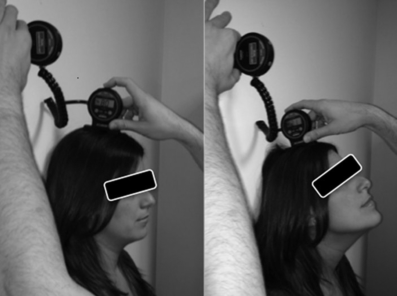
Suboccipital range of movement with a digital inclinometer.
Lumbar spine mobility
Lumbar mobility was evaluated with the sit-and-reach (SAR) test, and by means of measuring lumbar forward bending (LFB) with a DI. The SAR test is a common tool to measure maximum spinal mobility in flexion and HSM flexibility. 44 The subject was sitting with knees extended and both feet against the edge of a standard SAR box. Then, the participant was asked to bend slowly forward toward the toes as far as possible keeping knee extension and placing both hands on the box. 45 The final position was kept for 5 seconds. 44 This test has shown excellent intratester reliability, 46 and a moderate to high criterion-related validity, ranging from 0.46 to 0.89. 47 The SEM has been observed around 3 cm. 44
The LFB evaluates spinal mobility in flexion and it is an indicator of functional limitation. 48 Standard protocol requires the patient to be standing and to maintain knee extension while bending forward. 49 The DI was positioned with one of the sensors on the lumbar spine (L3–L5 segment) as the dynamic region, and the other was placed on the wall, as the static stable position. 50 The use of an inclinometer has been recommended as the preferred tool to measure lumbar mobility. 51 The accuracy level for trained evaluators has been established around 90% with absolute values of±3.4° for lumbar flexion. 52
Interventions
Intervention in the control group
In the CG, two techniques were performed in the following order.
1. Neuromuscular technique over the masseter muscles. With the subject lying supine, the therapist sat at the head of the table. One therapist's hand was placed on the zygomatic arch to fix the superior edge of the muscle, while the thumb of the other hand performed longitudinal myofascial strokes, of about 5–8 cm length (each of 4–6-second duration) (Fig. 3). Tissue restrictions were respected during the gentle slide movement and no pain was provoked. 20,53
2. Passive stretching of the HSM. With the subject supine, the therapist performed a pain-free hip flexion keeping knee extension until the edge of flexibility of the posterior muscle chain. 54 Then, the therapist underwent a dorsal flexion of the ankle and kept the position for 40 seconds. 54 The protocol was conducted in both lower limbs.
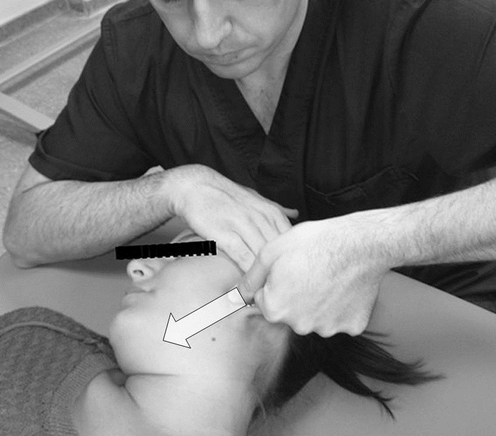
Neuromuscular technique over the masseter muscles. The arrow indicates the direction of the thumb pressure on the masseter muscle and from the mandibular angle to the chin.
Intervention in the experimental group
Subjects of the EG underwent the same protocol and in the same order as in the CG, but a new technique was included at the end of this, the suboccipital muscle inhibition (SMI) technique. 55,56 With the participant lying supine, the therapist placed both hands under the subject's head. While contacting the inferior edge of the occipital bone, the therapist exerted a constant and painless pressure in cranial and ventral direction until suboccipital soft tissue was relaxed. 56 The technique was performed for 4 minutes approximately. 55
Statistical analysis
Statistical processing of the data was done with the PASW Statistics tool, version 18.0 (SPSS Inc., Chicago, IL), and was conducted considering significance at a p-value<0.05. The Kolmogorov–Smirnov test was used to assess the normality of the data. The mean, standard deviation, and 95% CIs were calculated for quantitative variables, while categorical data were expressed in terms of percentage frequencies. Baseline characteristics were evaluated with the chi-squared (χ 2) test for qualitative variables and the Student's t-test for independent samples for quantitative variables. The inferential analysis of variance for repeated measures (ANOVA test), with the group as the between-subjects variable and with the time as the within-subject variable, was used to assess the between-group differences.
Results
Table 1 lists the physical and clinical characteristics of participants in the baseline measurements. The between-group comparison showed no statistical significance in any variable (p>0.05).
Data are reported as mean±standard deviation or in percentages (%). PPT values are expressed in kg/cm.2
Statistical significance of the between-group differences using independent t-test.
As determined by chi-squared test.
CG, control group; E, extension; EG, experimental group; F, flexion; L, left; LUMB, lumbar spine; M, masseter muscle; PPT, pressure pain threshold; R, right; ROM, range of motion (°, degrees); SAR, sit-and-reach test; SUB, suboccipital; V1, supraorbital nerve; V2, infraorbital nerve; V3, mental nerve; VMO, vertical mouth opening.
Intra- and between-group comparison
Table 2 includes the baseline and final values of the outcome measures in both groups, and the mean differences in the intra- and between-group comparisons.
Data are reported as mean±standard deviation or 95% confidence level. PPT values are expressed in kg/cm.2
Indicates no statistically significant between-group differences (p>0.05).
Indicates statistically significant intragroup differences (p<0.05).
In the intragroup analysis, the EG showed statistical significance for suboccipital flexion (p<0.001; F 1,29=14.47; R 2=0.33) and lumbar mobility as measured with the SAR test (p=0.009; F 1,29=7.89; R 2=0.21). There were no significant differences for the rest of outcome measures, even though results were close to it for VMO (p=0.057). The CG only showed statistical significance in the SAR test (p=0.003; F 1,29=10.81; R 2=0.27).
With regard to the between-group analysis, although the EG found better results than the CG for VMO, lumbar mobility, and suboccipital flexion, no differences were found in any outcome measure (p>0.05). The findings were close to statistical significance for suboccipital flexion (p=0.055) and LFB (p=0.057).
Discussion
The inclusion of an MYO technique in the multimodal protocol used in the CG combining local (neuromuscular technique over the masseter muscles) and distal treatment (hamstring stretching) to the craniomandibular area found no impact in the outcome measures in the comparison between groups. For ethics reasons, subjects in the CG also underwent an intervention. It is plausible to presume that the present findings may have been different otherwise.
Assuming that human fascia is able to contract and to influence biomechanical behavior, 57 it has been suggested that MYO techniques should be evaluated in TMD to assess their impact in clinical practice. 20 There is a fair level of successful evidence in the short-term for a multimodal manual approach of TMD generally combined with exercise. 6 No exercise was included in the protocol of the present study, which may explain the observed findings.
As regard with VMO, it has been reported that a difference of 6–9 mm should be observed after treatment in a painfully restricted TMJ to conclude a clinically relevant improvement. 38 In this trial, the mean improvement in VMO in the EG was almost 2 mm and hence close to statistical significance in the intragroup comparison (p=0.057), but insufficient to be meaningful. However, this score change is within the 1.4–2.5 mm average improvement that has been found in previous research after applying HSM stretching, 54 neuromuscular techniques, 23 and/or postisometric relaxation over the masseter muscles. 35 Nevertheless, all these studies were made on participants with tenderness in the masticatory muscles, but not diagnosed as having TMD.
Although a wider range of VMO has been found in individuals with normal disc and condylar translation movements, 58 our study subjects had restricted condylar mobility. The lack of positive findings in this trial is in line with the results reported by several studies. An MYO protocol over the masticatory muscles, 20 the strain–counterstrain technique in the latent myofascial trigger points of the masseter muscle, 38 and the fascial manipulation technique 59 have showed no immediate impact on VMO. On the contrary, the combination of intraoral myofascial therapy, home therapeutic exercises, and a self-stretching protocol has been concluded to have a positive effect on mouth opening in the medium- and long-term. 21 Likewise, a positive, but not clinically significant, trend was found in VMO after fascial manipulation technique 59 and intraoral myofascial therapy alone, 60 with mean improvements of 0.4 and 3 mm, respectively.
A restricted cervical mobility and an abnormal head posture (forward head posture) have been linked with a reduced VMO, both in healthy subjects and in TMD patients. 61,62 Although head position was not measured in this study, the SMI technique has been purported to improve cervical range of movement, 63 and to modify craniocervical posture, 55 which may explain that results were better in the EG. It has also been suggested that a combined therapeutic approach should show better results on VMO and PPT of the masticatory muscles than a single technique in subjects with TMD. 64 These observations have been confirmed after a multimodal treatment combining dry needling, spinal manipulation, and mobilization. 65 Similar findings were reported after different manual therapy techniques and/or conservative treatments using muscle stretching, relaxing exercises, and electrotherapy. 66 However, the present observations do not seem to support this claim. Different protocols have been carried out in the previous trials, which makes difficult to compare between studies.
Local and distal treatments have proven to positively affect orofacial PPT in pain-free subjects or neck pain patients with tense bands in the masticatory muscles. A better mechanosensitivity response has been found after several treatments, such as the strain–counterstrain technique or neuromuscular strokes over the masseter muscles, 23 spinal manipulation alone, 67 or in combination with the SMI technique, 68 HSM stretching, 54 and MYO maneuvers in the suboccipital region 55 and over the masticatory muscles. 20 In regard to TMD, dry needling, 69 cervical mobilization and stabilization exercises, 70 and posture training, 71 among others, have led to an immediate- and short-term increase in PPT over the masticatory muscles. These positive results have been explained based on the effect of manual therapy on the activation of the descending inhibitory pathways, which has not been demonstrated yet in TMD. 70 The lack of changes in PPT values in this study may be explained because this is the first trial in which inclusion criteria have also taken into account restricted mobility of C1 and mandibular condyles. Both regions have an anatomical link with the caudal nucleus of the trigeminal nerve (C1–C3), 72 which seems to have influenced the algometry results in the trigeminal nerve and masseter muscles.
Finally, turning to suboccipital and lumbar mobility, the duramater and the neuromuscular chains have been proposed as explanations to interrelate the suboccipital and HSM. 26 The myodural bridge establishes a direct relation between the suboccipital musculature and the duramater. 73 Hence, a dysfunction of the suboccipital muscles may have an impact on HSM flexibility and vice versa. This is the first study to evaluate the possible benefit from releasing tension in both muscles in individuals with TMD.
Lumbar flexion improved in both groups. A difference from baseline scores after intervention higher than 18% in the SAR test or 20% in LFB may indicate a real change in HSM flexibility. 74 Lumbar mobility results were better in the EG, although the improvement was only of 7.92% in the SAR test and of 6.79% in LFB. As to suboccipital ROM, an improvement of 6.48° in suboccipital flexion was observed in the EG. In this sense, the SMI technique has already been found to improve suboccipital ROM in flexion. 75 Changes in soft-tissue tension of the extensor muscles after the SMI technique may enhance the flexibility of this region when a passive stretching of extensor muscles is required to perform suboccipital flexion.
Study limitations
The results have assessed only immediate changes. A therapeutic program, including exercise, during several sessions, and a medium- to long-term follow-up would be of interest in future research. Likewise, although suboccipital muscle dysfunction has been linked with TMD, 76 the participants were not previously assessed to find tense bands or tenderness in this musculature.
Conclusions
The cumulative effect of combining local and distal techniques (myofascial induction, neuromuscular treatment, and HSM stretching) has no impact on mouth opening, orofacial mechanosensitivity, and suboccipital and lumbar mobility in subjects with TMD.
Footnotes
Author Disclosure Statement
No competing financial interests exist.