
Abstract
Abstract
Recent research indicates that electroacupuncture could help patients suffering from Alzheimer's disease. Indeed, certain acupoints, when stimulated by electric pulses, activate particular parts of the brain, the details of which have been revealed by magnetic resonance imaging and positron emission tomography. Observed stimulated regions of the brain of Alzheimer's patients can be compared with the images obtained by fluorescence technique with a proper dye in Alzheimer's brain sections. This article provides support based on Western medical science for the treatment of Alzheimer's with electroacupuncture. Electroacupuncture acts in different and efficient ways. First, the positive charge of the electric pulses helps to detach from the neuron or the neuron membranes β amyloid peptides, which have been associated with the pathogenesis of the disease. In addition to this, the presence of the carboxyl group of β amyloids suffers a torque in the presence of an electric field and creates a serious deformation. Lastly, the moderate shaking originating from the electric pulses facilitates the partial disintegration of the β sheet, a frequent conformational change of the β amyloid peptide.
Introduction
AD 9 causes severe impairments related to memory, language, social abilities, mental faculties, judgment capacity, disorientation in time and space, limitations in visual processing, and general coherent behavior. It is an inexorable chronic and progressive disease characterized by the loss of neurons in certain parts of the brain, which starts decades before the appearance of symptoms. At the present time, it cannot be prevented or cured. Unfortunately, it is a disease that worsens with age. In the United States, 13% of people aged 65 and older and 43% of people aged 85 and older suffer from this disease. 10 Its progression is slow, with death occurring around 10 years from the beginning of the clinical symptoms, and patients will spend most years in the most severe stage of the disease. 10 Therefore, it is essential to look for more alternative treatment approaches. In this context, it is worth mentioning that experimental data reveal that electroacupuncture treatment7,8 does provide a hope for AD patients, although a modern scientific basis for this important observation has not yet been provided. The purpose of the present investigation, therefore, is to elucidate how and why electroacupuncture works for the treatment of AD.
Electroacupuncture and Alzheimer's Disease
Electroacupuncture has been used to treat patients with several different conditions, particularly, torticollis, 3 high blood pressure, and chronic bronchitis, 4 as well as AD.7,8,11,12 In electroacupuncture, the points selected for treatment are the same as for traditional acupuncture, but the electrical stimulation is achieved by passing an electric current in the form of pulses. The magnitude of the current can be varied from 0 to 100 mA (1×10⁻³ A). However, it is possible to increase the current strength up to 400 mA. With the help of specially designed equipment, the form and frequency of the pulse can also be adjusted according to the abnormality to be treated. In many cases, electroacupuncture is found to be more effective than traditional acupuncture, and data confirm this, particularly for AD.7,8,11,12
In the electroacupuncture process, an additional parameter is introduced. It is not only the application of an electric field or the pulses of the fields that is important, but also the magnetic field associated with it. Hence, a mechanical force of substantial magnitude can be originated from electromagnetic fields.3,13 In electroacupuncture, therefore, mechanical oscillations are created, and they become the key parameter for some diseases. This has already been proven for the treatment of torticollis 3 and chronic bronchitis. 5 In AD, an additional parameter is introduced because of the specific features of the β amyloid; namely, its structure has a pair of ions and it gives rise to a repulsive force that tries to separate from neurons or its membranes. Moreover, the structure has a carboxyl group, and because of its presence, the probability of transformation to a monomeric coil structure is increased.14,15 These details will be discussed later.
Beta Amyloid Peptide and Alzheimer's Disease
This relationship has been extensively studied over the last decades,14–17 and it is now known that AD originates from a misfolding of the amyloid precursor protein (APP). Certain enzymes divide this protein into smaller fragments (proteopathy), and in so doing some conformational changes take place and the β amyloid peptide (β AP) is formed, which has a sheet structure instead of the usual helix structure, and it consists of 35–43 amino acid residues. 15 This abnormal protein has the predisposition to aggregate and form extracellular deposits known as plaques. The presence of the β AP is detected experimentally by using circular dischroism spectroscopy, 14 and its formation in the plaques structure in the brain can be examined with the help of fluorescence technique by using a dye such as thioflavin-S in the brain section. 17 In the living brain, Pittsburg compound (PiB) 18 and positron emission tomography (PET) have also been used to examine the precise location of the plaques.18,19 The latter technique has been widely employed over the past few years, as it provides a wealth of information related to amyloid deposition in vivo. 18
Figure 1 shows the typical image of an AD human brain cortex obtained with confocal fluorescence imaging technique by using a thioflavin dye. 17 The brain tissue is obtained following the rapid autopsy protocol. 20 Plaques (outside neurons) and neurofibrillary tangles (inside neurons) are clearly visible here, confirming the anatomopathological diagnosis of the disease. From Figure 1, it is clear that in the advanced stage of the disease, a substantial part of the brain is affected. Aggregated β AP and damaged neurons are deposited in extended regions of the brain. In such circumstances, help from pharmacological products cannot be expected. All experimental evidence points to the fact that abnormal β AP is one of the main attributed causes of AD pathogenesis.
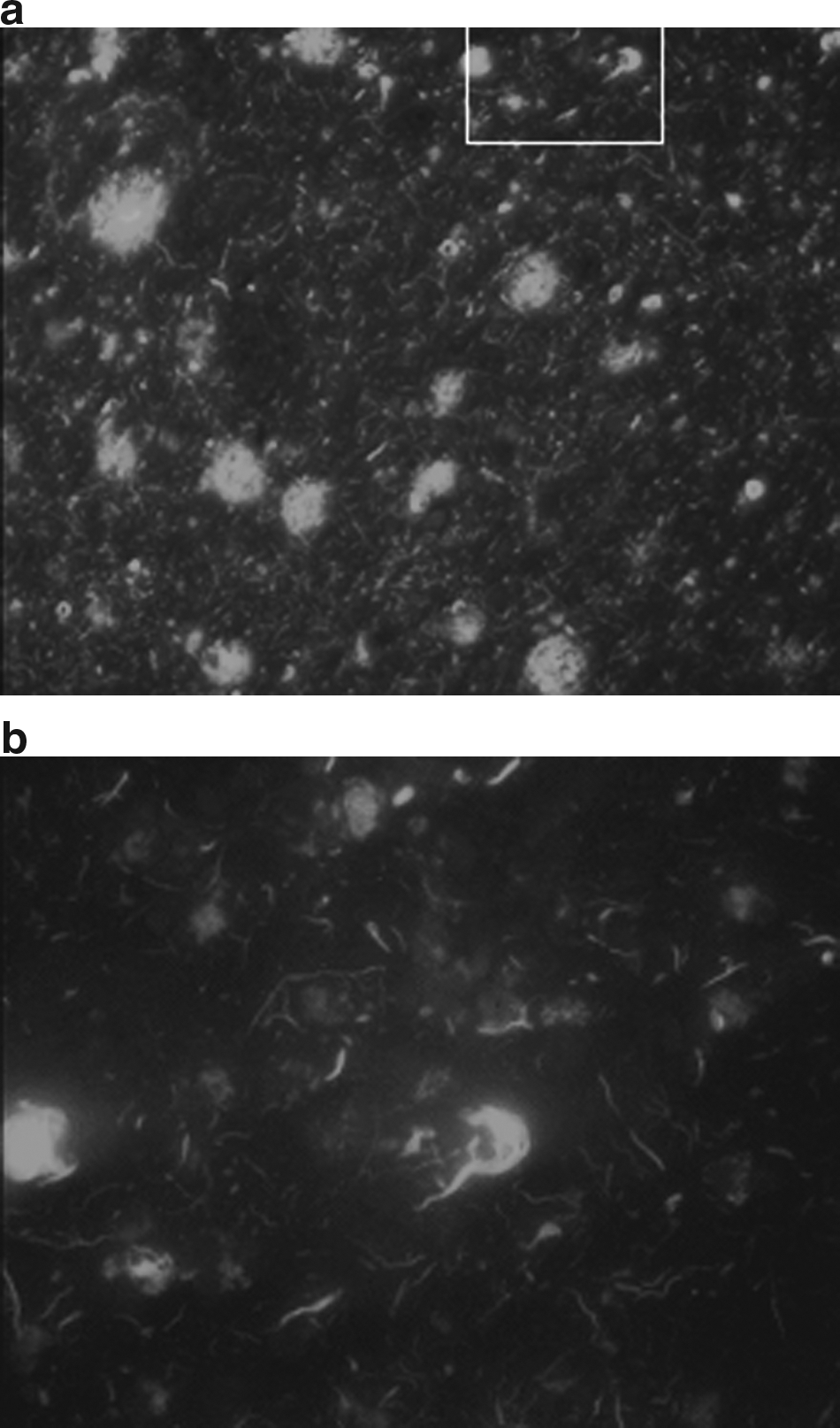
A section of human brain of an AD patient with severe disease, stained with thyoflavin (15).
Division and folding of the APP protein and its conversion in β AP sheets is a complex process, and not all the parameters involved in it are known. Most people affected by AD are known as sporadic cases where familial genetic mutations have not been demonstrated. However, in a minority of AD cases, it has been reported that there are mutations of certain genes that code proteins like APP, PS1, and PS2. 21 The knowledge of familial mutations has allowed the study of the disease in transgenic animal models. Carriers of the apolipoprotein E4 (APOE 4ɛ) gene are considered to have a higher risk of AD. APOE comes in several different forms (alleles), but statistical data reveal that APOE 4ɛ plays a crucial role, and extensive investigation in this direction has been carried out 22 in the last two decades with findings indicating that a higher concentration of APOE 4ɛ is strongly correlated with amyloid deposition. These investigations have helped us to understand pathogenic cascades in several neurodegenerative diseases such as AD. However, the details of the folding mechanism and the sheet formation processes are not clearly understood.
All the above-mentioned aspects are significant in the process of forming plaques, but in the context of removing or eliminating them, they do not provide key parameters. Hence, we will not address these issues in the present discussion and analysis.
Neuroimaging Techniques and Alzheimer's Disease
Recent developments in neuroimaging technology include functional magnetic resonance imaging (fMRI)10,11 and PET, 18 which allows the mapping of the functional activation of a specific part of the brain in a noninvasive manner. fMRI is particularly sensitive to any slight change in the blood oxygenation in a specific part of the brain, indicating that the neuronal activity is altered during a given task. This technique is very accurate for the majority of patients, and hence it is precise in locating the exact part of the brain that is being activated. Extensive investigation has therefore been carried out with the help of this approach.10,11 The role of Korean acupoints has been determined in detail, and it is now possible to know which point activates which part of the brain. 8 Therefore, neuroimaging techniques combined with acupuncture and electroacupuncture procedures can provide unique information about the actual role of this traditional therapy in the brain-activation practice and will afford a foundation for the treatment for AD and other neurogenerative diseases.
It is worth pointing out that Western science has not neglected the possibility of the effect of short-term nerve stimulation by electric pulses. It has been found that there is, indeed, a positive effect resulting in an improvement for AD patients.23,24 Scherder et al. 23 have reported that transcutaneous nerve stimulation improves memory-related abnormalities. Moreover, patients undergoing this treatment are more cheerful, more active, and have improved social behavior. In addition to these outcomes, global deterioration is reduced considerably. 24 Thus the improvements in AD patients as a result of an electric field are reported both by acupuncturists and by Western medical practitioners.
However, the main question still remains unanswered. How and why do acupuncture and electroacupuncture therapy seem to work? What could be the mechanism behind it? Will it be a very effective therapy? These aspects will be addressed in the light of the electric field effects on β sheets.
Discussion
The pathways for sensory information to reach the central nervous system are through cranial nerves such as the vagus and spinal nerves (Figs. 2 and 3). For example, the vagus nerve is particularly used to stimulate the brain electrically for patients suffering from epilepsy. 25 This indicates that the vagus nerve is a direct and efficient path for an electric current to reach the brain. Therefore, it follows that there are two or three biological pathways (two or three meridian systems, according to Traditional Chinese Medicine) by means of which electrical stimulation or energy flow can reach specific parts of the brain. Commonly used acupoints for this purpose are located on the heart, bladder, and stomach meridians, and earlier experimental work confirms that the points HT 7, ST 36, ST 40, KI 3, GV 14, GV 20, and BL 20 are very effective for AD treatment. 7 Shou and Jia 7 have carried out research work in this area, and using fMRI technique they have observed that by applying electrical stimulation through some of these points, specific parts of the brain are activated, for example, the right-hand side gyri (superior and middle temporal and hippocampal and posterior cerebellar). On the left-hand side, a considerable portion is also activated, including the superior parietal lobule, insula, and middle occipital gyri. Extensive and systematic work has been carried out in the last decade, and a recent review 8 reveals that a specific part of the brain is activated by specific acupoints or set of acupoints. Data reveal that acupoints ST 36, BL 60, and BL 67 stimulate the inferior frontal gyri, while GB 34 and GB 39 stimulate the superior and medial frontal gyri. Further details can be found in Table 1 of Shou and Jia. 7 In addition to this, in some cases, the frequency at which electroacupuncture is more effective has also been reported, with the most favorable results being obtained at approximately 2–5 Hz.
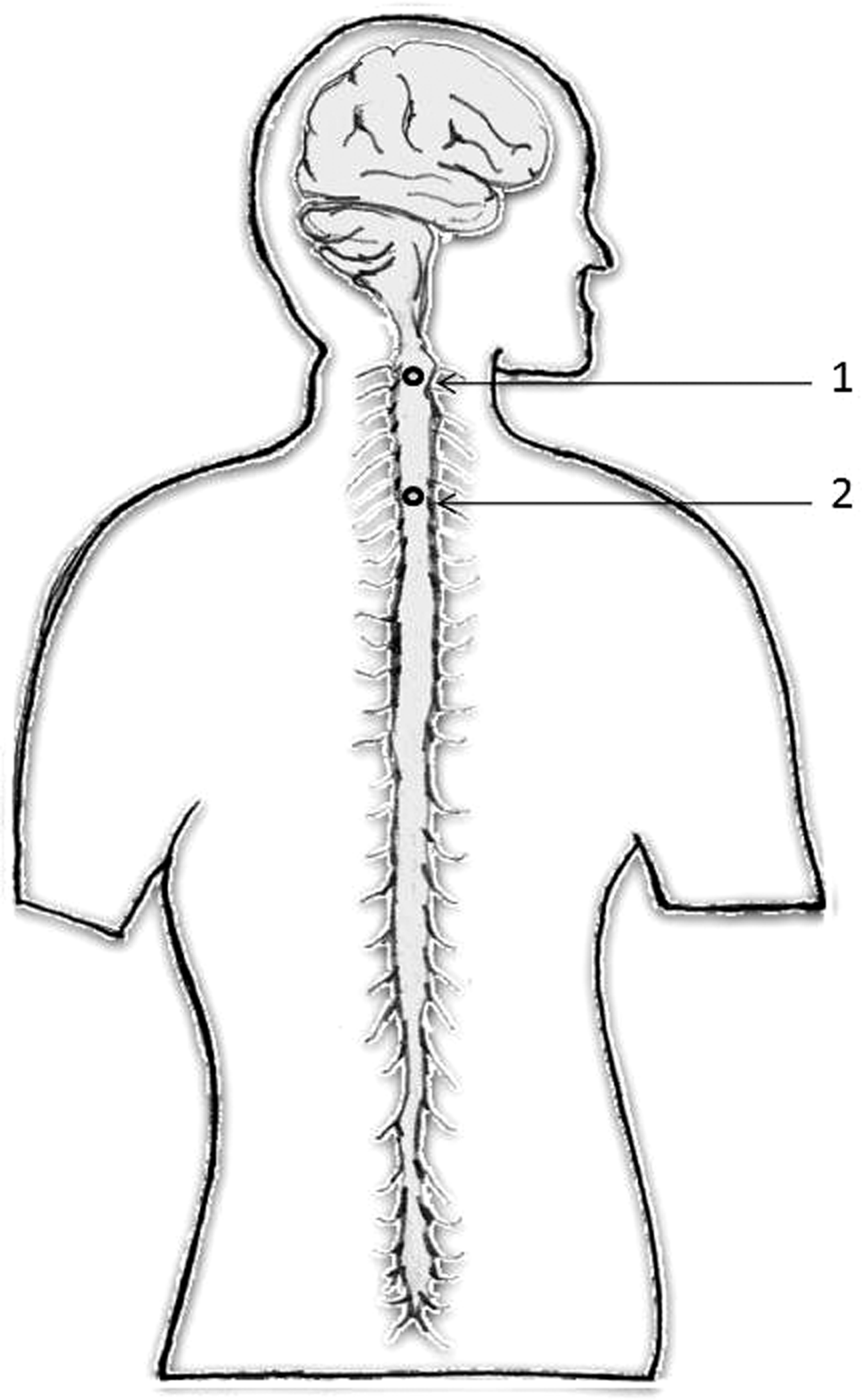
Spinal cord and the acupuncture points (1) GV 15 (Yamen) and (2) GV 14 (Dazhui).
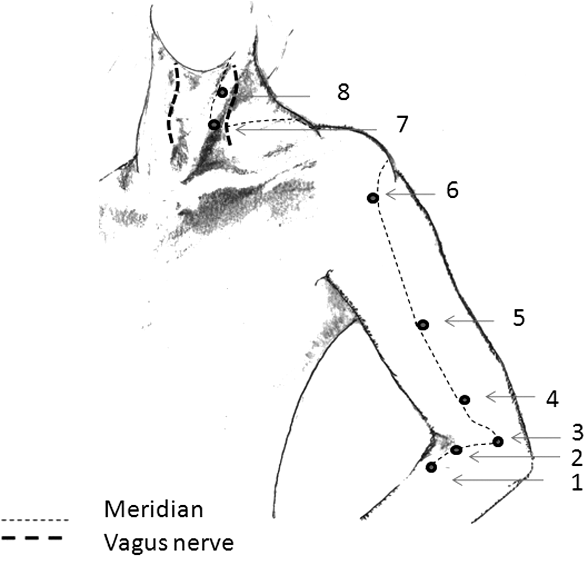
Connection between the large intestine meridian and the vagus nerve, which connects directly to the brain. (1) LI 10 (Shousanli); (2) LI 11 (Quchi); (3) LI 12 (Zhouliao); (4) LI 13 (Shouwuli); (5) LI 14 (Biano); (6) LI 15 (Jianyu); (7) LI 17 (Tianding); and (8) LI 18 (Futu).
It is worth mentioning that all these points lie on the stomach, heart, or bladder meridian system, and they are associated with the spinal cord or directly or indirectly the network of the vagus nerve system, which is both extensive and connected to the brain. For example, BL 20 lies on the spinal cord, and hence the electrical signal or pulses pass to the brain easily and activate certain parts. Similarly, GV 14 and GV 15 lie on the spinal cord (cervical region) and are obviously excellent points for passing electrical signals to the brain (Fig. 2). fMRI shows that GV 15 activates the right superior temporal gyri. Such observations provide strong experimental evidence that electroacupuncture can indeed activate specific parts of the brain that are affected by AD. Similarly, points LI 4, LI 10, LI 13, and LI 15 have been found to be very effective in AD treatment. These points lie on the large intestine meridian pathway, which is connected to the vagus nerve, as shown in Figure 3. In this case, meridian pathways are also in agreement with the anatomical trajectories or biological pathways confirmed by Western medical science. These trajectories are shown in Figures 2 and 3 along with the acupoints used for this purpose. These aspects together with fMRI create confidence in the use of the electroacupuncture technique for AD.
The above discussion suggests that acupoints ST 7 to ST 10 or BL 4 to BL 8 should be more effective, as they are near the vagus nerve or the spinal cord. However, electrical resistance near these points might be higher than the conventional experimentally confirmed points mentioned earlier. It should be noted that the electrical signal is forwarded to the brain via the vagus nerve using special apparatus that is used to treat epilepsy, The results of this are encouraging. 25 It is therefore recommended that positive pulses are sent through points ST 4 to ST 10 and BL 4 to BL 8 and that the signal is applied directly through the vagus nerve, as it is done for epilepsy treatment. 25 Previous experimental data7,8,10 show that there are a few sets of acupoints that give a favorable result with varying degrees of success. This is understandable, as the signal can be communicated to the brain through different channels like the spinal cord and the extensive circuit of vagus nerves.
In order to understand the details and the progress of AD, it is necessary to examine the biochemical aspects of the β AP, one of the implicated proteins in the neurodegeneration process.14,15 It is, therefore, necessary to look at the details of the structure, conformational changes, stability of the compound, and, most importantly in the present context, the effects of the electric field on the β sheet structure.
Beta amyloid is formed from the APP, and this change is conformational. The β sheets are shaped by alternating peptide strands. Strands are linked with another unit by a hydrogen bond, which is very weak and temperature sensitive. The β sheet linkage has the property of self-aggregation, and in due course it forms plaques from coil peptide. 9 They are fibrils in nature and believed to be neurotoxic. Amyloid is not a special case, but several proteins (e.g., prions, serpin, etc.) go in the same conformational transformation. This means that, in some parts, random coil↔β sheet transition takes place. Very often, conformational changes are reversible under certain conditions, and in the present case, it is reversible in aqueous solutions with specific pH values (4–5.5). One of the significant parameters that decides the irreversibility from β sheet to coil structure is the concentration of β sheet. Careful investigation in this area has been carried out using circular dichroism spectroscopy, 13 and it has been reported that when the concentration of coil sheet structure is reduced, the process becomes reversible, albeit slowly. Moreover, it has also been observed that the concentration near the membrane or neuron is higher compared with other parts. This is a serious impediment as far as a successful treatment is concerned. Some mechanisms have been suggested to explain why the β sheet is attached to the membrane or neurons. 14 One of the possibilities is the electrostatic attraction between the membrane potential, which is negative, to the positive components of the β sheet.
Pure peptide aggregates with time, forming fibrils with a β structure. The formation of a β sheet linkage is initially a really slow process, and it starts in the lower part of the hippocampus when it is considered an initial stage of AD. It then extends toward the cerebellum and the brain stem. Phospholipid vesicles present in the brain structure (particularly on the membrane) are formed by negatively charged phospholipids, and because of the electrostatic attraction, the process of aggregation is accelerated. 26 Soon, plaques and cerebrovascular amyloid deposition in the brain becomes unavoidable. At this stage, it is considered a moderate level. Following this, β amyloid sheets spread progressively to all parts of the brain. The disease then progresses in a rather accelerated manner, as the self-association process of β AP is predominant. 14 Figure 1 shows the wide extension of β AP deposition in the plaques. This is a severe case, and the patient becomes untreatable in every way.
The above discussion helps us to understand why it is difficult to find an adequate treatment, as any proper pharmacological product which might dissolve or separate the β amyloid sheets from the neurons membrane would have difficulties accessing some of the sites. Biochemical processes will not in any way help to detach β AP, but the electrostatic repulsion between the ion pair (Lys 28–Met 35) and the neural system may work for this purpose.
An extensive investigation in biochemistry14,15 and a model calculation have been carried out on folding, structural details, and the stability of β amyloids. The work reveals that an antiparallel β sheet structure is stable only under certain conditions, depending upon the concentration, the pH value of the solution, and, most importantly in the present context, the electrostatic charge distribution near the amyloid deposits. This is because at both ends of the β sheet, ion pairs are formed between Lys 28 and Met 35, 15 and there exist three amino acids dangling at the end of the β sheet. These structural details are very important, as this pair of ions help to attach the β sheet to the neurons or membranes, which have a negative charge.
Apart from the biochemical aspects, in the present framework, physical stimulation also plays an important role. This includes electric field and temperature-induced deformation in the β amyloid structure. The presence of an electric field around the β amyloid creates several alterations. The ionic interaction between ion pairs and negatively charged neurons plays a strong role in the stability of the β sheet. Moreover, β amyloid structure has a carboxyl group [−C(=O) OH]. Functional groups are active sites for field interactions in several ways.27,28 There is a difference in electro-negativity between the carbon and the oxygen atoms, and the structure shows electric field sensible behavior. Moreover, the carboxyl group is a polar component of β amyloids, and in the electric field, a torque—a measure of the turning or the rotating force—is originated. If the polar unit were free, it would rotate in the electric field, but in the present case, it is the part of the macro molecule, which is attached to the membranes or neurons. Obviously, it will not rotate, but it would cause a mechanical bending tendency and hence increase the instability of the structure. The protonation of [−C(=O) OH] or removal of its charge by any means shifts the equilibrium toward the coil monomeric state and means the β sheet has to be disintegrated. This suggests that if the pulses of positive voltage are supplied to the membrane or the neuron system, the possibility of the detachment of the β sheet from the neurons will be increased due to the repulsion of electrostatic forces, and also the probability of transformation of the β sheet in a monomeric coil form will increase.
It is worth mentioning that this conformational change is originated from hydrogen bonding—a weak bonding—and hence it is very temperature sensitive. For certain temperature and concentration of β sheet structure, it is possible to have equilibrium with the coil structure. Conformational changes, particularly in biochemistry, are very complex, 27 and careful investigation in this area is really necessary. As mentioned above, the combined effects of the electric field alters several interrelated parameters of stability, and the β sheet structure will try to disintegrate, even though the separate contribution of each parameter cannot be determined.
In addition to this, there is a vibrational effect. It has already been found that, in the electroacupuncture process, pulses of electric current create a magnetic field. According to the Biot–Savart Law, 13 the intensity of the magnetic field is proportional to the magnitude of the current and inversely proportional to the distance at which the field is created. Even though the magnitude of the current is small, of the order of a few mA, the distance between two neurons is very small—of the order of nanometers or a fraction of it. Therefore, the magnitude of the magnetic field is not negligible, and it is of the order of 2000 Gauss to 6000 Gauss depending upon the distance and the strength of the electric current (for details, see Fig. 2 of Reference 3). Electric and magnetic fields produce the mechanical force that creates mechanical vibrations in the system. The exact strength or the amplitude of vibrations depends upon the separation between neurons or the elements through which the pulses are passing. Earlier investigation shows that this force is not negligible, particularly when the separation is of the order of nanometers. The vibrations created by just a few mA can have a force of 0.1 dynes, and depending upon the mass of the vibrating elements, the amplitude will vary. The effects of mechanical vibrations in the electroacupuncture process have been discussed in detail, particularly for problems related to torticollis, asthma, and bronchitis. 5 In short, the vibrational shaking effect will take place in the membrane and in the region of the affected neuron. Thus the combined effect of electrostatic repulsion and shaking will help, to a certain extent, the β sheet to detach or distintegrate. This reasoning is valid only when self-aggregation of the β sheet or formation of plaques has just started.
As mentioned earlier, for AD therapy, low-frequency positive pulses are applied, and this is also understandable. This means that about 250 ms positive voltage is provided to the neuron membrane, and it is a sufficiently long period for the repelling β amyloid. High-frequency pulses might not work with the same efficiency, and further investigation in frequency dependence is really needed. If necessary, a device can be constructed specially for this purpose, which will provide a positive pulse of longer duration.
The application of the electric field has an additional effect, namely heating. A neural system with β amyloid sheets attached to it has a relatively high electrical resistance, and therefore the passage of a current of a few mA (say, 2×10⁻³ A) is enough to heat the system. It is not possible to estimate the exact magnitude, as the resistance varies from region to region. Amyloid sheets are formed by conformational change from helix structure by alteration of the hydrogen bond, which is a weak one. The heat energy might be enough to alter the bond direction and weaken the structure of the β sheets. Temperature effects on conformational changes are known, 29 and frequently even reversibility is achieved. In the present case, reversibility to coil structure may not be achieved, as it is observed in solution, but the stability of the β sheet structure will certainly be affected, and this could lead to its disintegration.
Recent investigation based on Western medical science also reveals the importance of the application of a high-frequency electric field to specific parts of the brain. Deep brain stimulation (DBS) therapy 30 for AD patients is found to be very effective, and results show that, in certain cases, it can even reverse the tendency. DBS therapy was applied for 12 months, and results were assessed with the help of glucose metabolism using PET to map the brain area and also examining cognitive functions. The results were encouraging. Investigation in this area is in progress.30,31 DBS and the electroacupuncture technique seem to help AD patients, and both techniques involve the effects of an electric field on amyloid sheets.
DBS, when exploited properly, is a powerful technique. However, electroacupuncture has certain advantages over DBS. In particular, the passage of an electric current creates magnetic fields that, at a short distance, have a significant magnitude and are detectable by magnetocephalography. Moreover, unlike DBS, fields (electrical and magnetic) are activated at specific points near the neurons and beta sheets. The directions of chemical bonds and functional groups of beta amyloids are altered in the presence of a magnetic field; hence, the instability is increased. 32 Therefore, these aspects need to be considered.
Both oriental and Western approaches are based on the effect of an electric field on specific parts of the brain. In the present investigation, we have provided support based on biochemical and physical aspects to explain how β amyloid sheets can be disintegrated in the presence of electric fields. Therefore, AD could be considered as a treatable disease, if these techniques are properly exploited.
The positive pulses used in the electroacupuncture process have multiple effects on β amyloid sheets attached to the nervous tissue in several parts of the brain. First, it causes repulsion due to the ion pairs formed at Lys 28 and Met 35. Second, the carboxyl group formed at the end of the β sheet is strongly affected by the presence of positive voltage and originates instability that might help to transform β sheets in a monomeric coil state transformation. In addition to this, the vibrational effect shakes the structure and accelerates the process of separation and disintegration. A slight increase in the temperature might accelerate the process of conformational changes.
Conclusions
Electric and magnetic fields together with effects of vibrations help to disintegrate β amyloid sheets, and hence electroacupuncture could be an additional effective approach to modify the course of AD without the risk of side effects.
Footnotes
Acknowledgments
We thank Professor Luis Hernández (Universidad de los Andes, Venezuela) for his discussion and valuable comments, and Dr. Larisa Poluetkova (Center for Neurovirology and Neurodegenerative Disorders USA) for her help in staining the brain sections.
Disclosure Statement
No competing financial interests exist.