
Abstract
Abstract
Background:
Acupuncture is an ancient medical technique of China. Clinical studies have found that acupuncture is an effective treatment modality for remitting stress disorder, anxiety, and depression.
Objective:
This study investigated the molecular mechanisms of acupuncture that have been demonstrated to be beneficial for treating depression.
Methods:
A depressive disorder model was induced by exposing Sprague Dawley rats to chronic unpredictable stress. Acupuncture stimulation and riluzole were used to treat the depressed rats. Physical and pathologic changes—including behavioral performance, body weight, hippocampal glutamate concentration, and glutamate–glutamine cycle related genes—were analyzed before and after therapy.
Results:
Acupuncture improved behavioral performance, reduced glutamate in the hippocampus, prevented damage to hippocampal neurons, reduced glutamate release, and increased glutamate recycling.
Conclusions:
Acupuncture is a safe and effective modality for depression therapy in rats. Acupuncture treatments may relieve depression and reduce glutamate content to normal levels. Additional research is needed to assess the effect of acupuncture on depression in human patients and to explore other possible mechanisms.
Introduction
D
Extremely high concentrations of glutamate have been found within the cells of the central nervous systems and a clinical study has revealed that depressive disorder is associated with increased intracortical elevated glutamatergic activity. 4 Glutamatergic neurotransmission occurs predominantly within the confines of a tripartite synapse. The interactions among synaptic SNARE proteins, synaptobrevin (vesicle-associated membrane protein [VAMP]), SNAP-25, and syntaxin in plasma membranes play an important role in glutamate neurotransmission. 5 Glutamate is then recycled through the glutamate–glutamine (glu–glu) cycle by glutamate receptor in postsynaptic neuron. 6 Dysregulation of the glutamatergic system has been found to be an important factor in depressive disorder.7,8
Acupuncture is an ancient medical treatment modality from China and can be traced back at least 2500 years. Acupuncture is now widely used as a complementary and alternative medicine in many countries.9–11 Clinical studies have shown that acupuncture is an effective treatment modality for remitting stress disorder, anxiety, and depression.11–15 Electroconvulsive therapy could regulate the expression of distinct neurotrophic-signaling pathways, including metabotropic glutamate receptor (GluR1), neuron glucose transporter, and gamma aminobutyric acid–A receptor. 16 In addition, electroconvulsive therapy has been accompanied by stable structural changes in neuronal networks that affect synaptic plasticity in various regions of the brain. 17 Yamada et al. reported that repeated electroconvulsive treatment induced the expression of VAMP 2/synaptobrevin-2 in rat depression models. 18
Despite the effectiveness of acupuncture for treating depressive disorder, very few studies have investigated the underlying mechanisms. The aim of this study was to develop further understanding of the mechanism of acupuncture for treating depressive disorder. The results of this study indicated that, in rats, glutamate, synaptic vesicle proteins, and the glu-glu cycle system, might be involved in antidepression with acupuncture because of their important roles in neurotransmission.
Materials and Methods
Animals
Adult male Sprague Dawley rats (180–220 g) were used in this experiment. Animals were obtained from the Experimental Animal Center, Traditional Chinese Medicine University of Guangzhou, China. Both animal care and the study protocol were conducted according to the Helsinki Declaration Accords. In addition, the guidelines of the Committee on Care and Use of Laboratory Animals in the animal center of Guangzhou University of Chinese Medicine were followed. Animals were housed under a 12/12 hour light/dark cycle at a constant temperature room temperature of 23°C according to Traditional Chinese Medicine University of Guangzhou care and use of laboratory animal guidelines.
Depressive Disorder Model and Treatment
Depression disorder rats were induced by exposure to chronic unpredictable stress (CUS) for 21 days in 24 of the rats. One group of rats was not subjected to CUS or treatment—a blank group (n = 8). For the experimental rats, the CUS included a cold swim (C, 5 minutes), water deprivation (W, 24 hours), food deprivation (F, 24 hours), tail pinch (TP, 1 minute), tail shaking (TS, 1 time/second, 15 minutes), reverse day and night (R), electric shock (ES, 5 minutes). Only one of the CUS was conducted each day at a frequency of 21 times/day (Fig. 1).
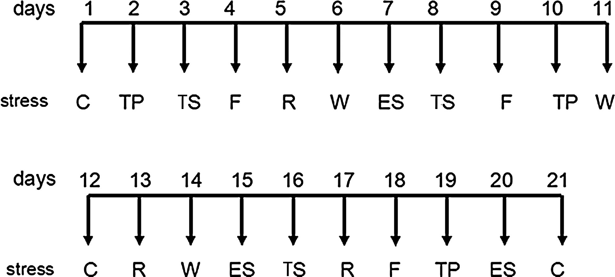
Sequence of chronic unpredictable stress in the 21 days of treatment. C, cold swim; TP, tail pinch; TS, tail shaking; F, food deprivation; R, reverse day and night; W, water deprivation; ES, electric shock.
After 21 days of CUS, the 24 model rats were randomized into 3 groups (8 rats in each group): (1) a negative control group in which no treatment was given to the rats; (2) an acupuncture group; and (3) a riluzole group. The acupuncture intervention was conducted each day for 3 consecutive weeks in the acupuncture group. The body acupuncture points used were Hegu (LI 4) and Taichong (LR 3) on both sides of the body (Fig. 2). The intensity was adjusted to induce slight muscle contraction of the hind limb. In the riluzole group, rats were given 1.8 mg/kg of oral riluzole at for 3 consecutive weeks.
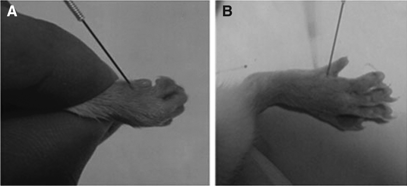
Body acupoints for acupuncture.
Behavioral Assays
Sucrose preference test (SPT)
On day 19, rats were habituated to 1% (wt/vol) sucrose solution (for 3 days) to prevent neophobia during testing. On day 22, following 2 hours of water deprivation, rats were presented with two bottles containing 1% sucrose or water and allowed to drink for 1 hour. Sucrose and water intake were recorded, and this test was performed for 3 consecutive days (days 22–24). Sucrose intake was averaged across experiments.
Open-field test (OFT)
Rats were subjected to an OFT test before CUS exposure and at 7, 14, 22 days after CUS exposure. The OFT was performed in a square iron box (80 × 80 × 40 cm) with a black base. The base was divided into 25 mini-squares (4 × 4 cm each). Each animal was tested in the open field for 4 repeats (3 minutes for each repeat). The crossing score was determined as the number of crossed squares during the test, and the rearing score was determined as the times of rat standing. The box was cleaned with 70% ethanol after each repeat.
Analysis of Glutamate in the Hippocampus
The rats were anesthetized with CO2 and decapitated. The hippocampus (both left and right) of each rat was dissected on ice and frozen, using liquid nitrogen rapidly. The samples were weighed and homogenized in 90% cold ethyl alcohol. The samples were then centrifuged at 1500g for 10 minutes, and the resulting supernatant was used for analysis. The precipitation was extracted for glutamate for thrice, and all the supernatant was collected in one tube. The supernatant was filtered with a 0.45-μm pore filter before loading (Millipore, Billerica, MA). Quantification of glutamate was performed by using a high-performance liquid chromatography system with fluorescence detection on a Hypersil octa decyl silane column (4.0 × 125 mm, 5 μm). The mobile phase consisted of 10 mmol/L Na2HPO4 (PB, pH 7, mobile phase A) and mobile phase B (PB:methanol:acetonitrile, 50:35:15, V/V). Mobile phase B was elevated from 0% to 100% within 25 minutes with a flow rate of 1.0 mL/min. The excited and emitted wavelengths were selected at 340 nm and 450 nm, respectively.
Hematoxylin and Eosin Staining
Each hippocampus was fixed in 4% paraformaldehyde–phosphate buffered saline (PBS) for 10 minutes and cut into 5-μm sections. Slides were stained as follows: 70% ethyl alcohol (EtOH) for 10 seconds; diethylpyrocarbonate-treated water for 5 seconds; hematoxylin with RNAase inhibitor for 20 seconds; 70% EtOH for 30 seconds; eosin Y in 100% EtOH for 20 seconds; followed by dehydration with a series of alcohol for 30 seconds each; and xylenes for 2 minutes.
Western Blot Analysis
The hippocampus was lysed in a radioimmunoprecipitation assay buffer. Protein concentrations were determined using a bicinchoninic acid protein assay. The protein extraction (30 μg/lane) was separated by 12% (w/v) gradient sodium dodecyl sulfate-polyacrylamide gel electrophoresis and transferred to nitrocellulose membranes (Millipore). Membranes were blocked with 5% (w/v) skimmed milk in 0.05% tris-buffered saline with Tween-20 at room temperature for 2 hours and incubated with rabbit polyclonal anti-V-GLUT1 (Abcam, 1:3000), V-GLUT2, syntaxin-1A, VMAP1, VMAP2, VMAP7 or SNAP-25 (Abcam, 1:2000) in PBS (0.25% Triton X, 1% BSA) overnight at 4°C. Membranes were then incubated with anti-rabbit horseradish peroxidase-conjugated secondary antibody (1:5000, Boster, Wuhan, China) for another 2 hours at room temperature. Blots were visualized by enhanced chemiluminescence and exposed on films. Blots were scanned with a ChemiDoc image analysis system (Bio-Rad Laboratories, Hercules, CA) and analyzed with ImageJ software (National Institutes of Health, Bethesda, MD).
Quantitative Real Time PCR
Total RNA was extracted from each hippocampus using an RNA Extraction Kit (Invitrogen) according to the manufacturer's instructions. The concentration of total RNA was quantified by measuring the absorbance at 260 nm. One and one-half μg of total RNA was used for cDNA synthesis using oligo dT primers and SuperScript II reverse transcriptase (Invitrogen), and subsequently was diluted with nuclease-free water to 10 ng/uL cDNA. qPCR was performed utilizing the hotstart SYBR-green based method (Invitrogen). Gene-fold changes were determined by utilizing the 2-ΔΔCt method using 18srRNA as normalization. DNA was amplified with an initial denaturation at 94°C for 3 minutes, followed by 35 cycles of 94°C (15 seconds) and 60°C (15 seconds). Primers are shown in Table 1. All experiments were performed in triplicate and repeated twice.
Primers were designed using Primer Express version 2.0 software. Primer specificity was confirmed using Primer-BLAST web software (National Centre for Biotechnology Information).
Statistical Analysis
The data were presented as the mean ± standard error and analyzed using the Statistical Package for the Social Sciences (SPSS), version 13.0, statistical software (SPSS Inc., Chicago, IL). The statistical significance of differences among the groups was analyzed with a Student's t-test or a one-way analysis of variance (ANOVA). p < 0.05 was considered to be statistically significant.
Results
Acupuncture Promoted Improved Behavioral Performance
All rats developed depression-like behaviors after 21 days of CUS exposure. Compared to the blank-group rats, those in the model groups had significant reductions of sucrose consumption (p < 0.05). Acupuncture therapy increased sucrose preference significantly, compared with the negative control (p < 0.05; Table 2). However, no statistical difference was found between the acupuncture and the riluzole groups (p > 0.05). The weight of the rats was consistent with the sucrose consumption test, which showed that acupuncture and riluzole produced great benefit for weight gain (p < 0.05; Table 2).
Data are the mean ± standard error of the mean (n = 3).
#, p < 0.05 versus blank group; *, p < 0.05 versus negative group.
OFTs were performed the day before treatment, and at 7, 14, and 22 days after treatment. Horizontal or vertical movements of rats in the model groups were much less than such movements in the control group before treatment. The movements of the model rats in the control group decreased in each test. However, rats who received acupuncture and riluzole showed increases in activity and this was restored to their original levels. The movements of rats in the acupuncture and riluzole groups were much better than the movements in the control group after 22 days of therapy (p < 0.05; Table 3).
Data are the mean ± standard error of the mean (n = 3).
#, p < 0.05 versus blank group; *, p < 0.05 versus negative group; ▲, p < 0.05 versus riluzole group.
Min, minutes.
Acupuncture Reduced Glutamate in the Hippocampus
Glutamate content in each model rat's hippocampus had increased greatly after 21 days. Glutamate content decreased to 1.07 ± 0.30 mg/g in the riluzole group and 1.09 ± 0.32 mg/g in the acupuncture group after 22 days of therapy, which were much lower than the content in the control group (p < 0.05; Fig 3).

Glutamate in hippocampus in each group. #p < 0.05 versus blank group, *p < 0.05 versus negative group.
Acupuncture Prevented Changes in Neurons in the Hippocampus
CUS induced obvious morphologic changes in neurons in the hippocampi of rats with depression. Neuron cells in the blank group were well-arranged and had normal nuclear structure. However, the number of neuron cells in the other three groups showed some decrement and there were many vacuoles in the nuclear material. Neuron cells are loosely connected and some were in disarray in the negative group; however, the acupuncture group and the riluzole group were better than the negative group (Fig. 4).

Morphologic changes of neurons in hippocampus of blank group
Acupuncture Reduced Glutamate Release and Increased Glutamate Recycling
As the glu–glu cycle plays an important role in glutamate cycling in hippocampus, the glu–glu cycle related genes were investigated before and after therapy. The mRNA levels of synapsin VGLUT1 and VGLUT2 were downregulated after 21 days of CUS exposure (p < 0.05), while the rats recovered after either riluzole or acupuncture treatments. SNARE complex–related genes, including SNAP25, VAMP1, VAMP2, VAMP7, and syntaxin1, were all upregulated significantly in the untreated groups (p < 0.05; Fig. 5A). The riluzole or acupuncture treatments reduced the expression of these genes in half; however, it was still much higher than in the untreated groups (p < 0.05). Expression of VAMP7 was lower in the acupuncture group than in the riluzole group (p < 0.05), and expression of syntaxin was higher in the acupuncture group (p < 0.05). Protein expressions were similar with these in mRNA levels (Fig. 5B).
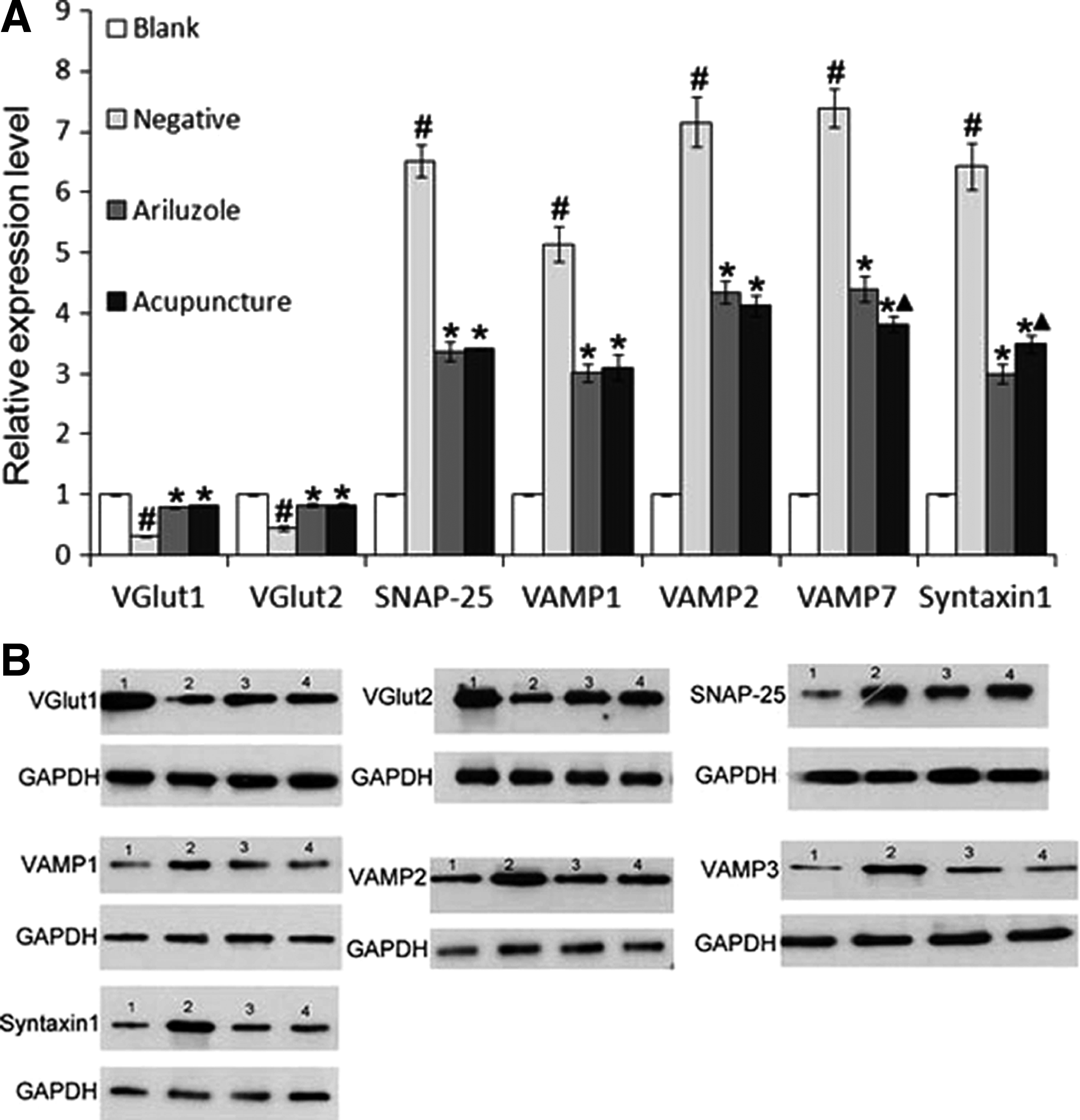
Expressions of glu-glu cycle related genes.
Discussion
Depressive disorder is a mental disorder characterized by persistent low mood, and is very difficult to cure. According to the World Health Organization HO report, the rate of the global depression incidence is ∼11%, and it has become one of the world's fourth major diseases. 1 However, the etiology of depression is not clear. Current advances and trends in the treatment of depression only focus on serotonin, norepinephrine, and dopamine. 2 The current authors' research suggests that glutamate, synaptic vesicle proteins and the glu-glu cycle system might be involved in an antidepression process by acupuncture in rats.
Glutamate is the major excitatory neurotransmitter in the brain. As glutamatergic neurotransmission occurs predominantly within the confines of a tripartite synapse, subtly controlling glutamate distribution within and between synapses was vital for normal neurotransmission. Glutamate transmission is mainly regulated by three processes, including presynaptic glutamate release, postsynaptic receptor trafficking, and transporter-mediated uptake and recycling through the glu-glu cycle.5,19,20
Glutamate release is sensitive to stress and glucocorticoids. Previous animal studies showed that acute stress exposure increased glutamate release in the hippocampus and prefrontal cortex rapidly, and CUS treatment also increased extracellular glutamate levels in the prefrontal cortex.21–24 Rats who received CUS treatment showed significant increases of glutamate and structural changes in their hippocampi in the present study. The antidepression drug riluzole and acupuncture treatments reduced this glutamate content to normal levels. Glutamate restoration was accompanied by enhanced behavioral performance, including on the OFT and SPT, indicating that acupuncture is an effective modality for depression therapy.
Stress-induced enhancement of glutamate release may be achieved by increasing assembling of synaptic vesicles that are docked to the membrane for release or by increasing the releasing of synaptic vesicles, or both of the two mechanisms.25–27 In the present study, the vesicle targeting/docking complex (SNARE) proteins, VAMP (VAMP1, VAMP2, VAMP7), SNAP-25 and syntaxin, were upregulated significantly after CUS exposure. The level of SNARE complexes was also found to be upregulated in prefrontal cortex neurons by introducing footshock-stress. 28 However, synaptic vesicles transporters (VGlut1 and VGlut2) responding for presynaptic vesicles docking were downregulated.
Thus, the current authors hypothesize that chronic unpredictable stress can enhance the capability of presynaptic vesicles release, which may be caused by increasing the expression of SNARE proteins. The resulting elevated glutamate level ultimately leads to a depression symptom. A 2014 study also proved that stress increased the readily releasable pool and releasing of glutamate vesicles in synaptic terminals of the prefrontal cortex. 29
In the present study, acupuncture and riluzole therapy showed great benefit for normal expression of SNARE proteins and vGLUT. As SNARE complex proteins mediate the interaction and fusion of vesicles with the presynaptic membrane, it is plausible to draw a conclusion that acupuncture therapy can eliminate depression symptoms by reducing the presynaptic glutamate release, and SNARE proteins are the targets. The expression of VGlut, a key factor involved in packaging of glutamate into synaptic vesicles, was inhibited in depressed rats, which was beyond the current authors' expectation.
The current authors hypothesize that the decreased expression of VGlut in this study could be negative feedback for balancing glutamatergic circuitry in the hippocampus. Previous studies also showed that repeated stress eliminated expression of vGLUT2, which was restored by administration of the antidepression drug tianeptine.30,31 Thus, the increase of SNARE proteins' expression and releasing of presynaptic vesicles might be the major cause of depression.
Conclusions
This study found that acupuncture is an effective modality for depression therapy and that glutamate in the hippocampus is involved in this process in rats. Further studies covering postsynaptic receptor trafficking, uptake, and recycling of glutamate are still needed for a deeper understanding of this antidepression mechanism of acupuncture. Additional research is indispensable to assess the effect of acupuncture on depressions in humans.
Footnotes
Acknowledgments
The authors would like to thank the staff of the Scientific Research Department of Guangdong Hospital of Traditional Chinese Medicine for technical support. The authors would also like to thank the staff of the Division of Epidemiology and Biostatistics, in the Mel and Enid Zuckerman College of Public Health, University of Arizona for data analysis.
Funding
This research was supported by: (1) a youth fund project of the Natural Science Foundation of China: Antidepressant Mechanism of Acupuncture Based on SNARE proteins modulating the release of glutamic acid in presynaptic neurons (81303041); (2) a class general financial grant from the China postdoctoral science foundation: Mechanism Research of Acupuncture on Hippocampal Tripartite Glutamatergic Synapses of Depressive Disorder Rats (2012M511784); (3) Science foundation of the outstanding young innovative personnel of department of education of Guangdong Province: Clinical Research of Acupuncture in the Treatment of Mild to Moderate Depression (2012LYM_0043); (4) special research foundation of the new teacher category for the doctoral program of higher school by National Ministry of Education: The Moderating Effects of Acupuncture for Hippocampal Tripartite Glutamatergic Synapses of Depressive Disorder Rats (20124425120005); (5) special financial grant from the China postdoctoral science foundation: The modulating effects of acupuncture for glutamic acid in hippocampal presynaptic neurons of depressive disorder rats (2013T60793); and (6) science foundation of the postdoctoral researchers in Guangzhou University of Chinese Medicine from Guangdong Provincial Department of human resources and social security fund: Clinical Study on Acupuncture and Moxibustion for Treatment of Depression Based on Patient Reported Outcome Indicators (BBK429122K19).
Author Disclosure Statement
The authors declare no potential conflicts of interest with respect to the authorship and publication of this article.