




Abstract
Human Na+, K+ ATPase, an ATP-driven ion transporter, is an emerging drug target for heart-related conditions. Three types of assays including purified enzyme, radiotracer flux, and cold Rb+ flux have been used to determine the activity of this transporter. As an alternative to primary cardiomyocytes, mouse embryonic stem cells-derived cardiomyocytes with functional expression of essential cardiac ion channels were used in the present studies. The results on its pharmacology with digitoxin and ouabain, the 2 well-known cardioglycosides, imply that these cardiomyocytes can be used as a predictive model for the identification of modulators of Na+, K+ ATPase in HTS format.
INTRODUCTION
Human Na+, K+ ATPase is the target for cardioglycosides such as digitoxin and digoxin, which are used in the treatment of congestive heart failure and related conditions; thus, it is emerging as an important drug target.1 The Na+, K+ ATPase pump generates electrochemical gradients that are used to drive the coupled transport of many ions and nutrients across the plasma membrane as it actively exports 3 Na+ ions with the concomitant import of 2 K+ ions hydrolyzing 1 ATP molecule in the process.
In non-cell-based assays, the activity of Na+, K+ ATPase has been determined by using purified enzyme preparations to hydrolyze ATP.2 In cell-based assays, the techniques such as patch clamping, fluorescence, H3–ouabain binding, and radiotracer (Rubidium86), and cold Rubidium flux assays have been used either in recombinant cell lines or in cells other than primary cardiomyocytes.3 –5 However, although primary cardiomyocytes would be the optimal cellular platform for these types of studies, it has so far not been possible to use them in developing cell-based assays in an HTS format as they lack homogeneity, sensitivity, and surface-binding properties.6
Standardized, pure genetically selected mouse ES-derived cardiac myocytes with functional expression of all essential cardiac ion channels have recently become available.7 We, therefore, validated the pharmacology of Na+, K+ ATPase in cultured cardiomyocytes using Rb+ uptake assay and Aurora Biomed’s ICR8000 instrument.
MATERIALS AND METHODS
Rb+ uptake by cultured mouse embryonic stem cells (ES)-derived cardiomyocytes was carried out as follows:
Culture and maintenance of cardiomyocytes: The cells endogenously expressing Na+, K+ ATPase were cultured as per the technical manual.8 On the fourth day of the culture, Rb+ uptake experiment was carried out.
Rb+ Uptake: Cells were washed once with 200 μL of Rb+ uptake buffer followed by incubation in the presence of 200 μL Rb+ uptake buffer at room temperature (∼22°C). The Rb+ uptake profile: The Rb+ uptake activity of the cells was studied by incubating the cells at specific time intervals in the presence of Aurora Biomed’s Rb+ uptake buffer. Dose response: The cells were incubated in the Rb+ uptake buffer containing appropriate dose of test compound. It was followed by incubation for 15 min at room temperature (∼22°C).
Wash: Residual Rb+ and compound were removed by 4 successive washes with 200 μL of SPA-Wash Buffer.
Cell Lysis: Intracellular samples were obtained by whole cell lysis with the application of 200 μL Lysis Solution.
Analysis: The level of Rb+ in the intracellular samples was measured by ICR8000 (Aurora Biomed Inc., Vancouver, BC, Canada) using flame atomic absorption spectroscopy. The intracellular Rb+ content (mg/L) of each test dose of a test compound was normalized [(actual uptake − basal uptake)/detection window of inhibitory activity of digitoxin × 100]. Further analysis of the normalized data was performed with Xlfit3 (IDBS, Guildford, Surrey, UK) to draw the curve fits, and to obtain information on different parameters like mean (n = 3), SEM, inhibition (%), and IC50 values of the test compounds.
RESULTS
The results on the Rb+ uptake by the cultured mouse ES cell-derived cardiomyocytes are described as follows:
1. Ready-to-use cells: The cardiomyocytes were observed to be ready-to-use cells for the assay after 3 to 4 days of maintenance in the incubator from the frozen stock in 96-well plates.
2. Expression of Na+, K+ ATPase isoform: a. The Rb+ uptake profile of these cells in the presence of Rb+ containing isotonic buffer over a duration of 160 min indicated an established expression of Na+, K+ ATPase in the cardiomyocytes (Fig. 1). This fact was verified by the sensitivity of the uptake to the presence of digitoxin (100 μM). A maximum uptake of Rb+ was taken up by the cells in about 80 min, with an exponential uptake seen from 0 to 50 min. Thus these cells can be employed for screening compounds in assays for 15 min to meet the demands of an HTS assay. As indicated by Figure 1, any duration from 5 to 20 min (on the exponential scale of Rb+ uptake) can be used in an assay. b. The Rb+ uptake pattern from these cardiomyocytes appears to be similar but slightly faster than that of Na+, K+ ATPase isoforms endogenously expressed in Chinese hamster ovary (CHO) cells and in human embryonic kidney (HEK) cells. However, the isoform in HEK cells presented a significantly faster uptake of Rb+ in the uptake medium containing 5.4 mM RbCl where the uptake takes plateau course at 50 min of incubation while it is about 80 min in both the mouse atrial cardiomyocytes and CHO cells (Fig. 2).
3. Pharmacology of Na+, K+ ATPase blocker: A complete block of Na+, K+ ATPase isoform was observed with 100 μM of digitoxin, a known cardiac glycoside. Similarly, a complete block of the Rb+ uptake was observed with 100 and 50 μM of digitoxin in the CHO cells and HEK cells, respectively. The digitoxin was determined to have an IC50 value of 6.82 μM (Fig. 3). The potency of digitoxin also displays a similar relation between mouse and Chinese hamster Na+, K+ ATPase isoform as CHO cell isoform has an IC50 of 6.94 μM. However, both of these isoforms are significantly less sensitive than HEK isoform where only α-subunits are expressed.9
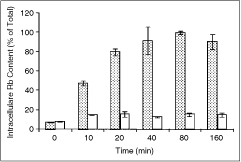
Rb+ uptake profile of the cardiomyocytes ATPase isoform (spotted bars), and inhibition by digitoxin (100 μM, open bars). Error bars represent standard error of n = 3 values.
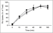
Rb+ uptake profile of the cardiomyocytes ATPase isoform (▪) in comparison to ATPase isoforms in Chinese hamster ovary (□) and in the human embryonic kidney (▵). Error bars represent standard error of n = 3 values.
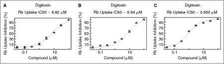
Pharmacology of digitoxin on the Na+, K+ ATPase isoform from the cardiomyocytes (
Ouabain, another cardiac glycoside and a well-known blocker of Na+-K+-ATPase, also inhibited the Rb+ uptake by these cardiomyocytes. A comparative picture of the pharmacology of ouabain on these 3 isoforms suggested that cardiomyocytes have higher sensitive to this drug than CHO isoform. However, both these isoforms were much less sensitive than HEK isoform as indicated by IC50 of 57, 94, and 1.2 μM (Fig. 4). The comparative sensitivity of the 3 isoforms for digitoxin and ouabain has been presented in Figure 5. The HEK isoform is known to be significantly more sensitive than other mammalian isoforms like CHO and mouse isoforms like ouabain.5
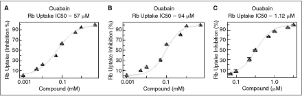
Pharmacology of ouabain on the Na+, K+ ATPase isoform from (

Comparative pharmacology of (
CONCLUSION
The data suggest that the mouse ES-derived cardiomyocytes (Cor.At cells) can be used as a predictive model for the identification of modulators of Na+, K+ ATPase in its original cardiac environment and that they are suitable for use in HTS formats of these assays.
Footnotes
AUTHOR DISCLOSURE STATEMENT
S.G., R.G., and S.L. are employees of Aurora Biomed Inc.