




Abstract

BIOLUMINESCENCE IMAGING OF TRANSGENE EXPRESSION AT THE WHOLE-MOUSE LEVEL AND IN THE MESENCEPHALIC TRIGEMINAL NUCLEUS
Dissertation by Daniel J. Hiler, Ph.D.
Bowling Green State University, Ohio, 2009
Advisor: Michael E. Geusz
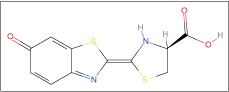
Bioluminescence imaging (BLI) of transgenic mice expressing the firefly luciferase gene luc has been used to monitor continuous changes in gene expression in cultures and in the whole animal. This dissertation describes new techniques for bioluminescence imaging of gene activity at whole-animal and cellular levels. To record gene expression at the whole-animal level, the luciferase substrate luciferin is typically injected into mice prior to imaging. To avoid the effects of handling and stress from injection on expression of the transgene, a new method for delivering luciferin orally was developed and tested. Orally administered luciferin was found to be readily absorbed from the digestive tract and produced levels of bioluminescence in the whole animal that were similar to results following injection. Imaging at the cellular level focused on identifying a new model system for analyzing circadian rhythms of vertebrate neurons. In the mammalian circadian system, multiple circadian pacemakers located throughout the body are synchronized to the external 24-h day by a molecular circadian clock. BLI has been used to detect rhythmic expression of mPer1 and mPer2 genes in the “master” clock of the suprachiasmatic nucleus (SCN) that regulates daily physiology and behavior and also sends timing cues to peripheral oscillators outside the SCN. Because SCN neurons are small and the SCN contains many neuronal phenotypes, subcellular imaging in identifiable neurons is challenging. As a result of the limitations of the SCN, larger neurons with similar circadian and molecular properties were sought, resulting in a focus on the mesencephalic trigeminal nuclei (Me5). The Me5 are a heterogeneous group of large (30–40 mm) pseudo-unipolar primary sensory neurons and multipolar interneurons that receive proprioceptive signals from spindle organs of the masseter muscles and periodontal ligaments of the teeth. Methods for Me5 cell culture and bioluminescence imaging of Me5 organotypic explants were developed using mPer1::luc and mPer2 luc transgenic mice. The period and phase of circadian rhythms in mPer1 and mPer2 gene expression in Me5 neurons were characterized. Most importantly, bioluminescence imaging and immunohistochemistry were used to provide evidence of a cell-autonomous circadian oscillator in Me5 neurons. The unique characteristics of the Me5 and its use as an alternative molecular model may provide opportunities to expand cellular studies of neural circadian pacemakers beyond the limitations of the SCN. Similarly, the ability to image bioluminescence after administering luciferin through drinking water could enable circadian rhythms in gene expressions to be monitored in various locations in animal models and more efficiently associate cellular circadian clock mechanisms with resulting animal behavior.
REAL-TIME IMAGING OF LIVE CELL ATP LEAKING OR RELEASE EVENTS BY CHEMILUMINESCENCE MICROSCOPY
Dissertation by Yun Zhang, Ph.D.
Iowa State University, 2008
Advisor: Edward S. Yeung
The purpose of this research was to expand the chemiluminescence microscopy applications in live bacterial/mammalian cell imaging and to improve the detection sensitivity for ATP leaking or release events.
We first demonstrated that chemiluminescence (CL) imaging can be used to interrogate single bacterial cells. While using a luminometer allows detecting ATP from cell lysate extracted from at least 10 bacterial cells, all previous cell CL detection never reached this sensitivity of single bacteria level. We approached this goal with a different strategy from before: instead of breaking bacterial cell membrane and trying to capture the transiently diluted ATP with the firefly luciferase CL assay, we introduced the firefly luciferase enzyme into bacteria using the modern genetic techniques and placed the CL reaction substrate
Then we started with this novel single bacterial CL imaging technique, and applied it for quantifying gene expression levels from individual bacterial cells. Previous published result in single cell gene expression quantification mainly used a fluorescence method; CL detection is limited because of the difficulty to introduce enough
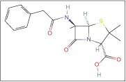
Penicillin G
Finally, we imaged ATP release from astrocyte cells. Upon mechanical stimulation, astrocyte cells respond by increasing intracellular Ca2+ level and releasing ATP to extracellular spaces as signaling molecules. The ATP release imaged by direct CL imaging using free firefly luciferase and
COMPARING THE RESPONSE OF SUSPENDED AND IMMOBILIZED WHOLE-CELL BIOLUMINESCENT BIOSENSOR PP F1G4
Dissertation by Marie-Helene Gravel, M.Eng.
McGill University Canada, 2008
Whole-cell bioluminescent biosensor Pseudomonas putida F1G4 (Pp F1G4) was developed to produce a bioluminescent response to a wide range of hydrophobic organic compounds (HOCs). The main objective of this study was to characterize the luminescence response of Pp F1G4 immobilized in silica gel and in inorganic membrane surfaces, and to compare the luminescence response to that for cultures of suspended Pp F1G4 cells.
Silica matrices were made by the sol–gel process using aqueous precursors, and Pp F1G4 cells were embedded in thin films of silica gel. The silica gels are chemically inert and optically transparent and proved to be a viable media for immobilizing Pp F1G4 cells. Upon comparison of the results obtained for suspended Pp F1G4 and Pp F1G4 immobilized in silica sol–gel matrix, it appears that immobilized and freely suspended cells displayed similar bioluminescence profiles in response to HOCs exposure, but immobilized bacteria yielded a lower response level. The intensity of luminescence was several-fold higher, ∼10 times, with use of suspended cells. In long-term experiments, the Pp F1G4 biosensor immobilized in silica gel also proved to be viable over a relatively long period of time (5 days).
Pp F1G4 cells were also immobilized by filtering culture solutions through a transparent inorganic membrane. Such filters can effectively retain the cells in close proximity to the luminescence detectors during online monitoring, but as it was seen with the immobilized biosensor in a silica matrix, the magnitude of the light signal was significantly lower, about 4 times, compared to the light emitted by freely suspended Pp F1G4 biosensor cells.
While it is clear that cells immobilization is a convenient method for preparation of relatively long-lasting and reusable biosensors, key challenges are to further improve the sensitivity of the system and the reproducibility of the experiments, particularly with filter-immobilized Pp F1G4 cells.
DEVELOPMENT OF EFFICIENT TOOLS FOR ANALYSIS OF THE HOST–PARASITE INTERACTION DURING THE LIVER STAGES OF MALARIA INFECTION
Dissertation by Agnes Mwakingwe, Ph.D.
Yeshiva University, New York, 2009
Advisor: Kami Kim
The morbidity and mortality due to malaria is rising as a result of the emergence and spread of drug-resistant parasites and the lack of an effective vaccine. Development of novel intervention strategies is hindered by the lack of efficient tools available to study liver stage parasites in vivo. Development of Plasmodium parasites in the liver is the first and obligatory step in its life cycle in the vertebrate host. Although a clinically silent infection, development of parasites in the liver is an immunologically active stage leading to protective immunity. It is thus imperative that we develop novel tools to study the biology of Plasmodium liver stages.
First, to study sporozoites–hepatocytes molecular interactions, we adapted transfection of mouse hepatocytes by hydrodynamic tail vein injection to deliver naked DNA to hepatocytes in vivo. Transfection by hydrodynamic injection can lead to creation of disease models and non-heritable transgenic research animals within a short time. With further optimization of experimental conditions, transfection by hydrodynamic injection is a promising invaluable tool to study molecular interactions between sporozoites and hepatocytes in vivo.
Second, for thorough screening of factors that affect liver stage parasite growth, we adapted bioluminescent imaging (BLI) of Plasmodium yoelii YM expressing firefly luciferase (PyLuc). BLI is a noninvasive and rapid technique that allows for real-time monitoring of infection in the same animal. Using BLI, for the first time, in vivo liver stage parasites were imaged after infecting mice with PyLuc sporozoites and effects of drugs and immunization on this stage were quantified. BLI allows for efficient analysis of effects of chemotherapeutic or vaccine candidates on liver stages.
Successful efforts to combat the devastation of malaria require understanding of how the parasite and host interact during infection. It is not entirely clear how the Plasmodium parasites establish infection, and how the host immune system responds to parasites. We have adapted transfection by hydrodynamic injection and bioluminescent imaging as important tools to study parasite biology in vivo. These important tools will help to better understand parasite biology thus leading to generation of effective intervention strategies to reduce the morbidity and mortality caused by malaria.
DYNAMIC ANALYSIS OF LIGAND-INDUCED EGFR PROCESSING AND REGULATION BY REAL-TIME BIOLUMINESCENCE IMAGING
Dissertation by Aparna Hemant Kesarwala, Ph.D.
Washington University in St. Louis, 2008
Advisor: David Piwnica-Worms
Epidermal growth factor receptor (EGFR), a member of the EGF superfamily of receptor tyrosine kinases, is a critical regulator of cell growth and proliferation and an important target for anticancer therapeutics. To further investigate the dynamics of EGFR regulation, we have developed a firefly luciferase (FLuc)-based reporter system to image ligand-induced processing of EGFR in real time.
The reporter was created by fusing EGFR to FLuc downstream of a CMV promoter. A flexible glycine–serine linker was engineered between the 2 proteins to minimize steric hindrance and allow for full function of both components; this fusion reporter was then used to create a stable HeLa cell line. Photon output from these cells was continuously monitored after treatment with EGF, which resulted in autophosphorylation and internalization of EGFR–FLuc. Reporter dynamics and pharmacokinetics correlated with the ligand-induced response of endogenous EGFR as determined by western blot as well as subcellular localization of an EGFR–green fluorescent protein (GFP) fusion protein. Pharmacological inhibition revealed the decrease in photon output to be contingent upon phosphorylation as well as proper intracellular sorting of EGFR. Thus, a novel FLuc fusion reporter allows continuous readout of EGFR activation in cells for temporal-based investigations of EGF-mediated signaling.
Further analysis showed proteasome inhibition with bortezomib and MG132 attenuated degradation of the bioluminescent reporter up to ∼50%. In cells expressing EGFR–GFP, treatment with proteasome inhibitors trapped essentially all of the receptor at the cell membrane and changed the ligand-induced localization of EGFR. Furthermore, potential regulatory partners and new drug targets in EGFR-mediated signaling pathways were identified by dynamic high-throughput screening of live reporter cells transfected with siRNA libraries targeting all human kinases and phosphatases.
Bortezomib

THE REGULATION OF INFECTION-INDUCED CARDIAC AUTOIMMUNITY AND NATURAL DISSEMINATION OF Trypanosoma cruzi IN EXPERIMENTAL CHAGAS HEART DISEASE
Dissertation by Kenneth V. Hyland, Ph.D.
Northwestern University, Illinois, 2008
Advisor: David M. Engman
Chagas heart disease, caused by infection with the protozoan parasite Trypanosoma cruzi, is still regarded as a major public health problem in Central and South America. The finding of cardiac-specific autoimmunity during infection in both humans and experimental animals has provided a basis for investigation of its potential role in disease pathogenesis for many years. However, the complex nature of Chagas disease has left questions of the true mechanisms of cardiac inflammation, in addition to an understanding of the natural course of infection, largely unanswered. My thesis research consisted of investigating aspects of parasite infection-induced cardiac autoimmune responses and the spatiotemporal dissemination of T. cruzi in an experimental model of Chagas heart disease. The infection of A/J mice with the Brazil strain of T. cruzi results in the development of acute myocarditis with both humoral and cellular autoreactivity by 21 days postinfection. To test the hypothesis that the magnitude of the autoimmune response is directly proportional to the amount of damage elicited by the parasite, I administered a trypanocidal drug, benznidazole, to mice to reduce the number of viable parasites following infection and determined that treatment not only decreased disease severity and eliminated mortality, but also significantly reduced cardiac myosin-specific DTH and antibody production. With a strong enough secondary cardiac insult, the autoreactivity and myocarditis could be restored, indicating the reestablishment of self-tolerance after the eradication of the parasite was not permanent in our disease model. Overall, an important link between the levels of live parasite and the presence of autoimmunity was provided, suggesting that treatments designed, such as vaccines, to specifically target the parasite will likely reduce or eliminate the induction of autoimmunity as well. To further understand the natural course of infection, I engineered firefly luciferase-expressing T. cruzi in order to noninvasively monitor the dissemination of parasites in mice over time using bioluminescence imaging technology. For a more in-depth analysis of parasite tropism during infection, the tissue distribution of T. cruzi was determined by imaging heart, spleen, skeletal muscle, lungs, kidneys, liver, and intestines ex vivo. This novel parasite line has already provided interesting results illustrating the natural dissemination of T. cruzi during infection and will continue to serve as a tool for studying a number of aspects of Chagas disease. In conclusion, these results not only provide encouragement for the future exploration of parasite-specific therapeutic strategies for Chagas disease by showing that elimination of T. cruzi is effective at reducing or eliminating autoimmunity, but also illustrate a novel tool that could be easily applied to the screening of such therapeutical agents via bioluminescence.
Benznidazole

PHOTOPROTEINS FROM JELLYFISH Aequorea victoria: NEW HORIZONS IN BIOANALYSIS
Dissertation by Emre Dikici, Ph.D.
University of Kentucky, 2008
Advisor: Sylvia Daunert
Photoproteins isolated from the jellyfish Aequorea victoria, namely green fluorescent protein and aequorin, have been used extensively in bioanalytical chemistry. They are indispensable tools because of theft versatility, sensitivity, and their availability. They have been used as labels in various assays. In this work, we demonstrate the use of green fluorescent protein fused to calmodulin as a label for the detection of tricyclic antidepressants. The same detection technology is applied to a whole-cell type assay. This way the time-consuming steps associated with the isolation and purification of fusion proteins are avoided.
The bioluminescent protein aequorin is one of the most widely used and studied proteins. Its sensitivity easily rivals that of radioactive labels, and unlike radioactivity it is not hazardous. The only drawback of using aequorin as a label comes from the fact like many other bioluminescent proteins isolated from the marine organisms it uses coelenterazine as a chromophore and thus the emission characteristics of aequorin is similar to other bioluminescent proteins. This drawback limits the use of the bioluminescent proteins into single analyte detection and the detection of multiple analytes within a single sample is not feasible.
In order to overcome the drawback associated with the bioluminescent labels, here, in this work we describe 2 different methodologies for the alteration of the emission characteristics of aequorin. In the first methodology, we have rationally designed and synthesized mutants of aequorin in a site-directed manner. Then these mutants were characterized and their usefulness in the area of multi-analyte detection was studied.
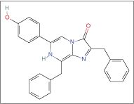
Coelenterazine h
In the second methodology, we have incorporated noncanonical amino acids into the aequorin structure by selective pressure incorporation methodology. Auxotrophs of Escherichia coli have been prepared to produce aequorin and the semisynthetic aequorins were prepared by growing these bacteria in minimal media in the presence of the non-natural amino acid analog. The resulting aequorins were also characterized with respect to their emission spectra and their usefulness is studied.
CHEMICAL GENETIC TOOLS TO MEASURE AND REGULATE CELLULAR KINASE ACTIVITY
Dissertation by James Christiansen Abellera Blair, Ph.D.
University of California, Berkeley, 2008
Advisor: Kevan M. Shokat
Protein kinases control most biological processes by regulating nearly every signal transduction pathway. Aberrant kinase activity causes many diseases including cancer and autoimmunity. Therefore, kinases are attractive targets for the regulation of these diseases by small molecule inhibitors. Since kinase activity plays such a fundamental role in biology, it is vital to our understanding of cellular signaling to both measure and regulate cellular kinase activity. Here, I describe new chemical tools to measure kinase activity in cells. In addition, I discovered the mechanism allowing a new class of multi-targeted inhibitors to potently inhibit distantly related kinases.
Using structure-guided design, I developed a chemical genetic system capable of measuring cellular kinase activity. I synthesized a series of 6-acrylamido-4-anilinoquinazoline irreversible kinase inhibitors that potently and selectively target rationally designed kinases bearing 2 selectivity elements that are not found together in any wild-type kinase: an electrophile-targeted cysteine residue and a glycine gatekeeper residue. Co-crystal structures of 2 irreversible quinazoline inhibitors bound to either EGFR or engineered c-Src show covalent inhibitor binding to the targeted cysteine. Based on these structures, we developed a fluorescent derivative—an affinity probe—to report the fraction of kinase necessary for cellular signaling. Using this probe, I quantitated the relationship between EGFR stimulation by EGF and its downstream outputs—Akt and Erkl/2.
6-Acrylamido-4-anilinoquinazoline

Kinase inhibitors hold great promise as anticancer therapeutics, but the first generation of selective kinase drugs have only modest clinical efficacy. To test whether inhibiting many kinases simultaneously overcome these modest effects, our lab developed a series of pyrazolopyrimidine inhibitors that inhibit both tyrosine kinases and phosphoinositide kinases in cancer cell lines. It was unclear how these inhibitors achieve dual potency, since tyrosine kinases and phosphoinositide kinases share little sequence and structural similarity. By solving the co-crystal structures of these inhibitors bound to c-Src and comparing these structures to complexes of inhibitor-bound phosphatidylinositol-3-OH kinase pilOg, I uncovered striking structural elements that allow these unique inhibitors to bind to 2 distantly related kinase families.
METABOLIC TARGETING OF CANCER CELLS: TWO MOLECULAR MECHANISMS INVOLVING GLUCOSE METABOLISM
Dissertation by Quintin Jose Quinones, Ph.D.
Duke University, North Carolina, 2008
Advisor: Salvatore V. Pizzo
Selective therapeutic targeting of tumors requires identification of differences between the homeostatic requirements of cancer and host cells. One such difference is the manner in which cancer cells acquire energy. Cancer cells often grow in an environment of local hypoxia; under these conditions tumor cells depend on glycolysis for energy, but are unable to perform oxidative phosphorylation. Many tumor cells, despite normoxic conditions, continue to perform glycolysis without oxidative phosphorylation. The net result of glycolysis without oxidative phosphorylation is 2-fold: the need to consume a greater amount of glucose than a noncancerous host cell, and the burden of increased intracellular lactic acid. The proteins responsible for the transport of lactic acid in and out of cells are known as the monocarboxylate transporters (MCTs). Monocarboxylate transporter 1 (MCT1) and monocarboxylate transporter 4 (MCT4) are the MCTs that play a major role in the transport of lactic acid. Tumor cells depend on MCT1 and MCT4 activity to excrete excess intracellular lactic acid to maintain neutral intracellular pH and homeostasis. Using human neuroblastoma and prostate cancer cell lines, this work demonstrates that tumor cells can be selectively targeted tumor under conditions of hypoxia or acidosis in vitro with the drug lonidamine, with a small molecule inhibitor selective for MCT1, or with RNA interference of MCT1. Inhibition of MCT1 activity in neuroblastoma cells under acidic extracellular conditions results in intracellular acidification and cell death. MCT1 mRNA is expressed in human neuroblastoma and positively correlated with clinical risk profile. Inhibition of MCT1 activity in hypoxic prostate cancer cells results in a reduction of lactate excretion, decreased intracellular pH, inhibition of ATP production, and subsequent cell death. MCT1 expression in sections of human prostate tumors has been demonstrated to validate MCT1 as a target in prostate cancer.
Through the Pasteur and Warburg effects, tumors have an increased demand for glucose. Some cancers store glycogen, but the reasons for this are largely unknown. It is hypothesized that tumor glycogen is used to promote tumor survival during transient hypoxia or low glucose, and that the mechanisms by which glycogen is stored is a potential therapeutic target in cancer. Tumors from human cell lines (WiDr, PC3, FaDu) have been grown in nude mice, sectioned, and stained to measure glycogen storage. Using consecutive frozen sections, levels of hypoxia, glucose, lactate, ATP, and CD31, an endothelial cell marker, have been determined. These sections have been employed to elucidate the “architecture” of tumor metabolism in terms of vessel distance. Additionally, PAS-stained EF5-labeled human tumor samples were used to obtain calibrated hypoxia measurements to correlate with PAS. These studies demonstrate a correlation between hypoxia and the formation of glycogen deposits in human tumors and nude mouse xenografts. In cell culture, formation of glycogen deposits after exposure to hypoxia has been demonstrated, in addition to expression of glycogen synthase in human cancer cell lines.
The development of novel selective cancer chemotherapeutics will require the identification of differences between cancerous cells and normal host cells to exploit as targets. Several differences in metabolism, including the need to excrete excess lactic acid and store glycogen under hypoxic conditions, are such targets. Novel therapeutics exploiting these targets should be effective against cancer cells and minimally toxic to host cells.
PHARMACODYNAMIC EFFECTS OF XENOBIOTIC ABC TRANSPORTERS IN PERIPHERAL TISSUES
Dissertation by Leslie W. Chinn, Ph.D.
University of California, San Francisco, 2008
Advisor: Deanna L. Kroetz
The ATP-binding cassette (ABC) superfamily consists of energy-dependent transporters that in many cases play crucial roles in physiological processes; indeed, mutations in several ABC transporter genes have been shown to cause inherited diseases such as cystic fibrosis. The involvement of ABC transporters in systemic xenobiotic protection has led to an examination of the influence of these transporters on drug pharmacokinetics (plasma levels), including a number of clinical studies in which transporter expression levels were associated with drug plasma levels in healthy subjects. Researchers have also demonstrated correlations between the presence of genetic polymorphisms in ABC transporters and altered drug pharmacokinetics. The impact of ABC transporters and their naturally occurring genetic variants on drug pharmacodynamics (pharmacological action) has been characterized to a lesser extent, in part because of the difficulties of quantifying the pharmacological actions of many drugs. In this dissertation, we focus on the pharmacodynamic effects of ABC transporter expression and function in peripheral, non-pharmacokinetic tissues such as adipose and lymphocytes, especially with respect to anti-HIV therapies.
First, we describe the expression of a putative splice variant of the ABCB1 (P-glycoprotein) transporter in lymphocytes. This half-sized protein functioned similarly to the classic full-size P-glycoprotein, but displayed altered immunoreactivity. ABCB1 RNA transcripts of approximately half the length of normal ABCB1 transcripts were found in lymphocytes from healthy subjects. The putative P-glycoprotein splice variant was detected in both HIV-negative and -positive subjects, indicating that it could influence the pharmacodynamics of anti-HIV drugs that have been classified as substrates of P-glycoprotein. We also investigated the effects of exposure to the HIV protease inhibitors atazanavir and saquinavir on the expression of lymphocyte P-glycoprotein. We found that in several subjects, the amount of P-glycoprotein expression increased substantially, suggesting that while there is no detectable generalized change in lymphocyte P-glycoprotein expression following atazanavir/saquinavir exposure, there may be genetic or environmental influences that affect the extent of lymphocyte P-glycoprotein induction in certain individuals.
We also examine the roles that polymorphisms in candidate genes play in patient response to anti-HIV medications. These genes include the drug-metabolizing enzyme cytochrome P450 2B6, the xenobiotic transporters ABCB1 and ABCC4, and the inflammatory cytokine TNF-α. We performed this study in HIV-infected populations in San Francisco and Uganda, and found no significant associations between any of the polymorphisms investigated and patient virologic or immunologic response to antiretroviral therapies. We also characterized the ancestral admixture of the San Francisco cohort and determined that the minor allele frequencies of several of the polymorphisms differed between ethnicities.
Atazanavir

Last, we describe the effects of the adipose RNA expression of the transporter genes ABCC4 and SLC29A1, which regulate cellular exposure to the nucleoside analog-based HIV reverse transcriptase inhibitors, on the development of lipodystrophy, a fairly common side effect of this class of anti-HIV therapies. Lipodystrophy is thought to result from mitochondrial toxicity, specifically the inhibition of the mitochondrial DNA polymerase. Indeed, we found that the nucleoside analog fialuridine inhibited mitochondrial DNA synthesis in vitro; the effects of transporters in this system remain inconclusive. While we did not see any correlation between adipose transporter expression and the occurrence of lipodystrophy, we identified a genetic polymorphism in ABCC4 that was significantly associated with the development of lipodystrophy following stavudine treatment in a San Francisco population of HIV-infected individuals. It is possible that screening for this polymorphism may help to predict which patients will develop lipodystrophy following nucleoside analog exposure.
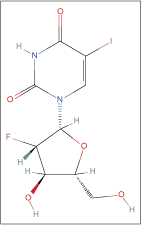
Fialuridine
In general, the results described in this dissertation indicate that ABC transporters in lymphocytes and adipocytes may have important pharmacodynamic functionalities, as they modulate the intracellular concentrations of certain drugs. In particular, we have focused on HIV antiretroviral therapies because they exert pharmacological effects (beneficial or detrimental) in these cell types. Finally, we show that genetic variation in ABC transporters may be an important factor that determines how a patient responds to an antiretroviral regimen. Future studies should expand on this work with mechanistic studies to further unravel the role that transporters play in antiretroviral action, as well as carefully designed clinical studies to ascertain the validity of the pharmacogenetic associations reported here.
DECIPHERING THE PROTEOLYTIC MECHANISM OF THE ATP-DEPENDENT PROTEASE LON USING FLUORESCENT PEPTIDES
Dissertation by Jessica Ward, Ph.D.
Case Western Reserve University, Ohio, 2008
Advisor: Irene Lee
Lon is an ATP-dependent serine protease that degrades damaged, misfolded, and certain regulatory proteins in the cell. The enzyme exists as a homo-oligomer with one ATPase domain and one protease domain on each subunit. Using our fluorescent model peptide substrate, lN89–98, as a tool, we have employed steady-state and pre-steady-state kinetic techniques to evaluate the proteolytic mechanism of Lon protease. Steady-state kinetic analysis of the peptidase activity of Lon with nucleotide triphosphates reveals that while nucleotide binding alone is enough to support peptide hydrolysis, the contribution of nucleotide hydrolysis toward the proteolytic activity is significant. Pre-steady-state experiments reveal a lag phase in the peptidase reaction and that ATP hydrolysis occurs before the peptide cleavage event. We propose that the slow step constitutes a peptide translocation event that contains 2 binding events in which the peptide is delivered from the initial peptide-binding site to the protease active site. I was able to measure this slow step using proteolytic inactive Lon mutants and a dansylated version of the LN89–98 peptide as well. Taken together, I propose a mechanism for the first round of peptide hydrolysis. Following initial nucleotide and peptide binding, a nucleotide-dependent conformational change occurs. Next, upon ATP hydrolysis, the proteolytic site is activated and peptide is delivered to the protease domain where peptide cleavage occurs. In addition to characterizing the kinetic mechanism of Lon protease, I have also utilized multiple peptide substrates to demonstrate the importance of the P3, P1, and P3′ positions in determining substrate specificity and provide evidence that the C-terminus of the LN protein is important for recognition by Lon.
MOLECULAR DETERMINANTS OF ATP BINDING TO P2X2 ION CHANNELS
Dissertation by Shlomo S. Dellal, Ph.D.
University of Michigan, 2008
Advisor: Richard I. Hume
ATP, in addition to its myriad roles in intracellular processes, is an extracellular signaling molecule. ATP serves as a neurotransmitter by binding to and opening ion channels known as P2X receptors. P2X receptors assemble as trimers of subunits. It is unclear if ATP binding occurs between or within subunits and if 3 ATPs are required to bind the receptor before the channel opens. To address these questions, we adopted 2 approaches, both based on introducing mutations that disrupt ATP binding at 1 or 2 of the 3 ATP-binding sites of P2X2 receptors. Some of these mutations (K69A and K308A) were sufficiently severe that homotrimers bearing them were nonresponsive to ATP. First, we characterized in Xenopus laevis oocytes several concatamers in which the open reading frames for 3 subunits were joined together to form a single coding unit with binding site mutations introduced at various positions. We obtained evidence that these concatamers could form a subpopulation of cross-assembly side products, which limited the conclusions we could draw. Second, we co-expressed subunits encoding ATP-binding mutants either together or with subunits that respond normally to ATP. From these experiments, we concluded that receptors with 1 wild type and 2 mutant-binding sites could respond to ATP and that binding is between receptor subunits. In a final set of experiments, we used 2 cysteine mutants, one at a critical ATP-binding residue (K69C) and another in a neighboring but nonbinding residue (I67C). K69C did not give rise to detectable ATP responses, but treatment with Alexa Fluor 546 C 5-maleimide (AM546), a thiol-reactive drug, caused the channels to be constitutively open, and to respond to the allosteric modulators zinc and acidic pH in the absence of exogenous ATP. Therefore, this mutant could serve as a specific biosensor of physiological zinc and pH. In contrast, AM546 caused the responses of I67C channels to decline to <10% of their initial amplitude. Additional experiments provided evidence that ATP binding was abolished at these receptors, therefore, this mutant will be useful in concatamer and co-expression based experiments to explore the requirement for ATP binding in receptor activation.
CARDIOMYOCYTE SIGNALING IN RESPONSE TO ATP DEMAND: THE ROLE OF ADENOSINE
Dissertation by Jason Leon Guichard, Ph.D.
Medical University of South Carolina, 2009
Advisor: Craig Beeson
During oxygen deprivation or ischemia, mitochondrial respiration decreases and glycolytic flux must subsequently increase to satisfy the basal cellular ATP demand requirements. Similarly, during an increase in oxygen demand or exercise, mitochondrial respiration increases and glycolytic flux must subsequently increase to satisfy the increased cellular ATP demand requirements. This ATP demand-driven glycolytic flux increase, in response to various stressors, demonstrates that mitochondrial respiration and glycolytic flux are coupled. Glucose transport into cardiomyocytes is regulated by the transmembrane glucose gradient and the content of glucose transporters (GLUTs) in the sarcolemma, mainly GLUT4 and GLUT1, both GLUT4 and GLUT1 are important in ischemia-mediated and demand-mediated myocardial glucose uptake in vivo. This ATP demand-driven glycolytic flux increase, in response to various stressors, demonstrates that mitochondrial respiration and glycolytic flux are coupled and we believe this coupling involves active signaling, namely phosphorylation cascades and protein translocation.
We have shown that the Seahorse Bioscience’s Extracellular Flux Analyzer (XF24) can measure extracellular fluxes for adult cardiomyocytes with time resolutions in the minutes and sensitivities in the mpH and pmol O2 for extracellular acidification and oxygen consumption, respectively. We have determined that A 1 R/A 3 R inhibition reduces ATP demand-driven glycolytic flux increase by ∼35%–45%, dominant-negative PKC [varepsilon] (dnPKC [varepsilon]) or dominant-negative p38 MAPK (dnp38 MAPK) overexpression reduces ATP demand-driven glycolytic flux increase by ∼35%–45%, and both A 1 R/A 3 R inhibition and dnPKC [varepsilon] or dnp38 MAPK overexpression additively reduces ATP demand-driven glycolytic flux increase by ∼75%–85%. In addition, we have demonstrated that PKC [varepsilon] is an upstream signaling partner of p38 MAPK signaling in ATP demand-driven glycolytic flux increase. We have determined that A 1 R/A 3 R inhibition or dnPKC [varepsilon] overexpression or dnp38 MAPK overexpression reduces glucose uptake to only ∼1.5-fold increase over basal, compared to a ∼3.5-fold increase over basal for controls. When A 1 R/A 3 R inhibition and dnPKC [varepsilon] overexpression or dnp38 MAPK overexpression are combined, glucose uptake is completely diminished and comparable to basal levels. We have determined that the A 1 R/A 3 R and PKC [varepsilon]/p38 MAPK-signaling pathways result in the translocation and activation of GLUT4 to the plasma membrane. We have determined that AMPK does not play a role in immediate, within 5 min, ATP demand-driven glycolytic flux increase. Additionally, we have determined that the A 1 R/A 3 R and PKC [varepsilon]/p38 MAPK-signaling pathways leading to increased glycolytic flux are insulin-independent. To our knowledge, previous studies have not linked the A 1 R/A 3 R and PKC [varepsilon]/p38 MAPK-signaling pathways to insulin-independent GLUT4 translocation and activation, due to ischemia or exercise, in resting and contracting adult cardiomyocytes.
USE OF A BILAYER MICROFLUIDIC DEVICE FOR CREATING AN ENDOTHELIUM MIMIC
Dissertation by Matthew K. Hulvey, Ph.D.
Saint Louis University, Missouri, 2008
Advisor: Robert S. Martin
Nitric oxide is synthesized in vivo and is involved in a myriad of biological functions as a messenger molecule in the circulatory and nervous systems. These functions include vasorelaxation, thrombosis, and neural activity to name a few. As important as this signaling molecule is to proper biological function, it is thought to be involved in the pathology of a number of diseases. An increasing number of publications are presenting work done to mimic biological systems in microfluidic devices. More specifically, there is a growing interest in developing a mimic of the blood–brain barrier to study the physiology of this physiochemical barrier as well as drug uptake and transport. Utilizing microfluidic technologies to create a general endothelium mimic would allow for a study of the NO· released from a layer of endothelial cells as well as lay a foundation for the creation of an accurate blood–brain barrier mimic. This dissertation describes the work done to create the basis for an endothelium mimic contained in a microfluidic device. Initial studies involved embedding carbon ink electrodes into a layer of PDMS and characterizing their performance as well as modifying them for selective detection of NO·. Second, the work done to assemble and characterize a 3-dimensional device, capable of simultaneous detection in separate layers, is described. Finally a microfluidic device containing bovine pulmonary endothelial cells is used to amperometrically monitor the release of NO· from the endothelial cells upon stimulation with adenosine triphosphate.
SPECTRAL-DOMAIN OPTICAL COHERENCE PHASE MICROSCOPY FOR QUANTITATIVE BIOLOGICAL STUDIES
Dissertation by Chulmin Joo, Ph.D.
Massachusetts Institute of Technology, 2008
Advisors: Johannes F. de Boer and Peter T.C. So
Conventional phase-contrast and differential interference contrast microscopy produce high contrast images of transparent specimens such as cells. However, they do not provide quantitative information or do not have enough sensitivity to detect nanometer-level structural alterations. We have developed spectral-domain optical coherence phase microscopy (SD-OCPM) for highly sensitive quantitative phase imaging in 3D. This technique employs common path spectral-domain optical coherence reflectometry to produce depth-resolved reflectance and quantitative phase images with high phase stability. The phase sensitivity of SD-OCPM was measured as nanometer level for cellular specimens, demonstrating the capability for detecting small structural variation within the specimens. We applied SD-OCPM to the studies of intracellular dynamics in living cells and the detection of molecular interactions on activated surfaces as a sensor application.
In the study of intracellular dynamics, we measured fluctuation of localized field-based dynamic light scattering within cellular specimens. With its high sensitivity to amplitude and phase fluctuations, SD-OCPM could observe the existence of 2 different regimes in intracellular dynamics. We also investigated the effect of an anticancer drug, colchicine and ATP-depletion on the intracellular dynamics of human ovarian cancer cells, and observed the modification in diffusion characteristics inside the cells. Based on the optical sectioning capability of SD-OCPM, quantitative phase imaging was performed to examine slow dynamics of living cells. Through time-lapsed imaging and spectral analysis on the dynamics at the vicinity of cell membrane, we observed the existence of dynamic and independent subdomains inside the cells that fluctuate at various dominant frequencies with different frequency contents and magnitudes.
SD-OCPM was further utilized to measure molecular interactions on activated sensor surfaces. The method is based on the fact that phase varies as analyte molecules bind to the immobilized probe molecules at the sensing surface and SD-OCPM can measure small phase alteration at any surface with high sensitivity. We have measured a ∼1.3 nm increase in optical thickness due to the binding of streptavidin on a biotin-activated substrate in a microfluidic device. Moreover, SD-OCPM was extended to image protein array chips, demonstrating its potential as a multiplexed protein array scanner. (Copies available exclusively from MIT Libraries, Rm. 14–0551, Cambridge, MA 02139–4307. Ph. 617–253-5668; Fax 617–253-1690.)
ADVANCING MICROARRAYS WITH ACOUSTIC DISPENSING TECHNOLOGY AND PROBING THE KINOME
Dissertation by Eric Yu-Yang Wong, Ph.D.
University of Pennsylvania, 2008
Advisor: Scott L. Diamond
We have developed a novel microarray platform for constructing enzyme bioassays. Utilizing acoustic dispensing technology, reactions were assembled on glass slides in a no-contact manner without cross-contamination, surface linkages, or wash steps. The method handles nanoliter reactions in the liquid phase (glycerol in the buffer and incubating under humidity minimized evaporation) with coefficients of variation under 10%, is compatible with radiolabel, fluorescence, or surface binding assays, and is suitable for enzyme–substrate, high-throughput, or compound library screening. Using proviral integration site-1 (Pim1) kinase, a 3-point dose response at 0.75, 1.5, and 3 mM with a known Pim1 inhibitor (hb1217) and radiolabeled ATP showed the feasibility of multicomponent microarray assembly through acoustic dispensing as Pim1 was increasingly inhibited. In the detection step, the arrays were blotted to phosphocellulose membranes to capture the phosphorylated substrate, and phosphor imaging was used to quantify the washed membranes. Signal-to-background ratios were as high as 165 and average CVs were ∼20%. CVs from dispensing typical working buffers were ∼5%. While most microarray approaches use solid pins for contact spotting, acoustic dispensing avoids the drawbacks of undesired physical contact with biological samples, difficulties in assembling multicomponent reactions, rigid modes of printing operation that lacks flexibility, and time-consuming wash steps. We demonstrated the utility of acoustic dispensing over pins by delivering cathepsin L in a spot-on-spot fashion into individual 50 nL nanodroplets to rapidly activate reactions, generating variable volume spots from 2 to 50 nL with less spot size variations, and linearly dispensing suspensions of fluorescent beads from source wells containing 0.1 to 10 × 108 beads/mL. Acoustic dispensing meets the needs associated with spatially addressed assembly of multicomponent enzyme reactions on a nanoliter scale, especially the need to re-address a previously spotted position. Finally, we have screened a series of ruthenium organometallic compounds whose structures are based on the indolocarbazole scaffold of staurosporine, a natural kinase inhibitor. Based on the information derived from profiling a library of 58 such compounds against a panel of 50 kinases, a series of structure–activity relationship studies were performed, resulting in the development of selective, nanomolar inhibitors for TrkA and Pim1 kinase.
Indolocarbazole

OPTIMIZING NEW CLASSES OF LUMINESCENT LANTHANIDE COMPLEXES
Dissertation by Amanda Patrice Surajhie Samuel, Ph.D.
University of California, Berkeley, 2008
Advisor: Kenneth N. Raymond
Emissive lanthanide(III) complexes possess distinct advantages as luminescent reporters in bioassays owing to their unique photophysical properties. Typical luminescent lanthanide complexes consist of Ln(III) ions chelated by chromophore-containing ligands that absorb light that they then transfer to the metal center. Raymond and coworkers (University of California, Berkeley) have demonstrated that 2-hydroxyisophthalamide (IAM)-based antenna ligands are highly efficient sensitizers of visible-emitting lanthanides while providing water-soluble complexes that are stable at the nanomolar concentrations required for practical use. The present study aims to investigate how synthetic modification of IAM-based antenna ligands impacts Tb(III) emission with the goal of determining what ligand attributes result in maximal Tb(III) luminescence. This was pursued through the synthesis of novel IAM ligands that incorporate systematic changes in both the chromophore and the ligand backbone that links the chromophores together. The photophysical properties of the ligands and their Ln(III) complexes were characterized using a variety of spectroscopic techniques, including absorption, emission, circular dichroism (CD), and circularly polarized luminescence (CPL) spectroscopies, as well as lifetime measurements.
(1) A series of substituted IAM ligands was prepared to probe how altering the electronics of the IAM chromophore impacts Tb(III) emission. In addition to spectroscopic characterization, time-dependent density functional theory (TD-DFT) calculations were performed on model systems, which were able to reproduce ligand singlet and triplet energies. The experimental and theoretical results serve as a predictive tool that can be used to guide antenna ligand synthesis. (2) The internal heavy atom effect (spin–orbit coupling) in Tb(III)–IAM complexes was investigated through the synthesis of an iodo-substituted ligand. It was found that the heavy atom effect caused by the halogens outweighs the heavy atom effect induced by the Ln(III) itself. (3) Chiral Tb(III) complexes were developed that exhibit circularly polarized luminescence (CPL) activity. The use of a rigid tetradentate scaffold was successful in yielding Tb(III) complexes that possess both high emission intensity and strong CPL activity. (4) The influence of the ligand backbone on the emission of Tb(III)–IAM complexes was studied using a series of tetradentate JAM ligands that incorporate a variety of backbones. Differences in emission were attributed to changes in ligand-to-Tb(III) energy transfer and in the intrinsic quantum yield of the Tb(III) ion brought about by differences in the coordination environments provided by each of the ligands. (5) Four new IAM-based antenna ligands were developed that incorporate combinations of these modifications that yielded the brightest Tb(III) complexes. The complexes formed with these ligands indeed show strong Tb(III) luminescence, though not as strong as seen previously for Tb(III) complexes with tetra- and octadentate IAM-based ligands.
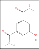
2-Hydroxyisophthalamide
A NOVEL DUAL-LUCIFERASE MONITORING APPARATUS
Dissertation by Thomas Andrews, M.S.
The University of Texas Health Science Center at San Antonio, 2008
Advisor: Peter J. Hornsby
Bioluminescence technology is developing as an important tool in biological research. Borrowed from species of jellyfish and firefly, luciferase genes are being used to create bioluminescence in cells not normally bioluminescent. This technology can be used in applications such as gene reporters, microarray analyses, and real-time monitoring of protein interactions. Two luciferases with a depth of characterization and widely used in cellular and molecular biology are firefly luciferase (Fluc), isolated from Photinus pyralis, and Renilla luciferase (Rluc), isolated from Renilla reniformis.
The emission wavelengths of Fluc and Rluc differ such that they are distinguishable using appropriate optical filters. Technology that is capable of monitoring light emission of 2 luciferases simultaneously has the potential to benefit a wide range of biomedical research areas. In this study, a novel apparatus has been tested to determine the capability and quality of monitoring light emission from Fluc and Rluc simultaneously. Assays were developed to serve as examples of potential uses for the dual-luciferase monitoring apparatus. Using cell lines modified with luciferase genes, it was possible to show that the apparatus could successfully distinguish between light emission from Fluc and Rluc, monitor luciferase activity in a 3D mimic of an in vivo environment, and quantify changes in a biosensor-type cell line expressing an additional luciferase as an internal control.
THE SYNTHESIS AND LUMINESCENCE PROPERTIES OF LANTHANIDE-BASED NANOPARTICLES FOR BIOLOGICAL APPLICATION
Dissertation by Mingzhen Yao, M.S.
The University of Texas at Arlington, 2008
Advisor: Wei Chen
Lanthanide-doped nanoparticles have great potential as a new kind of luminescent material. In this thesis, we report the synthesis of Cerium-doped LaF 3 nanoparticles in dimethyl sulfoxide (DMSO) using chemical reaction at different temperatures. We found that the emission of nanoparticles in DMSO depends not only on reaction temperature but also on reaction time. The emission of DMSO solution can be tuned by reaction time, from 490 to 650 nm. The formation of LaF 3:Ce3+ nanoparticles has been identified by X-ray diffraction (XRD) and transmission electron microscopy (TEM). The TEM results show that the average sizes of these nanoparticles are within 10–13 nm, which is consistent with the sizes we obtained from XRD measurement. The mechanisms for tunable emissions are being investigated. In most biological application, especially in vivo application, nanoparticles are required to be water soluble and biocompatible. Based on this concept, we have developed a simple chemical method for making high-quality, well-defined, water-soluble lanthanide-ions-doped LaF 3 nanoparticles. All the nanoparticles we synthesized are water soluble and emit very strong luminescence.
LUMINESCENT WATER-SOLUBLE QUANTUM DOTS: IMPROVED STABILITY THROUGH SURFACE FUNCTIONALIZATION
Dissertation by Manuela Aseye Ayele Ayee, M.S.
Iowa State University, 2008
Advisor: Aaron R. Clapp
Colloidally prepared semiconductor nanocrystal quantum dots (QDs) are inherently incompatible with biological systems because of the native hydrophobic molecules that coat their surfaces. Post-synthetic techniques such as ligand exchange and encapsulation are therefore employed to make them soluble in aqueous media while preserving their valuable properties such as bright photoluminescence and stability. Small hydrophilic organic molecules like dihydrolipoic acid and tris(hydroxypropyl)phosphine are used to displace the hydrophobic surface ligands, and derivatization by carbodiimide bioconjugation can enhance pH stability. Several QD encapsulation methods in polymer shells are covered, with the most promising one involving the use of a 4-stream vortex mixer to create QD-embedded polymer microspheres with a small size distribution and high fluorescence intensity. The ability of polyhistidine tags to self-assemble onto QDs via metal coordination interactions is also exploited to attach dye-labeled protein molecules onto QD surfaces so as to perform fluorescence resonance energy transfer investigations.
Dihydrolipoic acid

SPECTRAL BARCODING OF POLYSTYRENE MICROSPHERES USING LUMINESCENT SEMICONDUCTOR NANOCRYSTALS (QUANTUM DOTS)
Dissertation by Shyam V. Vaidya, Ph.D.
University of New York, 2008
Advisors: Alexander Couzis and Charles Maldarelli
The focus of this study is the development of optically barcoded polymer beads for use in high-throughput, multiplexed screening applications such as protein microarrays or flow cytometry. Luminescent semiconductor nanoparticles (or quantum dots [QDs]) with different emission wavelengths (colors) and incorporated in different compositions in polystyrene (PS) beads are used to define an optical barcode. The incorporation is undertaken by copolymerizing the PS beads with hydrophobically capped, core-shell, CdSe/ZnS QDs, using a spraying suspension polymerization procedure. Confocal laser scanning microscopy (CLSM) and transmission electron microscopy (TEM) images of the beads indicate that the QDs are segregated into inclusions distributed throughout the bead. The segregation of the QDs inside the polymer beads is due to enthalpy and entropy-driven rejection of the QDs from polymerizing loci as the polymerization proceeds. CLSM and fluorometer measurements of the emission spectra of PS beads embedded with 3 color QDs in varying concentrations are reported, which verify that distinguishable optical ratiometric barcodes derived from the spectral scans of the barcoded beads can be obtained by this technique.
A comparison of the emission profiles of the barcoded beads with that of the same QDs dispersed in styrene indicates luminescence energy transfer from the lower wavelength QDs to the higher wavelength QDs, providing evidence that the QDs are situated within nanometers of each other in the inclusions. The energy transfer limits our ability to obtain and a priori define a considerable number of ratiometric barcodes for multiplexing applications. We observed that the energy transfer could be reduced by separation of the segregating QDs from one another during the polymerization process. We used cross-linking between the polymer molecules during polymerization for separation of the QDs. Use of divinyl benzene (DVB) along with styrene for bead synthesis reduced the number of QD inclusions and dispersed the QDs to separate them by a distance of >10 nm as observed in the TEM images of the bead interiors. A comparison of the CLSM spectral scans of the beads prepared using DVB with that of the beads without cross-linking and a CLSM reference spectral scan—obtained from the QDs dispersed in styrene—indicates a significant reduction in the energy transfer and almost complete recovery of the emissions from the QDs similar to those dispersed in styrene.
MATHEMATICAL THEORY AND NUMERICAL ANALYSIS OF INTEGRATED BIOLUMINESCENCE TOMOGRAPHY AND DIFFUSE OPTICAL TOMOGRAPHY
Dissertation by Syed Hassan Kamran Kazmi, Ph.D.
University of Iowa, 2008
Advisor: Weimin Han
Bioluminescence tomography (BLT) is a recently developed area of molecular imaging. The goal of BLT is to produce a quantitative reconstruction of a bioluminescent source distribution within a small animal from bioluminescent signals measured at the surface of the body.
In most BLT studies so far, the optical parameters are assumed to be known exactly and the only unknown is the light source function. Values of these optical parameters are usually obtained from literature or diffuse optical tomography (DOT) and are not very accurate in general. In this thesis, we upgrade the conventional BLT framework to include the feature of self-adjustment of the optical parameters. In the upgraded BLT framework, we know the optical parameters only approximately, and reconstruct the light source and at the same time determine more accurate values of the optical parameters. We show the existence of a solution of the upgraded problem, develop numerical approximations, and prove the convergence of the numerical method. Numerical results are presented to illustrate the utility of our approach and evaluate its performance.
We also develop a mathematical model that combines DOT and BLT together so as to reconstruct the optical parameters and the bioluminescent source distribution simultaneously. We show the existence of a solution, introduce numerical schemes, and prove convergence of the numerical solutions. We also present numerical results to illustrate the utility and performance of our method.