
Abstract
In investigating ion channel pharmacology, the manual patch clamp is still considered the gold standard for data quality, notwithstanding the major drawbacks of low throughput and the need for skilled operators. The automated patch clamp platform CytoPatch™ Instrument overcomes these restrictions. Its modular fully automated design makes it possible to obtain scalable throughput without the need for well-trained operators. Its chip design and perfusion system reproduces the manual patch technique, thus ensuring optimal data quality. Further, the use of stably transfected frozen cells, usable immediately after thawing, eliminates the cell quality impairment and low success rates associated with a running cell culture and renders screening costs accurately calculable. To demonstrate the applicability of this platform, 18 blinded compounds were assessed for their impact on the cardiac human Ether-à-go-go related gene K+ channel. The IC50 values obtained by the CytoPatch Instrument using the frozen human embryonic kidney 293 cells showed a high correlation (R 2 =0.928) with those obtained from manual patch clamp recordings with human embryonic kidney 293 cells from a running cell culture. Moreover, this correlation extended to sticky compounds such as terfenadine or astemizole. In conclusion, the CytoPatch Instrument operated with frozen cells ready to use directly after thawing provides the same high data quality known from the manual voltage clamp and has the added benefit of enhanced throughput for use in ion channel screening and safety assessment.
Introduction
Nowadays, early hERG liability profiling of potential drug candidates has become an integral part of the drug development strategy in the pharmaceutical industry. The approaches taken range from in silico assessments and binding techniques (radioligand binding and fluorescence polarization) to cell-based assays using ion flux measurements, membrane potential-sensitive dye fluorescence signals, and voltage-clamp techniques. 10 Although most of these approaches offer high-throughput capabilities, their information content is limited because they are, at best, indirect measures of hERG K+ channel activity. The only method available to analyze ion channel function and modulation directly is the patch clamp technique, 11 which is why the manual patch clamp continues to be considered the “gold-standard” with regard to data quality. Unfortunately, this method is time consuming, requires an experienced operator, and is burdened by low throughput, thereby failing to match the needs of the pharmaceutical industry for a method that can be used in drug screening and profiling. In recent years, however, several companies have developed automated patch clamp platforms, which offer different throughput and data quality. 10,12 –17
What most automats have in common is that the sealing process takes place on a planar substrate featuring one or several holes per recording chamber. Unlike in the conventional patch clamp technique in which the patch pipette is conveyed toward the cell via micromanipulators, in automated instruments a cell suspension is pipetted into the recording chamber and the cells are placed by suction onto the patch aperture(s). For this reason, only suspended cells are used in automated platforms. Following cell capture, negative pressure provokes the building of the giga seal, a tight contact between the surface of the recording chamber and the cell membrane. Once the seal has formed, electrical access to the cytosol is established either by rupturing the membrane patch above the aperture (whole-cell configuration) 13,16,17 or by perforating the membrane patch using pore-forming substances (perforated patch). 14 Once one of these configurations is established, the cell can be voltage clamped as in the conventional patch clamp technique. 10
A pivotal step for successful patch clamp recordings is the seal process: only a tight seal in the gigaohm range guarantees that the cells can be properly voltage controlled, and a tight seal is also a prerequisite for the long-term stability in the recording situation necessary for determining slowly developing compound effects.
The present article therefore aims (i) to introduce the CytoPatch™ Instrument in operation with frozen human embryonic kidney (HEK) 293 cells stably expressing the hERG ion channel and usable directly after thawing and (ii) to demonstrate its suitability for safety pharmacology studies. For this purpose, a set of 18 compounds provided by Bayer Schering Pharma (BSP) was tested in a blinded manner for their impact on hERG channel activity. The IC50 data obtained using the CytoPatch Instrument were compared with the data obtained using the manual patch clamp at BSP. We demonstrate that the automated patch clamp platform CytoPatch Instrument is well suited for investigating drug effects on the hERG K+ channel. Whole-cell patch clamp recordings using frozen cells that express the hERG ion channel and can be used immediately after thawing generate data quality similar to that associated with the manual patch clamp.
System Description
CytoPatch Instrument
The CytoPatch Instrument is a fully automated patch clamp platform utilizing a planar glass chip for whole-cell voltage-clamp recordings. In contrast to other automated planar patch clamp platforms, the CytoPatch Chip features a real glass patch pipette. The chip consists of two components: the chip core made of quartz glass and a silicon packaging in which the chip core is embedded. The chip core contains two microfluidic channels: first, the patch channel, which is connected to the patch pipette and filled with intracellular (IC) buffer, and second, the Cytocentering Channel, which is connected to a concentric opening surrounding the patch pipette 18 (Fig. 1e). In the packaging, a third channel is embedded, known as “transport channel.” This channel runs over the chip core surface and the pipette (Fig. 1f, g) and is used to perfuse the voltage-clamped cell with extracellular (EC) buffer or compound.
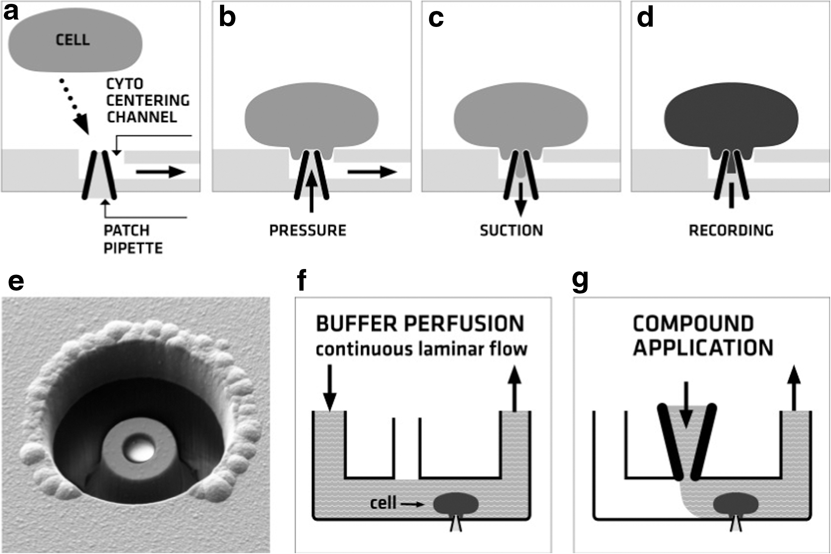
Whole-cell voltage clamp with the CytoPatch™ Chip. The chip contains a real patch pipette, which is connected to the patch channel and surrounded by the Cytocentering Channel
It is a common finding from the manual patch clamp that only clean patch pipettes result in true giga seal formation. In this issue, the Cytocentrics technology mimics the conventional patch clamp process and results in the same high data quality because of its chip design. By applying negative pressure to the Cytocentering Channel, a single cell is captured and positioned on top of the patch pipette. During this process, a positive pressure is applied to the patch channel. This generates an outflow of IC buffer and keeps the patch pipette clean until a cell is captured and the sealing process is initiated by the application of negative pressure to the pipette. The whole-cell configuration is then established via the application of negative pressure pulses to break open the membrane patch. Because of this technology, unlike other automated voltage-clamp platforms, the CytoPatch Instrument does not require fluoride in the IC buffer to obtain giga seals and stable whole-cell recordings. Once a stable whole-cell configuration has been established, the cell is continuously perfused with EC buffer to stabilize it during the recording. To investigate the effect of drugs on ion channels, test compounds can be applied into the transport channel for up to 20 min by means of constant perfusion. Alternatively, precisely triggered rapid perfusion, as required for the proper activation of rapidly desensitizing ligand-gated ion channels, for instance, is also possible with this system. As in the conventional patch clamp, each chip is used for only one cell. This guarantees a clean pipette, and thus, a good seal performance. Carry-over of compounds from one chip to another is ruled out, because chip core and packaging are discarded after every measurement.
A key feature of the CytoPatch Instrument is its modular concept: up to 20 patch clamp units can be connected to form one multichannel CytoPatch Instrument, and each single CytoPatch Instrument can also be run independently as a stand-alone device. Each individual instrument is equipped with its own integrated patch clamp amplifier, data acquisition system, liquid handling system, dispenser needle, chip supply, and cell reservoir (a device to keep the cells usable for the recordings in a vital and nonclustered condition). The modular design minimizes delay times and even makes it possible to run different assays involving different cell types in parallel. Further, the number of channels can be easily increased if required. The individual instruments are connected by the software, which requires only standard intranet infrastructure and the network capabilities provided by the Microsoft Windows operating system.
The CytoPatch Software (CPS) package consists of two main components, the central Workflow Manager (WFM) and the CPS. The WFM is used to plan, edit, and schedule patch clamp studies and to collect, analyze, and archive the data. The CPS runs on each independent CytoPatch Instrument and is used to start assays and monitor the recordings. In the WFM, the compounds and concentrations to be tested and the assay to be run are defined as jobs, which are then directed to the individual channels of the CytoPatch Instrument. After receiving the jobs, the CPS controls the respective, entirely automated patch clamp processes running on a single CytoPatch Instrument channel: chip filling, cell application, the sealing process, and the establishment of the whole-cell configuration are all executed automatically. Electrical capacitances and serial resistance are compensated by the CPS-controlled patch clamp amplifiers. Test compounds are applied to the cell as required by the study, which is designed using the WFM software. Raw data and assay results are written to local hard disks, where they can be accessed by the WFM for further analysis and data archiving (Fig. 2).

The modular concept of the CytoPatch Instrument: The Workflow Manager software is used to import compound lists, to plan (1) and schedule studies, and to direct the samples to be measured to the respective CytoPatch Instruments (2). The voltage-clamp recordings are carried out on the individual CytoPatch Instruments, which are controlled by the CytoPatch Software (3). Once an assay has been finalized the results are sent back to the workflow manager. Here, the data can be analyzed further (4) and exported to the clipboard (5).
Frozen HEK 293 Cells Usable Immediately After Thawing
A crucial factor in obtaining reliable data in automated patch clamp screening are the cells used for the recordings. In most cases, Chinese hamster ovary-K1 or HEK 293 cells, which stably express the relevant ion channels, are used. A constant high throughput and success rate can be achieved only under well-controlled cell culture conditions. The handling of the cells and the number of passages play a critical role in channel expression, seal performance, and whole-cell stability. Ensuring that cells are available in the quantity and quality necessary for automated patch clamp screening therefore requires a highly standardized cell culture and a cultivation time after unfreezing of some days prior to the recordings. 10
To overcome these limitations and to ensure constantly high cell quality, Cytocentrics developed frozen Instant Cells. 19 Stably transfected HEK 293 cells expressing the hERG ion channel are cultured in large batches of up to 5 billion cells, frozen, and stored in liquid nitrogen in up to 500 aliquots of 10 million cells each. Because the cells are pooled shortly before freezing, each aliquot of an Instant Cells batch contains cells of comparable cell quality. For quality assurance purposes, at least two aliquots of each cell batch are unfrozen. Using the conventional patch clamp, ion channel expression is verified and the success rates for giga seal formation and attainment of the whole-cell configuration are checked. Cells must be stable in the whole-cell configuration in terms of current amplitude and membrane resistance over a time period of 25 min. Cell batches with a low success rate are discarded.
For use, the cells are thawed in EC buffer at room temperature, centrifuged, and resuspended in EC buffer. They are ready to use in patch clamp studies within <15 min. The cells are stored in the Cytocentrics Cell Reservoir, which preserves them for use in automated and manual patch clamp recordings for up to 4 h.
Methods
Automated Patch Clamp
HEK 293 cells were stably transfected with cDNA encoding the hERG K+ channel. 20 The cells were frozen as Instant Cells by Cytocentrics AG, quality controlled, and stored in liquid nitrogen. For use, an aliquot of frozen cells was thawed in EC buffer, spun down for 2 min at 100 g, resuspended in EC buffer, and kept in the Cytocentrics Cells Reservoir as a cell suspension at a density of 2.0×10⁶ cells/mL at room temperature. The cells were used for 4 h after thawing.
Automated whole-cell patch clamp recordings were performed using the CytoPatch Instrument. hERG outward tail currents were measured by executing the following pulse protocol every 10 s: from a holding potential of –70 mV, cells were voltage-clamped for 100 ms to –50 mV and then for 2 s to+40 mV. To evoke outward tail currents, a 2 s step to –50 mV followed. The peak tail current was corrected for the leak current determined during the first short voltage step to –50 mV. The cells with a whole-cell membrane resistance of >500 MOhm and an hERG tail current amplitude of at least 200 pA were then continuously perfused with EC buffer for 10 min to establish stable predrug recordings. After this control period, test compounds were continuously applied to the cell via the transport channel for 12 min. Up to two increasing concentrations of the same compound were applied to one cell. HERG tail currents were averaged over 50 s at the end of the control phase and the end of each application phase. If the whole-cell membrane resistance decreased to <500 MOhm during compound application, the hERG tail current was only averaged if a clear steady state developed upon drug wash-in before cell loss. Current inhibition was calculated by dividing the mean tail current in the presence of the drug by the mean tail current of the control phase. All measurements were taken at room temperature.
The EC buffer used for automated patch clamp recordings and for thawing and storage of the frozen cells was composed of the following ingredients (in mM): 140 NaCl, 2.5 KCl, 2 MgCl2, 2 CaCl2, 10 HEPES, 10 glucose, and 15 sucrose. pH was adjusted to 7.4±0.1; the osmolality was 320±5 mOsm/kg. The buffer was stored at 4°C, degassed, and heated up to RT prior to use. The IC buffer used for automated patch clamp recordings was composed of the following ingredients (in mM): 100 K-Gluconat, 20 KCl, 1 CaCl2, 1 MgCl2, 10 HEPES, 11 EGTA-KOH, 4 MgATP, 3 phosphocreatine-Na2-H2O, and 9 sucrose. pH was adjusted to 7.2±0.1; the osmolality was 295±5 mOsm/kg. Aliquots were stored at –20°C, thawed prior to use, and used for maximum 4 h.
Manual Patch Clamp
HEK 293 cells stably transfected with cDNA encoding the hERG K+ channel were cultured as previously described. 21 The single electrode whole-cell voltage-clamp method was applied at room temperature using an EPC-9 amplifier and TIDA 5 software (HEKA Elektronik) as previously reported. 21
The clamp protocol consisted of stepping the command voltage to+20 mV (duration: 1,000 ms), followed by a hyperpolarizing step to −120 mV (500 ms) and a step back to the holding potential of –80 mV (cycle length: 12 s). The inward tail current elicited by stepping from+20 to –120 mV was used to quantify hERG K+ current. The EC buffer was composed of the following ingredients (in mM): 146 NaCl, 4 KCl, 2 CaCl2, 2 MgCl2, and 10 HEPES (pH 7.4; 300–310 mOsm/L). The IC buffer (electrode filling solution) was composed of the following ingredients (in mM): 135 KCl, 2 MgATP, 10 HEPES, and 10 EGTA (pH 7.4; 290–300 mOsm/L). The composition of the buffers and the voltage protocols used in manual and automated voltage-clamp recordings mainly differ for historical reasons and are supposed to have no significant influence on the pharmacological data compared in this study. In particular, no significant difference was observed for the IC50 values of 10 compounds (8 of them included in this study) when using this inward tail protocol or a standard outward tail protocol similar to the one used with the CytoPatch Instrument (Himmel, unpublished data).
Compounds
All compounds (Table 1) were obtained from commercial suppliers or are proprietary structures synthesized at BSP. Compounds were dissolved in dimethylsulfoxide (DMSO) and aliquots were stored at –20°C. The stock solutions were further diluted in DMSO and then diluted 1:1,000 in EC buffer, yielding a final compound concentration of 0.1% DMSO if not otherwise stated. Compound dilution procedure and final DMSO concentration were identical for manual and automated patch clamp recordings. Dilutions of compounds were used for no longer than 3 h after preparation. Leftovers of thawed stocks were discarded at the end of a working day.
Physicochemical Properties and Their Effect on the Human Ether-à-go-go Related Gene K+ Current of Test Compounds as Tested Using a Manual Patch Clamp Setup and the Automated CytoPatch Instrument
1% Dimethylsulfoxide required.
Prodrug containing bis[(aminopropanoyl)aminopropanoate] moiety; loss of seal at 10 μmol/L; ∼20% inhibition at 10 μmol/L; IC50>10 μmol/L .
∼13% inhibition at 100 μmol/L, IC50>100 μmol/L .
N-oxide; stability in extracellular solution, <2 h.
MW, molecular weight (Dalton); clogP, logarithmic partition coefficient of the molar n-octanol/water distribution of a compound calculated using the fragment summation method CLOGP (version 4.3 with version 23 of its associated fragment database as implemented in Sybyl version 8.0; Tripos, Inc.); tPSA, topological polar surface area is defined as the sum of the topological surface area of all nitrogens and oxygens plus any attached hydrogens in Å2 (calculation according to Ref. 23 , with sulfur and phosphor excluded); stock solutions were prepared in dimethyl sulfoxide and stored at−20°C; inward tail measured by means of manual voltage clamp at BSP; outward tail measured by means of automated voltage clamp at Cytocentrics.
The concentration dependence of effects was modeled with a standard four-parameter logistic equation: effect=min+(max/(1+10^[(logIC50−logX)×n H)]), the parameters being minimal and maximal effects (min, max), half-maximal inhibitory drug concentration (IC50), drug concentration (X), and Hill slope (n H). Minimal and maximal effects were usually treated as constants (max=100 and min=0) and IC50 and n H as variables (GraphPad Prism 3).
Results
Biophysical Characterization
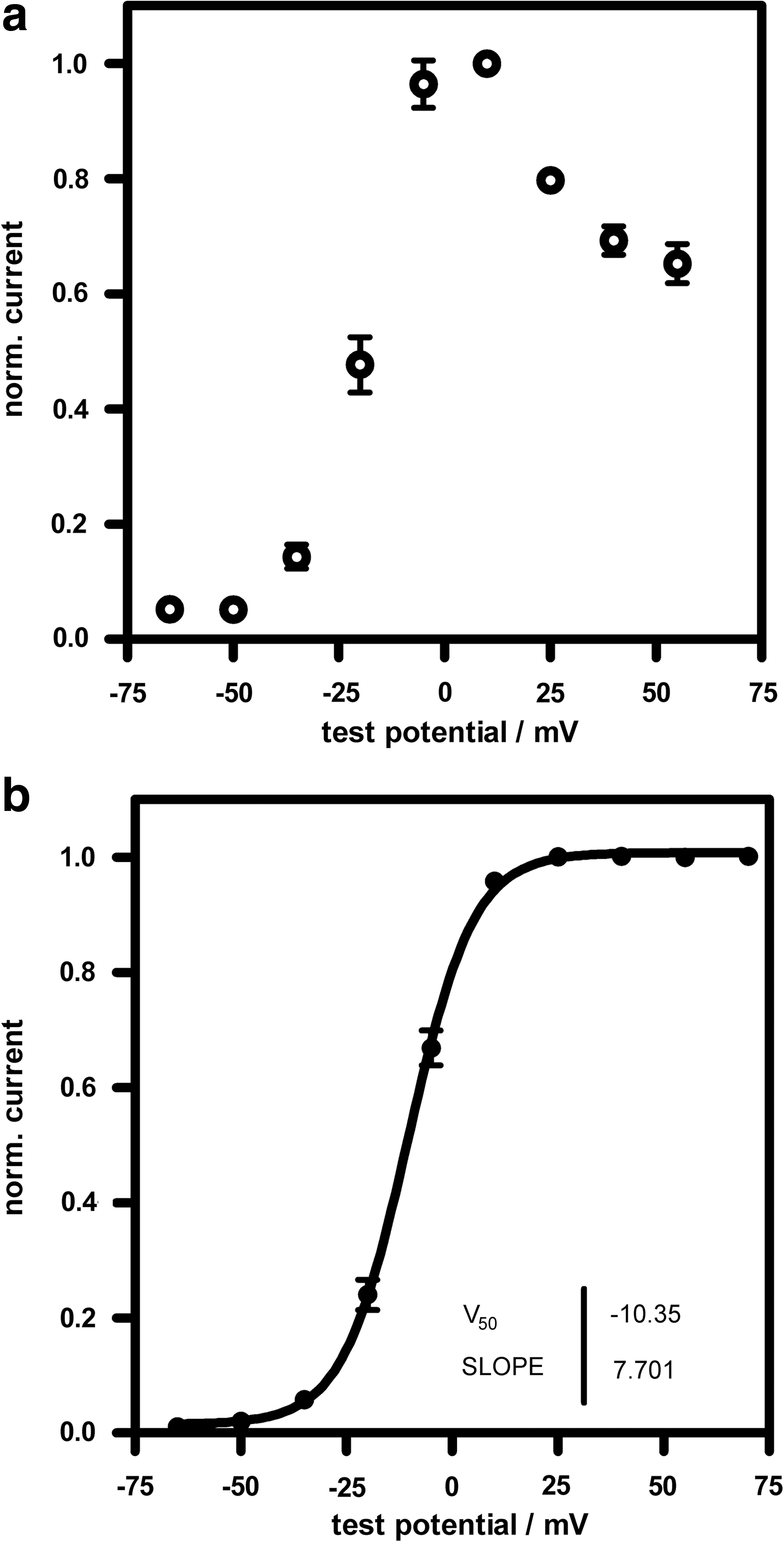
Frozen hERG-expressing HEK 293 cells ready to use directly after thawing were used in conjunction with the CytoPatch instrument. The electrophysiological properties of these cells include the typical hERG characteristic of slow channel activation upon depolarization. Channel activation is superimposed by rapid inactivation. Stepping the test potential back to –50 mV (“deactivation”) elicits the typical large hERG outward tail currents (Fig. 3a, b). The current–voltage relation of the isochronal current at the end of the depolarizing voltage pulse is bell shaped (Fig. 4a). The voltage dependence of the tail current amplitude (“deactivation”) can be fitted by a Boltzmann equation, which results in a half maximal activation voltage of −10.35±0.46 mV. Channel deactivation reaches a steady state at activation voltages of 20 mV and higher (Fig. 4b).
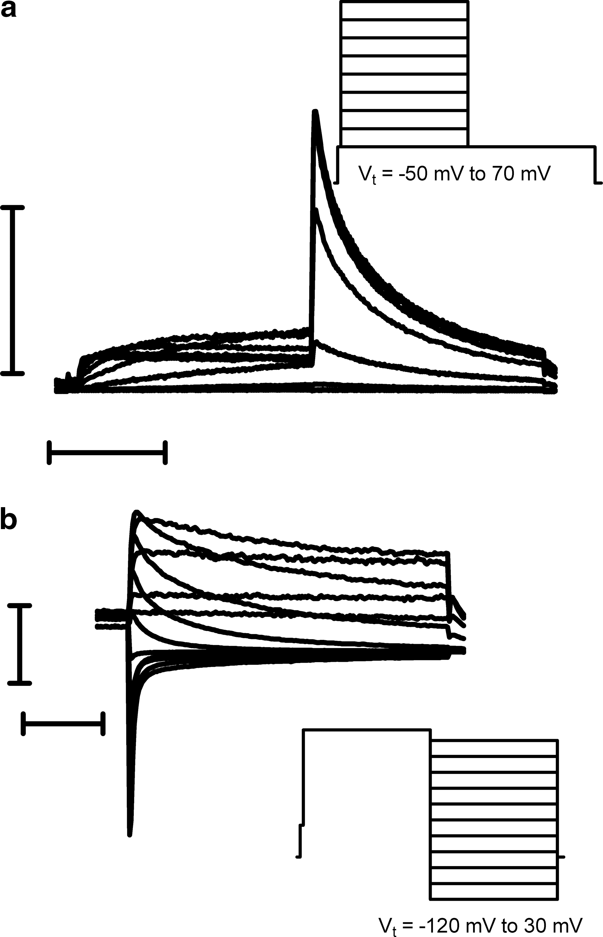
Representative whole-cell patch clamp recordings of hERG-expressing HEK 293 frozen Instant Cells obtained using the CytoPatch Instrument.
Voltage dependence of hERG currents recorded from frozen HEK 293 cells using the CytoPatch Instrument:
Of 234 cells, which formed a stable whole-cell configuration with a membrane resistance higher than 350 MOhm, 223 showed a hERG tail current of at least 200 pA (95.3%). The median tail current amplitude of these cells was 638 pA. One hundred thirty-two successful recordings including at least one successful compound wash-in for 12 min were obtained using these cells (56.4%).
Pharmacological Characterization
The hERG blocking potency of a set of 18 different compounds was assessed by means of whole-cell patch clamp recordings using frozen hERG HEK 293 cells in conjunction with the CytoPatch Instrument and by means of conventional patch clamp recordings using stably transfected cells from a running cell culture. For each compound, 3 different concentrations were tested in triplicates, as was 0.1% DMSO as a negative control.
On the basis of the CytoPatch Instrument and manual patch clamp datasets, IC50 values were calculated and compared as described in the Methods section. The compounds and concentrations tested and the results of the patch clamp experiments are listed in Table 1. The spectrum of effects which the tested compounds had on hERG activity was broad. IC50 values ranged from 5 nM for the potent hERG blocker astemizole to 120 μM for the weak hERG inhibitor metoprolol. The IC50 values obtained using the manual patch clamp and using the CytoPatch Instrument correlated very closely over the whole concentration range (R 2 =0.928; Fig. 5). Interestingly, the compound BAY_11 led to cell loss at 10 μM, a phenomenon that was observed in both the automated and manual methods. This molecule exhibited a markedly larger topological polar surface area than all the other tested compounds (Table 1). The lack of hERG K+ current inhibition by the compound Bay_16 was identified in both assays.
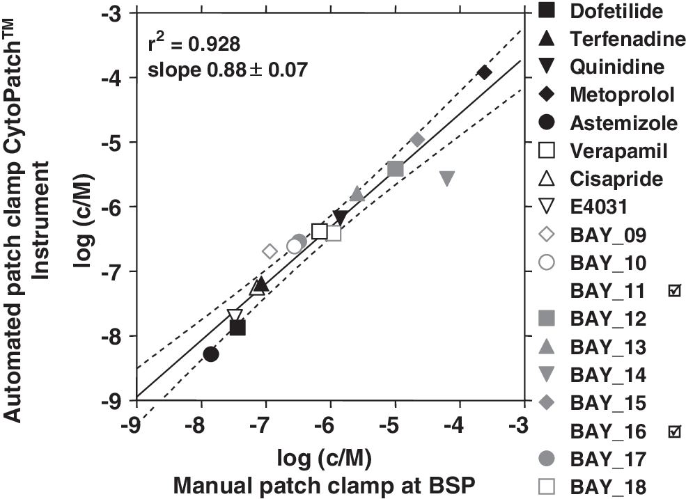
Correlation of IC50 values determined using the automated CytoPatch instrument and those determined using the manual patch clamp. IC50 values obtained with the CytoPatch instrument are plotted against the IC50 values obtained with the manual patch clamp at BSP as described in the Methods section. Linear regression (solid line) is displayed with a 95% confidence interval (dotted lines). For details on BAY_11 and BAY_16, please refer to Table 1.
Discussion
In recent years, automated patch clamp was routinely applied in the laboratories of the pharmaceutical industry. 10,12 –17 The approach to parallelize the patch clamp process has made it possible to multiply the number of data points per day. However, the data quality and the flexibility of the perfusion schemes of the manual patch clamp could not be completely reproduced by any of the automated devices presented so far. Here, we demonstrated for a set of 18 compounds in a single blind study that the automated patch clamp device CytoPatch Instrument is capable of achieving the same data quality as manual patch clamp recordings. The observed IC50 values show a very good correlation with those from manual measurements, indicating that the data quality delivered by the CytoPatch system is superior or at the very least comparable to that reported for other automated voltage-clamp systems. 12 –14,22 This is due to the CytoPatch Chip design with its dedicated patch pipette that is kept clean until sealing, resembling the manual patch clamp process. Unlike in other automated patch clamp devices, the patched cell is continuously perfused with buffer or compound, meaning that cell stability is greater than it would be in a stagnant system. This results in very stable whole-cell recordings, which last long enough to quantify the inhibitory effects of several types of compounds and correspond to a high degree to manual patch clamp recordings. A highly skilled operator is needed to perform manual patch clamp recordings, whereas the CytoPatch Instrument can be operated by a nonelectrophysiologist. Once cells, chips, buffers, and compounds have been supplied, the assay is started and patch clamp recordings are performed fully automatically. Frozen cells, ready to use immediately after thawing, are produced in large batches and pooled shortly before freezing. Therefore, the cell quality within a batch is comparable. The culture condition of the cells is optimized for the CytoPatch Instrument and the batches of the frozen cells are quality assured. The frozen cells do not differ from the running culture in terms of IV curves, current characteristics, or IC50 values. Using these quality-assured cells offers some advantages: no culture is needed and the cells are stored in liquid nitrogen and prepared within 15 min. The simple preparation minimizes quality fluctuations and study design is rendered more flexible as the cells do not need to be cultivated for days in advance before screening can begin. A collapse in data point generation caused by a drop in the quality of the running culture is ruled out from the start. This allows the costs and timeline of a study to be calculated with precision. Further, genetic changes or cell changes caused by differences in culture conditions are also ruled out, and because of the long storage life of the cells in liquid nitrogen, compound effects are reproducible even after several years.
Of all planar substrate-based systems, the members of the IonWorks family (Molecular Devices) generally offer the highest throughput, although they also bring with them certain disadvantages including the inability to form gigaohm seals, discontinuity in voltage clamp because test substance application and recording cannot occur simultaneously, and nonspecific adsorption to the plastic surfaces of lipophilic compounds, resulting in the underestimation of the inhibitory potency of the test compound. 10 Other systems, for example, PatchXpress (Molecular Devices), QPatch (Sophion), and PatchLiner (Nanion), are capable of both gigaseal formation and continuous voltage clamp and offer experimental flexibility with regard to voltage protocols, test compound application, and washout. Therefore, these systems provide a data quality of recordings that almost resembles the manual voltage clamp. 10 However, limitations in the correct determination of IC50 values, particularly for hydrophobic compounds, have been reported for the PatchXPress. 22 Nevertheless, because of its chip design the CytoPatch Instruments' real Gigaohm seals are achieved without the need of fluoride in the IC buffer. Through the flexible perfusion system, not only continuous compound application is feasible over minutes but also the fast and precise application of agonists is possible, which is required for the investigation of fast desensitizing ligand gated ion channels. For example, rise times (10%–90%) for the nicotinic acethylcholine receptor α7 of <5 ms were observed without the need to modulate the viscosity of the applied compound solution (Scheel et al., in preparation). In contrast to any other automated patch clamp platform in the market, the CytoPatch Instrument is GLP compliant as the software package features a complete audit trail, user administration, and sample history. In summary, the CytoPatch Instrument is a very flexible patch clamp platform, referring to the possibility of evaluating multiple ion channels as well as to the option to easily enhance the throughput because of the modular concept.
Disclosure Statement
O.S., G.R.-E., and T.K. are employees of Cytocentrics AG, which manufactures the CytoPatch Instrument and CytoPatch Software.