
Abstract
Low-volume dispensing of neat dimethyl sulfoxide (DMSO) into plate-based assays conserves compound, assay reagents, and intermediate dilution plate cost and, as we demonstrate here, significantly improves structure–activity relationship resolution. Acoustic dispensing of DMSO solutions into standard volume 384W plates yielded inconsistent results in studies with 2 cell lines because of apparent effects on the integrity of the cell monolayer (increased intracellular Ca++ levels as indicated by elevated basal dye fluorescence after acoustic transfer). PocketTip-mediated transfer was successful at increasing apparent potency on a more consistent basis. Notably, the correlation coefficient among fluorescence imaging plate reader (FLIPR):electrophysiology (EP) across a representative ∼125 compound collection was increased ∼5× via conversion to a PocketTip direct dispensation, indicating a triage assay more predictive of activity in the decisional patch-clamp assay. Very importantly, the EP-benchmarked false-negative rate as measured by compounds with FLIPR EC50 more than the highest concentration tested fell from >11% to 5% assay-wide, and the relative FLIPR:EP rank-order fidelity increased from 55% to 78%. Elimination of the aqueous intermediate step provided additional benefits, including reduced assay cost, decreased cycle time, and reduced wet compound consumption rate. Direct DMSO dispensing has broad applicability to cell-based functional assays of multiple varieties, especially in cases where limit solubility in assay buffer is a recognized impediment to maximizing interassay connectivity.
Introduction
Understanding and optimizing away from solubility-mediated potency frame-shifts among sequential triage assays is essential to maximally effective assay design and prosecution of an optimal screening tier. For optimal performance in a plate-based assay, test compounds must be fully solubilized in assay buffer and resistant to and/or have limited exposure to nonspecific surface binding (e.g., tips, plates) within the assay. Here, the objective was twofold: (1) to identify and describe the primary determinants of an effective multiassay ion channel lead optimization triage and (2) to identify best practices in maximizing compound solubilization in the context of a fluorescence imaging plate reader (FLIPR)–patch–clamp sequential triage.
Solubility factors are omnipresent in plate-based assays and a very active area of investigation with regard to in vitro assay design. 1 –3 Low limit solubility in aqueous phase assay buffer is a primary cause of potency frame-shifting false negatives and can negatively impact the delineation of structure–activity relationships. Targets within the central nervous system (CNS), which naturally predispose medicinal chemistry efforts toward lipophillic compounds, are at particular risk of solubility-associated assay interference. 4 This is among many factors that can hinder the forward path for CNS targets, which underscores the need for assay innovation in this preclinical space to mitigate assay interference associated with incomplete solubilization.
A number of technical approaches in test compound prep and dispensing may mitigate solubility considerations by eliminating the need for aqueous intermediate dilutions, such as touch-free acoustic dispensing of neat dimethyl sulfoxide (DMSO) stock compound solutions, and nanoliter volume disposable tips. Here, we describe the implications of test compound solubilization for the apparent potency and cross-assay connectivity of compounds targeted for an ion channel target.
Materials and Methods
Preparation of Test Compound Master Plates and Assay-Ready Daughter Plates
Using a fixed-probe TECAN Genesis liquid handler, the appropriate amount of DMSO (99.9% EMD Chemicals, Inc.) was dispensed (100 μL/s speed, 100 μL/s break-off speed, z-dispense height) into 1-dram vials to dissolve compounds to 20 mM. Each vial was capped and inverted to ensure the entire vessel was coated with DMSO. The vial was then manually vortexed (7 speed, minivortexer; VWR Scientific Products) and visually inspected for complete dissolution of the compound. If needed, sonication in a heated water bath (50°C, 1–5 min, VWR B2500A-DTH) was performed to aid compound solubilization. If the sample was determined to be insoluble, then the compound was rejected from further processing.
A constant volume (60 μL) of the dissolved compound as well as four reference compound solutions (previously dissolved to 2, 20, and 200 mM as described earlier) was transferred (aspiration: 10 μL preair gap, 5 μL trailing air gap, 50 μL/s speed, 200 ms delay, z-max +0.04 mm aspiration height; dispensation: 30 μL/s speed, 200 50 μL/s breakoff speed, 100 ms delay, z-max dispense height) into column 1 or 11 wells in a 384-well plate (No. 230490-104, REMP). DMSO from TECAN's system fluid (40 μL) was added (100 μL/s speed, 100 μL/s break-off speed, z-dispense height) to the wells in columns 2–10 and 12–20. Columns 21 through 24 were left empty. The plate was then centrifuged (30 s, 900 RPM, Centra CL3, Thermo IEC).
Using a 16-channel VPrep (Agilent) equipped with 30 μL tips (No. 11484-202gilent), the solutions in column 1 were mixed (0.5 μL air preaspiration, 30 μL, 6 cycles, 0.5 μL blow-out, 2 mm from bottom of plate, aspiration and dispensing parameters: 100 μL/s, 50 μL/s2, 0 s delay). Compound solution (20 μL) was aspirated (0.15 mm from bottom, 0.5 μL air preaspiration, 5 μL/s, 10 μL/s2, 500 s delay) from column 1 and dispensed (0.15 mm from bottom, 0.5 μL blow-out, 5 μL/s, 10 μL/s2, 500 s delay) into column 2. This pattern of mix and transfer steps was repeated for columns 2 through 10. The tips were changed and the mix and transfer routine was performed for columns 11 through 20. The plate was then sealed (1.5 s, 160°C, No. 24210-001; PlateLoc, Agilent) and stored in the dark in a dessicator (ambient conditions).
To create assay ready plates, the compound plate was unsealed and centrifuged (2 s, 720 rpm, 100% acceleration and breaking; VSpin, Agilent). Using a 384-channel VPrep (Agilent) equipped with 30-μL tips, a 10 μL DMSO plug was aspirated (3 μL air preaspiration, 5 μL trailing air gaps, 3 mm from reservoir bottom, 20 μL/s, 10 μL/s2, 200 s delay). Then, 8 μL of solution was aspirated (0.2 mm from plate bottom, 1 μL/s, 5 μL/s2, 1,000 s delay) from the compound plate. After dispensing (0.5 mm from plate bottom, 0.5 μL/s, 2.5 μL/s2, 1,000 s delay, 2 mm rise height, 1.95 mm horizontal tip touch) 2 μL back into the compound plate, three separate 1 μL dispenses (0.15 mm from plate bottom, same dispensing settings as compound plate) were made into the REMP assay plates. The remaining 3 μL of compound was dispensed back into the compound plate (same compound source dispense parameters). The tips were emptied into the waste and a wash cycle was performed (water wash: 3 μL air preaspiration, 32 μL, 7 cycles, 3 μL blow-out, 0 mm from bottom of 384-microwash [Agilent], 50% inflow, 75% outflow, −23 mm, 2 mm rise height and 0.7 mm horizontal distance for tip touch; 30 μL DMSO aspiration [3 mm from reservoir bottom, 20 μL/s, 10 μL/s2, 200 s delay] and dispensation to waste; water wash; DMSO aspiration and dispensation; water wash). The source and assay plates were then sealed.
Aqueous Compound Transfers
Assay-ready daughter plates were stamped as described earlier into SV384W REMP plates and sealed. Prior to the assay, the seal was removed, and 100 μL of assay buffer was added to each well via Multidrop liquid dispenser to yield a 5× aqueous stock of test compounds. The assay ready plate was then transferred to the BioMek FX384, wherein 10 μL was transferred from each well into the cell plate to yield a 1× final assay concentration in a total assay volume of 50 μL prior to reading on the FLIPR after a 10 min incubation.
PocketTip Compound Transfers
Test compounds were dissolved in DMSO at 20 mM. On the day of the experiments, compounds were diluted into the cell plates using Thermo Scientific PocketTips (catalog No. FX384P30-250) using the Beckman Biomex FX384 liquid handling robotic system. Thermo Scientific PocketTips feature a unique capillary pocket on the internal surface to allow delivery of nanoliter amounts of sample using Beckman Biomex FX384 liquid handling robotic system. The PocketTips draw the compound sample into the tip after mixing and then transfers 250 nL of compound sample into the destination cell plate. Tow hundred fifty nanoliters transfers were made from the DMSO compound source plate to the cell plate via PocketTip on the Biomek FX384. Cell plates were then transferred to the FLIPR stacker and read after a 10 min compound incubation interval.
Echo Acoustic Droplet Ejection Compound Transfers
Assay ready daughter plates were stamped into Echo low dead volume (LDV) shallow 384W Echo source plates (3 μL/well) and sealed. Prior to the assay, 250 nL acoustic transfers were performed at the center of wells in the dye-loaded cell plates. Within the DMSO dispersion, droplet size was defined at 2.5 nL. Compounds were dispersed from the Echo source plate to the destination cell plate in a 1:1 transfer (1 source well per corresponding destination plate well). Prior to dispensation, each well in the LDV source plate was surveyed for meniscus height and relative H20 content (because of the hygroscopicity of DMSO, excessive H20 absorption deflects the precision of acoustic dispersion). In the present setting, meniscus height was constrained to the range between 3 and 7 μL, and H20 content in the LDV source plate was set at a minimum threshold of 30% H20. Total 1:1 dispense time for a full 384W transfer was ∼4 min.
Preparation of Fluorometric Imaging Plate Reader Assay Plates
Two days prior to conducting the assay, HEK cells stably expressing a ligand-gated ion channel were seeded into poly-
Measurement of Intracellular Ca2+ Using the FLIPR
The FLIPR enables a whole well-based measurement of changes in intracellular calcium in 384-well formats. Assay-ready cell plates were prepared as described earlier. After dye incubation and removal, test compounds were added using the PocketTips on the FX liquid handler as previously described. Excitation was performed at 485 nm, and the emissions read-out was at 518 nm. Assay quality assessment was achieved by the implementation of the Z-factor: Z=[1−3SD sample+3SD control]/[mean sample−mean control]. Baseline fluorescence was collected for 10 s in the presence of test compounds alone, followed by agonist addition and an additional 2-min collection of fluorescence signal (equivalent to evoked calcium responses). The concentration response data are expressed in terms of percentage of a control agonist response. Calculations using in-house data software package determine relative fluorescence units (RFUs), and EC50 values were generated by nonlinear regression analysis.
Generation of Stable Cell Lines
HEK293 cells were transfected with the ion channel gene of interest using the Invitrogen Lipofectamine Plus Protocol (Invitrogen Lipofectamine Reagent catalog No. 18324-101 and Plus Reagent catalog No. 11514-015) according to manufacturer's instructions. Briefly, 3 μg of “Subunit X” in pIRES neo2 and 3 μg of “Subunit Y” in pIRES hygro2 were incubated with 20 μL of Plus Reagent and 30 μL of Lipofectamine reagent. Cells were incubated with the transfection mixture for 5 h and then fed growth medium. Cells were placed under antibiotic selection using 150 μg/mL hygromycin B (Invitrogen catalog No. 10687-101) and 500 μg/mL geneticin (Invitrogen catalog No. 10131). Cells were cloned by limiting dilution, and clones were evaluated by Ca++-dependent fluorescence measurements using the FLIPR and by electrophysiology.
Results
Factors Impacting Apparent Potency In Vitro
To triage compounds for a lower-throughput kinetic assay such as patch clamp, a higher-throughput prescreen, which is similarly run in kinetic mode (as opposed to nonkinetic binding or second-messenger accumulation assays), is optimal. This is a common motif in ion channel programs where compounds tested in patch-clamp SAR driver assays are staged via FLIPR kinetic functional assays. The conundrum that this arrangement presents is that FLIPR assays commonly incorporate aqueous intermediate compound dilution plates. If compounds are solubility limited, this format can reduce apparent potency in the FLIPR assay and thus deflect connectivity with subsequent electrophysiology measurements. To minimize solubilization-mediated confounds on interassay connectivity, aqueous dilution steps can be eliminated in favor of dispensing compound solutions in neat DMSO directly into the assay plate via tip-based or acoustic transfer of DMSO compound solutions. Pros and cons of both options will be explored here.
To explore the relationship between compound solubilization in assay buffer and apparent potency in plate-based assays, 125 small molecule activators of a ligand-gated ion channel were tested in an SV384W plate-based Ca++ microfluorometry assay read on the FLIPR and then tested in a confirmatory manual electrophysiology (EP) assay. Parallel nephelometry was employed to identify the concentration representing the upper limit of solubility of test compounds in FLIPR assay buffer. When data coherence between the FLIPR and EP assay was examined in context of upper limit solubility, interassay coherence was stronger among compounds with reasonable solubility in FLIPR assay buffer. As depicted in Figure 1, the subset of compounds that displayed a mean EP:FLIPR potency ratio of <5 (e.g., the fold-shift in potency between the 2 assays) had a significantly higher upper limit of solubility in assay buffer than the subset of compounds with FLIPR:EP fold-shift of >5 (39.2±6.2 μM vs. 20.5±4.3 μM, P<0.01).
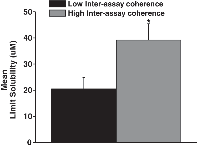
Solubilization in assay buffer is central to coherence of data across assays. Compounds were triaged for progression into a second-order functional assay (in this case, manual patch-clamp electrophysiology) via FLIPR functional plate-based assay. Retrospective analysis of connectivity between the two assays taken in the context of solubilization in the FLIPR buffer demonstrated a significant positive correlation between solubilization in the triage assay buffer and assay coherence. Gray bars signify high-interassay coherence, as defined by EP:FLIPR potency ratio <5. Compounds that met this criterion displayed a significantly higher upper limit of solubility as determined by nephelometry in assay buffer than the subset of compounds with fold-shift >5 (39.2±6.2 μM vs. 20.5±4.3 μM, P<0.01). Bars indicate SEM. EP, electrophysiology; FLIPR, fluorescence imaging plate reader.
A second factor that deflects assay sensitivity is the loss of solubilized compound to nonspecific binding, which can range from moderate to considerable depending on the nature of the compounds, solvent concentration, and the particular composition of the labware involved. Figure 2 depicts an experiment performed to identify the source of a potency decrement in an in vitro functional assay with a multistep compound preparation process. The percent remaining solubilized compound relative to the primary DMSO stock was measured via liquid chromatography/mass spec after each successive dilution and transfer step, a process that revealed a substantial depot binding of compound in the primary aqueous dilution plate (Fig. 2A). Systematic substitution of labwares of varying constitution mitigated this loss of solubilized compound only slightly (Fig. 2B).
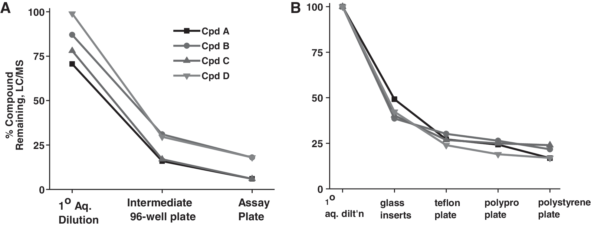
Exposure to multiple iterations of assay plastics via aqueous dilution can reduce the final available solubilized compound and deflect apparent potency.
Acoustic Versus Tip-Based Direct DMSO Dispensing to Improve Solubilization
Techniques that increase the predictive value of plate-based assays by maximizing solubilization and minimizing exposure to potential binding surfaces are at a premium. Eliminating aqueous intermediate dilution plates by dispensing directly from a neat DMSO compound source plate into the assay plate allows the primary DMSO stock to be prepared at a lower concentration by eliminating the successive dilution steps associated with the intermediate plate. Direct DMSO dispensing also reduces the overall assay cost/well by eliminating consumption of transfer plates and the tips and buffers associated with their use. Lower starting DMSO concentration both conserves compound and reduces the likelihood of precipitation upon introduction into the aqueous assay solution (Fig. 3A, B). Figure 3 depicts representative plate flows for all plate-based experimental modes and configurations described herein.
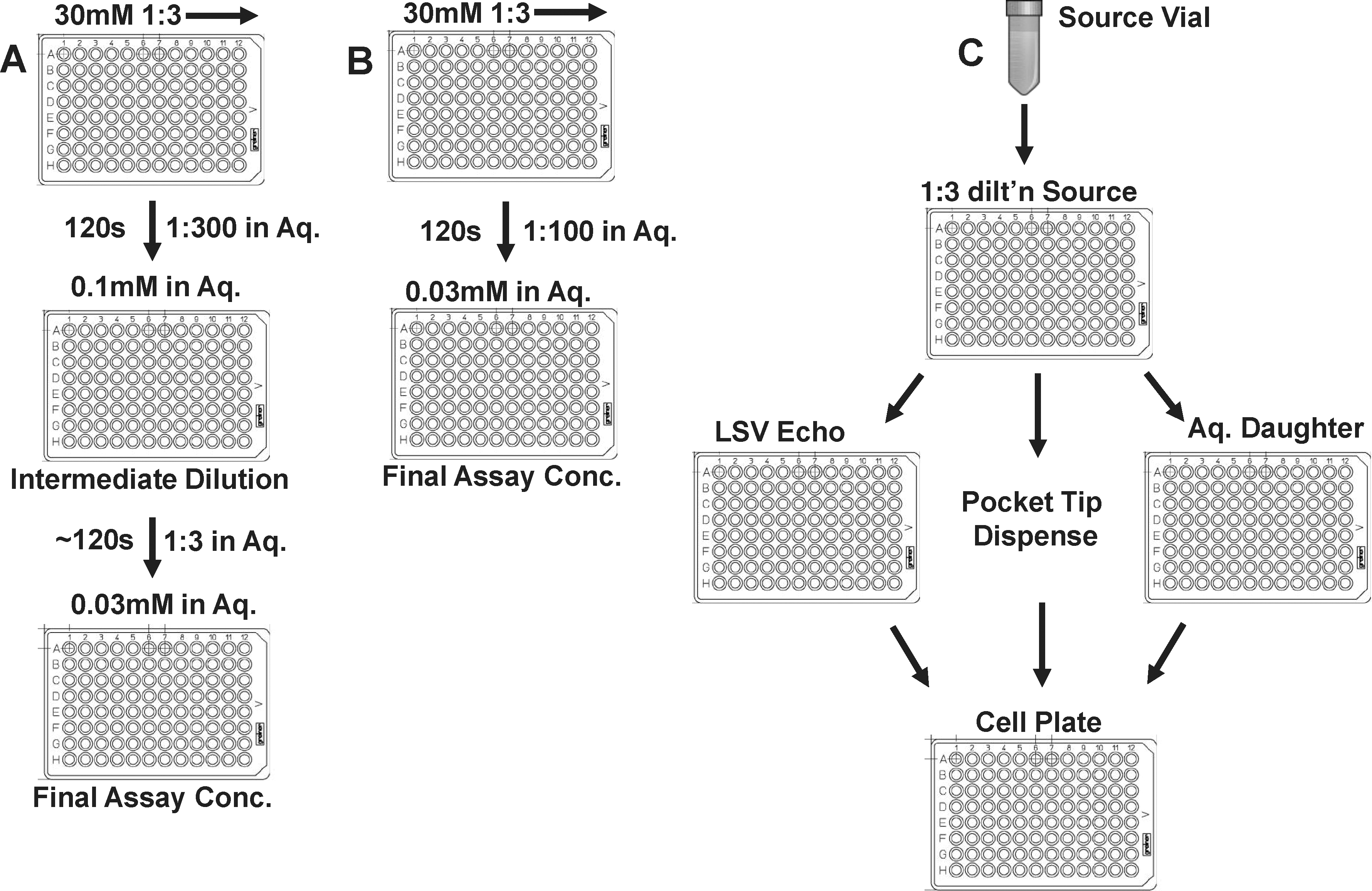
Elimination of aqueous intermediate solutions of test compounds promotes resource avoidance, reduces assay cycle time, and eliminates transfer steps associated with progressive compound loss to adsorption or precipitation.
Among the current compound toolset, potency underestimation was proportional to compound solubilization in FLIPR assay buffer. To mitigate potency shifting across the full solubility spectrum of tool compounds, we sought to eliminate the aqueous-intermediate dilution in the FLIPR assay by exploring two different techniques for nanoliter volume direct DMSO dispensation directly into the FLIPR assay plate. In the first, 200 nL/well of a direct DMSO primary CRC was introduced center-well into the dye-loaded FLIPR assay plate by acoustic dispersion (LabCyte Echo 550). Two distinct HEK293 cell lines, one expressing the ion channel of interest and one coexpressing a ligand-gated ion channel and an enzyme, were used in these tests.
As depicted in Figure 4A, following center-well acoustic dispersion of 200 nL neat DMSO solution into 30 μL/well of assay buffer to a final DMSO concentration of 0.75%, visible disruptions to the cell monolayer were apparent in ∼50% of wells examined (most typically, monolayer detachment). Correspondingly, basal Ca++-sensing dye fluorescence was elevated in the affected wells, indicating elevated basal intracellular Ca++ levels relative to control (no Echo transfer) wells. The assay window as determined by the cellular response to a characteristic agonist was also significantly degraded in the Echo cohort (Fig. 4B) relative to conventional aqueous-intermediate tip-based transfer to an identical final DMSO concentration (Fig. 4C). As a secondary strategy to mitigate cell damage, we attempted quadrant dispersion of DMSO into four corners of the well, 50 nL/transfer. This approach substantially increased the full-plate Echo transfer time relative to center well dispensing, but did not appreciably offset the adverse effects noted above.
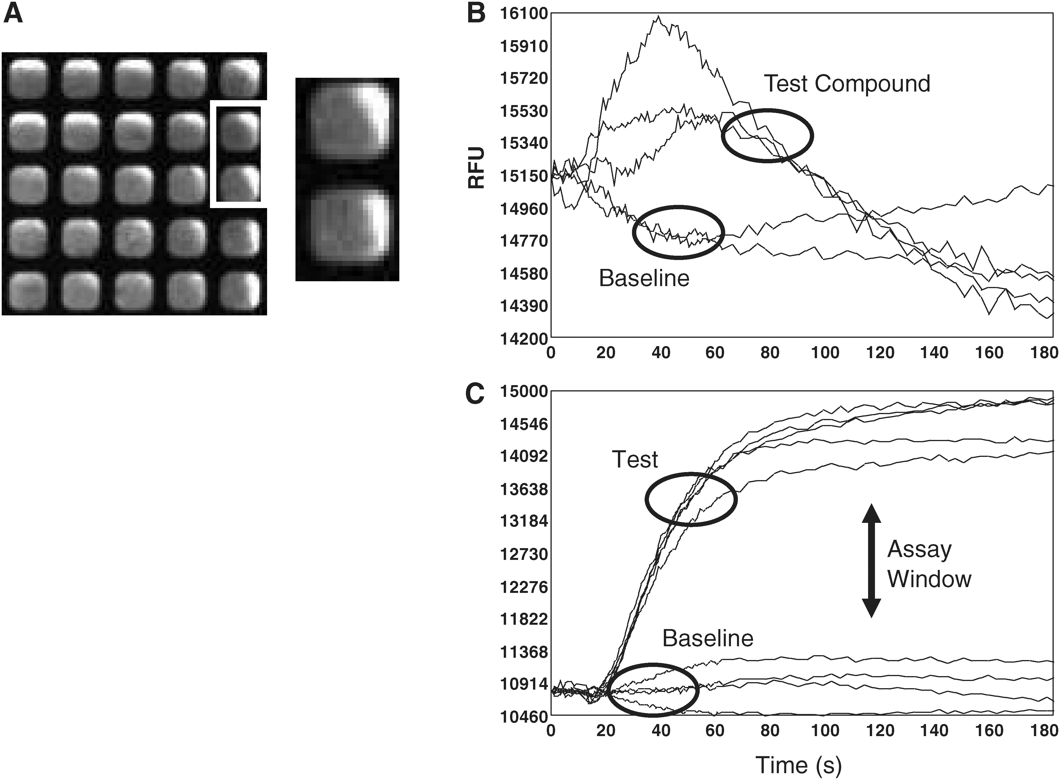
Acoustic transfer of DMSO to a final concentration <1.0% into an SV384W plate containing HEK293 cells caused substantial damage to the cell monolayer of two distinct HEK cell lines each heterologously expressing unique proteins, leading in both cases to elevated basal Ca++ counts and a reduced assay window as measured by the peak agonist-mediated Ca++ response versus baseline.
We next explored nanoliter transfers via ThermoFisher PocketTips (Document 0108, 2009). Following DMSO transfer via 250 nL PocketTips, mean basal fluorescence counts (6875±629 vs. 6566±404), agonist-induced fold change in RFUs (7.5±0.34 vs. 7.7±0.2), and the ensuing full-plate Z-prime values (0.53±0.03 vs. 0.52±0.09) were very similar to values obtained via aqueous intermediate transfer yielding an identical final assay DMSO concentration (Fig. 5A–D, all determinations n=2 with ≥5 replicates). In addition to degrading assay window, acoustic transfer can only be used in FLIPR assay formats requiring preincubation of test compounds, for example, antagonist or modulator assays. Agonist mode additions are not possible because of the need of instantaneous fluorescent read after acute addition of agonist test compounds. Table 1 depicts the time and material implications of replacing a standard volume aqueous disposable tip transfer of test compounds from intermediate plate to assay plate with a direct DMSO transfer, as well as the contrasting limitations of the two direct DMSO transfer techniques. Assay cycle time, cost/well, and compound consumption rate were notably reduced (for transfer volumes and detailed protocols associated with PocketTip transfers, see Materials and Methods section).
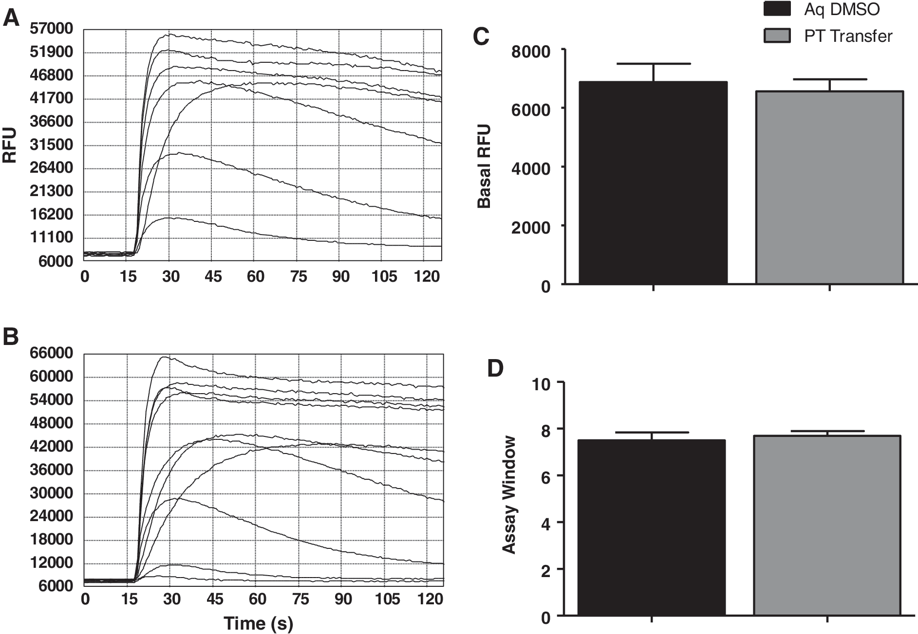
Base assay parameters are unaffected by introduction of DMSO directly into the assay plate via PocketTips.
Direct Dimethyl Sulfoxide Dispensing Was Associated with Reductions in Overall Assay Cost/Well
PocketTip dispensing shows clear advantages in the SV384W format in terms of conserving assay window and versatility of assay format.
DMSO, dimethyl sulfoxide; N/A, not applicable.
Impacts of Direct DMSO Dispensing on Interassay Connectivity
A representative set of test compounds were screened in a FLIPR functional primary assay employing both the standard aqueous intermediate test compound dilution and a direct compound transfer into the assay plate via PocketTips (see Materials and Methods section). Subsequently, compounds were screened in a manual patch-clamp second-order SAR assay. Direct DMSO compound addition had a pronounced impact on the predictive value of the primary FLIPR functional assay relative to the downstream EP assay. FLIPR EC50 values were derived from 10-point CRCs diluted 1:3 starting at 125 μM, whereas 7-point CRCs were obtained in the EP assay over a similar concentration range. Final assay concentrations of test compounds in the FLIPR assay were identical in both assay configurations, as were compound incubation times, final DMSO concentration (<1%; see Materials and Methods section), and overall assay volumes.
When direct DMSO compound transfer was substituted for aqueous intermediate, FLIPR:EP potency correlation was increased substantially (Fig. 6; R 2=0.09 in the aqueous dilution condition, vs. R 2=0.64 with the direct DMSO dispensation method). The gains in FLIPR:EP coherence were a function of substantial increases in apparent potency in the FLIPR assay associated with PocketTip direct DMSO dispensing. The leftward potency shift range between aqueous and direct DMSO dispensation across the compound collection as a whole ranged 5–50-fold. In certain instances in the direct DMSO group, the FLIPR potency substantially exceeded the potency as determined by EP, which was observed in a comparatively limited number of instances under the aqueous transfer condition.

Bypassing an aqueous intermediate compound dilution in favor of neat DMSO direct DMSO dispensing substantially improves the predictive ability of a FLIPR functional triage assay for an ion channel target. Approximately 100 test compounds derived from multiple chemotypes were screened in a manual patch-clamp second-order SAR assay and in a FLIPR functional primary assay employing both the standard aqueous intermediate test compound dilution and a direct compound transfer into the assay plate via PocketTip.
Table 2 highlights compounds from the representative toolset displaying very notable shifts. These compounds in particular would not have been escalated to the second-order EP assay based on apparent FLIPR potency under the aqueous dispensing condition. In all, the risk of compound attrition due to false negatives was markedly decreased by direct DMSO dispensing (12% false-negative rate with aqueous transfer versus 5.0% with direct DMSO transfer; Fig. 7A). For the purposes of this comparison, a false negative was identified as a compound that in either FLIPR assay configuration displayed an EC50 greater than the highest concentration tested (typically 125 μM) and that was subsequently determined to be active in the second-order EP assay.
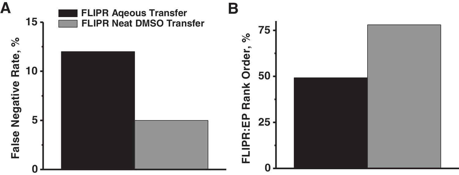
False-negative liability was markedly decreased by direct DMSO dispensing.
Direct Dimethyl Sulfoxide Dispensing Was Associated with Pronounced Shifts in FLIPR Potency, Which Increased Coherence Between the First- and Second-Order Functional Assays
FLIPR, fluorescence imaging plate reader.
In addition to reducing the frank false-negative rate, direct DMSO dispensing increased the utility of the FLIPR assay as a compound prioritization tool. In the aqueous intermediate transfer FLIPR assay, 49.2% of test compounds rank ordered by potency within±5-fold of their corresponding EP values. In the direct DMSO transfer FLIPR assay, this figure increased to 78.0% (Fig. 7B). In testing the two FLIPR assay configurations, compounds were run from identical master DMSO stocks. All assay determinations were run to an n=2 using two inter-run replicates per condition.
Conclusions
The present report examines considerations around maximizing test compound solubility in a FLIPR triage assay as well as the broader process considerations around lead optimization in modern ion channel programs. The overall effectiveness of a multiassay triage is dependent upon the coherence of early higher-throughput assays with the more granular, secondary assays, which succeed them. Poor predictive value of plate-based triage screens will focus subsequent assay support resource on the wrong compounds (false positives) or, perhaps worse, fail to detect activity when it is present (false negatives). A common cause of potency disconnects among assays is differing degrees of compound solubilization.
An evolving strategy for optimizing away from compound solubility considerations in plate-based assays is low-volume direct DMSO dispensing. In this manner, the loss of solubilized compound that attends incremental dilution of DMSO-solubilized compound in an aqueous-phase intermediate plate, followed by a pipette-based transfer of the intermediate solution into the final assay plate, can be omitted. Instead, DMSO-solubilized compound is transferred directly into the assay plate. In addition to solubility benefits, this approach allows for proportional reduction in the final assay volume with associated reduction in test compound and reagent consumption and the elimination of the aqueous-intermediate plate itself. Assay cycle time is also typically condensed by the elimination of a transfer step into and out of the intermediate plate, which for large compound collections produces substantial time savings.
Although Echo preprinting of compounds into 384-well cell-free assay plates via acoustic dispensing is a highly effective strategy, especially in light of the ready integration of Echo systems into automated multi-instrument assay platforms, acoustic dispensing of compounds into cell-attached SV384-well assay plates is less straightforward. As we report here, this strategy has achieved variable results in 384W plates in which typical DMSO dispensing volumes range from 150 to 250 nL. Acoustic transfer in this volume range has the potential to disrupt the cell monolayer and/or disrupt cell homeostasis (such as by elevating intracellular Ca++) in a manner that significantly degrades the assay S/B window.
It bears mention that Echo acoustic dispensing has been leveraged to considerable effect in a variety of 1,536-well cell and biochemical assay applications. 5,6 Moreover, low-volume acoustic dispensing has enabled considerable reductions in the burn rate in large static compound collections such as industrial compound decks used to support modern HTS campaigns. 7,8 However, the assay volumes typically required of SV384-well plate-based applications (typically, 20–50 μL), coupled with the typical DMSO solubility range of drug-like small molecules, introduces a few constraints on acoustic dispensing into assays that are best suited to SV384-well cell-attached configurations.
Although the present technical observation of potency gains via enhanced solubilization in a cell-based assay is valuable, it is important to examine the larger context of this process optimization. The current setting involves a plate-based functional assay, which is employed as a prescreen for a second-order lower-throughput assay (in this case, patch-clamp electrophysiology). So-called triage assays serve critical dual functions. Obviously, “weeding out” compounds with negligible activity will reduce the burden on the second-order assay and provide instant binary (e.g., “active” or “not active”) SAR feedback to medicinal chemistry in advance of the more granular (and less rapidly forthcoming) determinations from the second-order assay. As important, but more subtle, is an ability of the first-order assay to provide a useful potency rank-ordering of test compounds. This allows selective screening of the more active compounds preferentially in the second-order assay, focusing the more time-precious assay resource on the compounds of presumptive greatest interest.
The current strategy of eliminating an aqueous intermediate dilution and transfer step improves substantially versus both of the above functions. The global false-negative rate was lowered, reducing the probability of excluding active compounds from secondary testing. With regard to compound prioritization, the rank-order alignment between first and second-order assays improved substantially with direct DMSO dispensing. In assays wherein primary compound dilutions are performed manually (as most common in electrophysiology assays), it is possible to maximize solubilization through vortexing, heating, and sonication of individual compound vessels. These techniques are of limited practicality, at best, in plate-based assays. Enhancing compound solubilization in the first-order assay plate closes this methodology gap and drives interassay coherence.
In the ion channel space, the throughput of “gold standard” patch-clamp assays is a universal bottleneck, which necessitates multiassay triage to focus precious resource on the most critical compounds. Here, we explore both the optimal format for plate-based triage assays and the practical considerations that drive lead evaluation in modern ion channel research. Coupled with the ancillary benefits to compound consumption, cycle time, and assay cost, direct DMSO dispensing into cell-based functional assays clearly lies along the critical path to scientifically rigorous, resource-conscious lead evaluation of ion channel targets.
Footnotes
Disclosure Statement
No competing financial interests exist.