
Abstract
Heat shock protein-90 (HSP90) is an ATP-dependent molecular chaperone with intrinsic ATPase activity. HSP90 is required for the stability and function of client proteins, many of which are involved in oncogenesis. Thus, identification of HSP90 inhibitors would potentially lead to the discovery of cancer therapeutics. Here, we present a high-throughput screening campaign utilizing two geldanamycin (GM)-labeled probes in a fluorescence polarization (FP) assay. For the primary screen, a previously reported green BODIPY-labeled GM (GM-BODIPY) was used to evaluate a library collection of about 400,000 compounds. From this screen, 3058 compounds showed >30% inhibition. To distinguish true positives from compound interference, a confirmatory screen was deemed necessary. Accordingly, a red-shifted FP binding assay was developed using GM labeled with red BODIPY. This tool enabled reliable identification of promising HSP90α inhibitors.
Introduction
The HSP90 family has several isoforms based on their localization, including HSP90α, HSP90β, GRP94, TRAP1/HSP75, and HSP90N. 6 Structurally, these proteins are made up of three domains: an NH2-terminal ATPase domain, a middle domain involved in client protein binding, and a COOH-terminal dimerization domain. 6 Geldanamycin (GM), the first discovered natural product inhibitor of HSP90, has been reported to bind to the N-terminal domain, thereby inhibiting the ATPase activity. 7 In vivo studies, however, revealed hepatoxicity issues associated with the reactivity and lability of this compound. 8 To alleviate these undesirable properties, researchers prepared analogs of GM, including 17-allylamino-17-demethoxygeldanamycin (17-AAG) 9 and 17-dimethylaminoethylamino-17-demethoxygeldanamycin (17-DMAG). 10 17-AAG, the first HSP90 inhibitor to enter clinical trials, demonstrated improved inhibitory activity and decreased hepatoxicity than GM but its cumbersome formulation hindered evaluation of true maximum tolerated dose. 11 Recently, the more water-soluble 17-DMAG progressed into clinical evaluations in patients with advanced solid tumors. 12
To identify novel small molecule inhibitors of HSP90, several groups have reported binding assays using different technologies. These include fluorescence polarization (FP), 13 –15 time-resolved fluorescence resonance energy transfer, 16 surface plasmon resonance, 16 and AlphaScreen. 17 The FP assays employed a derivatized small molecule based on the crystal structure of a lead compound, 13 a red-shifted fluorescently labeled GM (GM-Cy3B), 14 or a green BODIPY-labeled GM. 15 Assay systems that measure the ATPase activity of HSP90 have also been reported. 18 –21
In this study, we utilized an FP assay with the green BODIPY-labeled GM previously published by Kim et al. 15 (Fig. 1) as the ligand for our initial high-throughput screening (HTS) campaign. The approach was straightforward but the results were complicated by interference from fluorescent compounds and precipitated compounds in the compound library. It has been shown that one strategy to circumvent this issue is to utilize a red-shifted fluorophore in the FP assay, such as Cy dyes or a polar screen far-red–labeled tracer. 22,23 In this study, a novel red-shifted BODIPY-labeled GM (Fig. 1) was synthesized and used in a confirmatory assay with FP format. The BODIPY fluorophores possess excellent spectral properties, including high photostability, high molar absorptivities, high quantum yields, and strong narrow-wavelength emission maxima in the visible region. 24 These dyes are unusual in that they are relatively nonpolar and the chromophore is electronically neutral. Such properties tend to minimize dye-induced perturbation of conjugate functional properties. BODIPY dyes are, therefore, often the preferred choice for labeling nucleotides, amino acids, and other low-molecular-weight ligands. The red BODIPY-GM has excitation and emission wavelengths that are farther red shifted than those for the published Cy3B-GM; thus, it should provide advantages in overcoming interference from autofluorescent and insoluble compounds in the library. Data are presented herein.
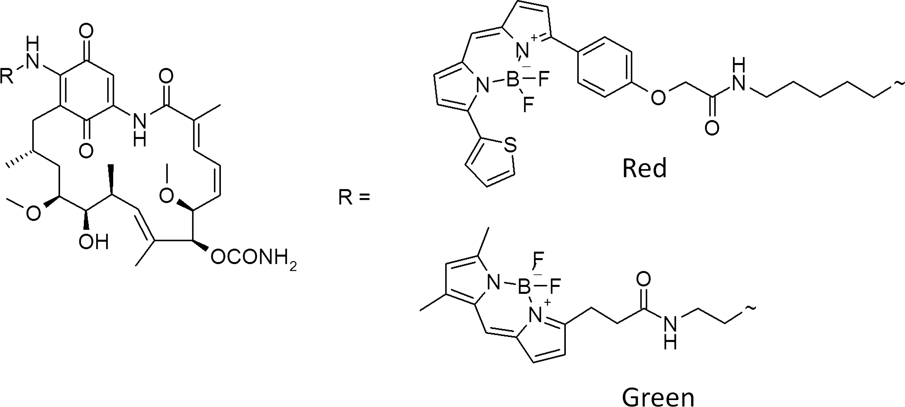
Structures of green BODIPY-labeled GM and red BODIPY-labeled GM. GM, geldanamycin.
Materials and Methods
Materials
Green BODIPY-labeled GM (Fig. 1) was prepared according to Llauger-Bufi et al. 25 GM and 17-AAG were purchased from A.G. Scientific (San Diego, CA) and Stressgen Bioreagent (Ann Arbor, MI). Black 384-well flat-bottomed polystyrene plates (Packard OptiPlates #600727) were purchased from PerkinElmer Life Sciences (Boston, MA). HEPES was purchased from AMRESCO (Solon, OH), KCl from TEKNOVA (Hollister, CA), MgCl2 and Na2MoO4 from Sigma-Aldrich (St. Louis, MO), NP-40 detergent from EMD Chemicals (Gibbstown, NJ), bovine gamma globulin (BGG) from Invitrogen (Carlsbad, CA), and dithiothreitol (DTT) from Fisher Scientific (Pittsburgh, PA).
Instrumentation
FP assays for the primary and secondary screens were read on an EnVision™ 2100 or 2102 (PerkinElmer). The excitation/emission filter wavelengths were 485 nm/535 nm for the green BODIPY-labeled GM and 555 nm/632 nm for the red BODIPY-labeled GM (Fig. 1). The plate height, dimension, and gain were optimized for each assay. The FP binding assays were conducted in 384-well black polystyrene flat-bottomed plates. Liquid-handling instruments (Biomek FX; Beckman Coulter, Fullerton, CA) and Multidrop (Thermo Labsystems, Vantaa, Finland) were used to add compound, protein, and BODIPY-labeled GM for the FP binding assays. Data were analyzed using XLFit 4 (ID Business Solutions, Guildford, United Kingdom), GraphPad PRISM® 5 (GraphPad Software, La Jolla, CA), and Spotfire® (TIBCO Software, Palo Alto, CA).
Synthesis of Red GM-BODIPY
A solution of BODIPY® TR cadaverine (Invitrogen) (3.26 mg, 0.0060 mmol), GM (5.00 mg, 0.0089 mmol), and diisopropylethylamine (6 μL) in dichloromethane (1.0 mL) was stirred at room temperature for 24 h. The reaction mixture was then loaded onto a silica gel column (4 g) and eluted with a 5:1 mixture of dichloromethane and acetone. Product was isolated as a purple solid (1.90 mg, 31%). Liquid chromotography/mass spectrometry (m/e: 1060 M+Na).
HSP90α Expression and Purification
The sequence-verified cDNA encoding full-length human HSP90α protein was generated from PCR amplification of human uterus and kidney cDNA (5′ end; Clontech, Mountain View, CA) and the I.M.A.G.E. Consortium Clone ID: 4404328 (3′ end; American Type Culture Collection, Manassas, VA). This was ligated into the pFastBac™-1 plasmid (Invitrogen) that had been engineered to express the HSP90α as a chimeric protein with an NH2-terminal glutathione S-transferase (GST)/rhinovirus 3C protease cleavage site.
Recombinant baculovirus was prepared by transposition in E. coli (Bac-to-Bac® System; Invitrogen), confirming expression by immunoblot analysis using a human HSP90α mouse monoclonal antibody (Affinity BioReagents™, Golden, CO). Large-scale protein production was performed using a 10-L Wave Bioreactor® (GE Health Care, Piscataway, NJ). Sf21 insect cells grown in Excell 420 serum-free media (SAFC BioSciences, Lenexa, KS) were infected at 2×10⁶ cells/mL with a multiplicity of infection of 2.0 and harvested at 66 h. Cells frozen at −80°C were suspended in Harvest buffer (20 mM HEPES, pH 7.4; 20 mM NaCl; 1 mM ethylenediaminetetraacetic acid [EDTA]; and protease inhibitors). Cells were lysed by the addition of 0.1% Triton X-100 with gentle rocking for 30 min on ice. Nuclei and large debris were removed by low-speed centrifugation, followed by centrifugation at 100,000 g for 1 h. The clarified extract was then adjusted to 150 mM NaCl and 1 mM DTT for binding to Glutathione Sepharose™ 4B resin (GE Healthcare) overnight at 4°C with gentle rotation. The extract–resin slurry was poured into a column and the resin was washed extensively with wash buffer (50 mM HEPES, pH 7.4; 150 mM NaCl; 1 mM EDTA; and 1 mM DTT). GST-HSP90α was eluted with wash buffer containing 20 mM reduced glutathione (pH 7.4). Pooled GST-HSP90α fractions were then dialyzed overnight at 4°C against storage buffer (50 mM HEPES, pH 7.4; 100 mM NaCl; 1 mM EDTA; 1 mM DTT; and 10% glycerol). Protein aliquots were flash-frozen and stored at −80°C until use. The yield was ∼1 g of high-purity GST-HSP90α from a 10-L culture.
FP Assay for Primary Screen with Green BODIPY-Labeled GM
The FP assay using green BODIPY-labeled GM was previously described by Kim et al. 15 and utilized here with some modifications. Briefly, both GST-HSP90α and green BODIPY-labeled GM were diluted in assay buffer containing 20 mM HEPES (pH 7.2), 50 mM KCl, 5 mM MgCl2, 20 mM Na2MoO4, 0.01% NP-40 (v/v), 0.1 mg/mL BGG, and 2 mM DTT. 15 GST-HSP90α (15 μL) and 1.25 μL test compounds (final concentration of 10 μM) were first added to 384-well black Optiplates using the Biomek FX liquid handler, immediately followed by addition of 35 μL of green BODIPY-labeled GM using Multidrop to bring the total reaction volume to 50 μL (final concentrations of GST-HSP90α and ligand were 50 nM and 10 nM, respectively). The assay plates were shaken at room temperature for 16 h before reading on the EnVision 2100 or 2102 plate reader (PerkinElmer) at excitation wavelength of 485 nm and emission wavelength of 535 nm. The format of the assay plate allowed for 0% inhibition (dimethylsulfoxide [DMSO]) and 100% inhibition (1 μM GM) to be added to columns 1, 2, 23, and 24 for internal control.
FP Assay for Secondary Screen with Red BODIPY-Labeled GM
The FP assay for the red BODIPY-labeled GM (Table 1) was performed similar to the one described previously for the green BODIPY-labeled GM. First, GST-HSP90α (15 μL) and 1.25 μL test compounds (final concentration of 10 μM) were added to 384-well black Optiplates using the Biomek FX liquid handler. Then, 35 μL of red BODIPY-labeled GM was added to the assay plate using Multidrop to bring the total reaction volume to 50 μL (final concentrations of GST-HSP90α and ligand were 100 nM and 20 nM, respectively). The assay plates were shaken at room temperature for 5 h before reading on the EnVision 2100 or 2102 plate reader (PerkinElmer) at excitation wavelength of 555 nm and emission wavelength of 632 nm. The format of the assay plate allowed for 0% inhibition (DMSO) and 100% inhibition (10 μM GM) to be added to columns 1, 2, 23, and 24 for internal control.
Red Fluorescence Polarization Assay Protocol Table
1. 384-well black Optiplates, the Biomek FX liquid handler.
2, 3. Columns 1, 2, 23, 24: rows A–F, DMSO; rows G–J, 40 nM GM; rows K–P, 10 μM GM. All were added with the Biomek FX liquid handler.
4. Multidrop dispenses reagent to all wells.
5. Plates were lidded until read.
6. Excitation filter (555/38 nm), emission filter (632/45 nm).
DMSO, dimethylsulfoxide; FP, fluorescence polarization; GM, geldanamycin, GST, glutathione S-transferase; HSP90, heat shock protein-90.
Data Analysis
Signal-to-noise (S/N) ratio was calculated based on the following equation:
Results and Discussion
FP Assay Development for Secondary Screen with Red BODIPY-Labeled GM
To eliminate false positive results due to autofluorescent compounds or insoluble compounds in the compound library, a secondary screen was designed using red BODIPY-labeled GM as the ligand for the FP assay. It has been demonstrated that using a red-shifted tracer in FP is a valid strategy to overcome interference due to autofluorescent compounds or light scatter from precipitated compounds. 22,23 The difference in structure between green and red BODIPY-GM is depicted in Figure 1. The red BODIPY has increased conjugation relative to the green BODIPY, thus making the red fluorophore more hydrophobic than the green fluorophore.
Both the BODIPY TR and BODIPY TMR amines were available as starting materials for synthesis of the probe but the TR dye was chosen over the TMR dye due to its longer fluorescence emission wavelength (617 nm for TR vs. 574 nm for TMR). The optimal concentrations of HSP90α protein and red BODIPY-labeled GM were first evaluated in the red-shifted FP assay as shown in Figure 2A. The amount of GST-HSP90α was varied from 0 to 500 nM against different concentrations of red BODIPY-labeled GM (2.5, 5, 10, 20, and 40 nM). The mP values increased with increasing GST-HSP90α concentration and appeared to plateau at ∼200 nM. For the screen, 100 nM protein and 20 nM ligand were utilized to obtain a reasonable dynamic range (∼100 mP; Fig. 2A), without losing sensitivity to weak hits. 27 Red-shifted fluorophores are usually hydrophobic or “sticky,” so the polarization values are high in free label state, causing decreased assay windows in FP. 23 To compensate for that, higher protein and label concentrations in the red-shifted secondary assay were chosen than in the green-labeled, primary assay. The K d of the red BODIPY-GM is 8.2 nM, close to that of the green label (12 nM), consistent with the reported value (6.6 nM). 15 In addition, assay stability was evaluated since it directly affects the throughput for the HTS campaign. The assessment of the optimal incubation time and signal stability of the HSP90α reaction was performed over a 10-h period. The highest dynamic range was observed between 4 and 7 h (Fig. 2B); a 5-h incubation time was selected for the actual screen. The performance of the red FP assay was evaluated in assay window, S/N ratio, and Z′-factor using twenty 384-well assay plates, each containing 24 “0% inhibition” or negative control wells (100 nM HP90α with 20 nM red BODIPY-GM and DMSO) and 24 “100% inhibition” or positive control wells (100 nM HP90α with 20 nM red BODIPY-GM and 10 μM GM) (Fig. 3). The average mP (±SD) values for the negative and positive controls were 170±6 mP and 77±8 mP, respectively, resulting in an assay window of 93 mP (Fig. 3A). As shown in Figure 3B, the S/N ratios were ≥8 (9±1; mean±SD), which was higher than the minimal acceptable S/N ratio in the FP assay (S/N≥5 is acceptable, S/N≥10 desirable). 28 The Z′-factor for the assay was 0.60±0.05, indicating that it was a robust assay (Fig. 3B).

FP assay development for secondary screen with red BODIPY-labeled GM.
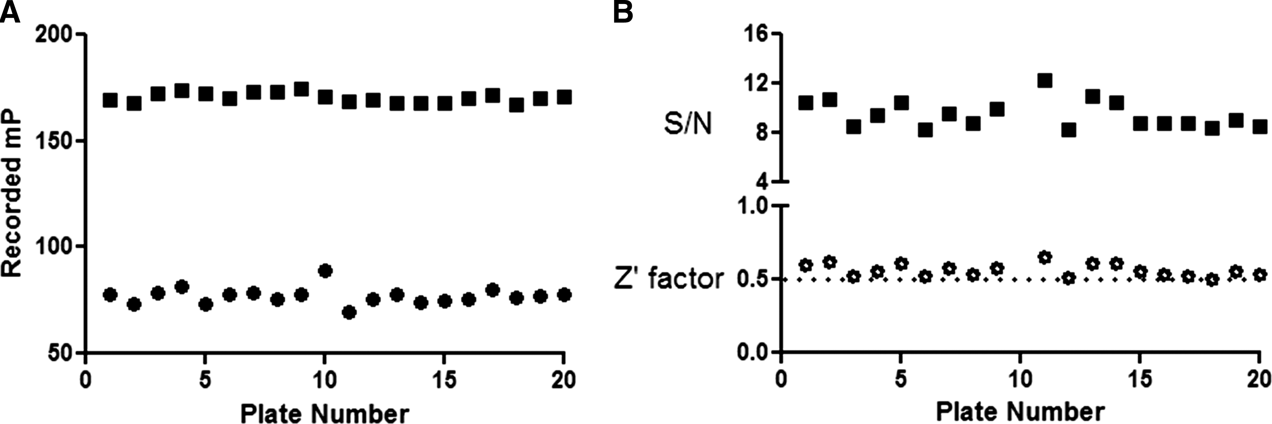
Evaluation of HSP90α FP assay performance with red BODIPY-labeled GM.
To further validate this assay, the concentration response for two commercial compounds was performed with red-shifted tracer and half-maximal inhibitory concentration (IC50) values were determined to be 45±17 nM (in-house HSP90α) for GM and 74±22 nM (in-house HSP90α) for 17-AAG (Fig. 4), consistent with the published data that used Stressgen Bioreagent HSP90α (77 nM for GM 15 and 110 nM for 17-AAG 25 ). All these IC50 values were the average of at least three determinations with triplicates for each determination.

Inhibitory activity of GM against red BODIPY-GM binding with HSP90α. About 50 μL of 100 nM HSP90α was mixed with 2.5 μL of GM that was serially diluted in half log intervals, followed by addition of 50 μL of red BODIPY-GM at 20 nM.
This sensitive and robust FP-based assay for binding of red BODIPY-labeled GM to HSP90α was automated and used to screen for novel HSP90α inhibitors.
Primary Screening Results
The primary screen was carried out by screening >1,200 assay plates in 384-well format at 10 μM final compound concentration. More than 400,000 compounds were screened and the Z-factor for each assay plate was calculated. Z-factor is similar to Z′-factor except that it uses the samples and not just controls in the calculation. 29 Only those plates with Z-factor >0.5 passed the quality control (QC) standard and had their results returned to the database. This QC standard was applied to the FP assay for red BODIPY-labeled GM as well. For the green assay, the Z′-factor was 0.60±0.08, S/N was 11±3, and the assay window was 144 mP. The mean percent inhibition of the compounds from the primary screen was −12, and the SD was 15. The number of compounds with SD (σ) of the percent inhibition from less than −6σ to more than+6σ are shown in Figure 5. A 30% cutoff criteria was used, which was above three times of the SD, thus above the noise level. From the primary screen, 3,058 compounds showed inhibitory activities >30%; thus, the hit rate was 0.76%. Those compounds that showed>200% of fluorescence intensity of negative controls in S and P channels were excluded and flagged as autofluorescent compounds. 30 In a mock screen using fluorescein-labeled casein, fluorescent compounds were found to enlarge, reduce, or not affect polarization signals. 31 Also, compounds that absorb probe fluorescence either reduced or did not affect polarization signals.

Histogram of the primary screen of HSP90α binding to green BODIPY-labeled GM. The number of compounds with SD (σ) of the percent inhibition from <−6σ to >+6σ are shown.
Secondary Screening Results
To identify the true positives from the HSP90α screen, the 3,058 compounds were further screened in the red-shifted FP assay. The red BODIPY-GM is more highly conjugated than Cy3B-GM in structure. 32 It has the excitation/emission wavelength of 555 nm/632 nm, respectively, farther red shifted than the published Cy3B-GM, which has excitation/emission wavelength of 530 nm/580 nm, respectively. 32 The longer excitation and emission wavelengths of red BODIPY-GM should help to overcome the autofluorescent compound interference, as well as light scattering caused by insoluble compounds. 22,23 The mean percent inhibition of the compounds from the secondary screen was 7.5. When using the same 30% inhibition cutoff criteria for the red FP assay as for the green FP assay, 340 compounds were identified as positives. As for the primary screen, those compounds that showed >200% of fluorescence intensity of negative controls in S and P channels were excluded and flagged as autofluorescent compounds. 30 With these criteria, 748 compounds were flagged as autofluorescent compounds, even though 127 of these compounds met the 30% inhibition threshold. Therefore, the true hits from the secondary screen comprised 213 compounds. Consequently, the false positives from the primary assay totaled 2,845 compounds (3,058 – 213), indicating a false positive rate of 93% for the green FP assay. These results highlighted the advantage of the red probe. Apparently, the red FP assay has its own false positives, demonstrated by 127 compounds that met the 30% cutoff criteria but were flagged as autofluorescent compounds. Structures of three select HSP90α inhibitors confirmed by the red BODIPY-labeled GM FP assay are shown in Figure 6 with their IC50 values (0.4, 3, and 6 μM).
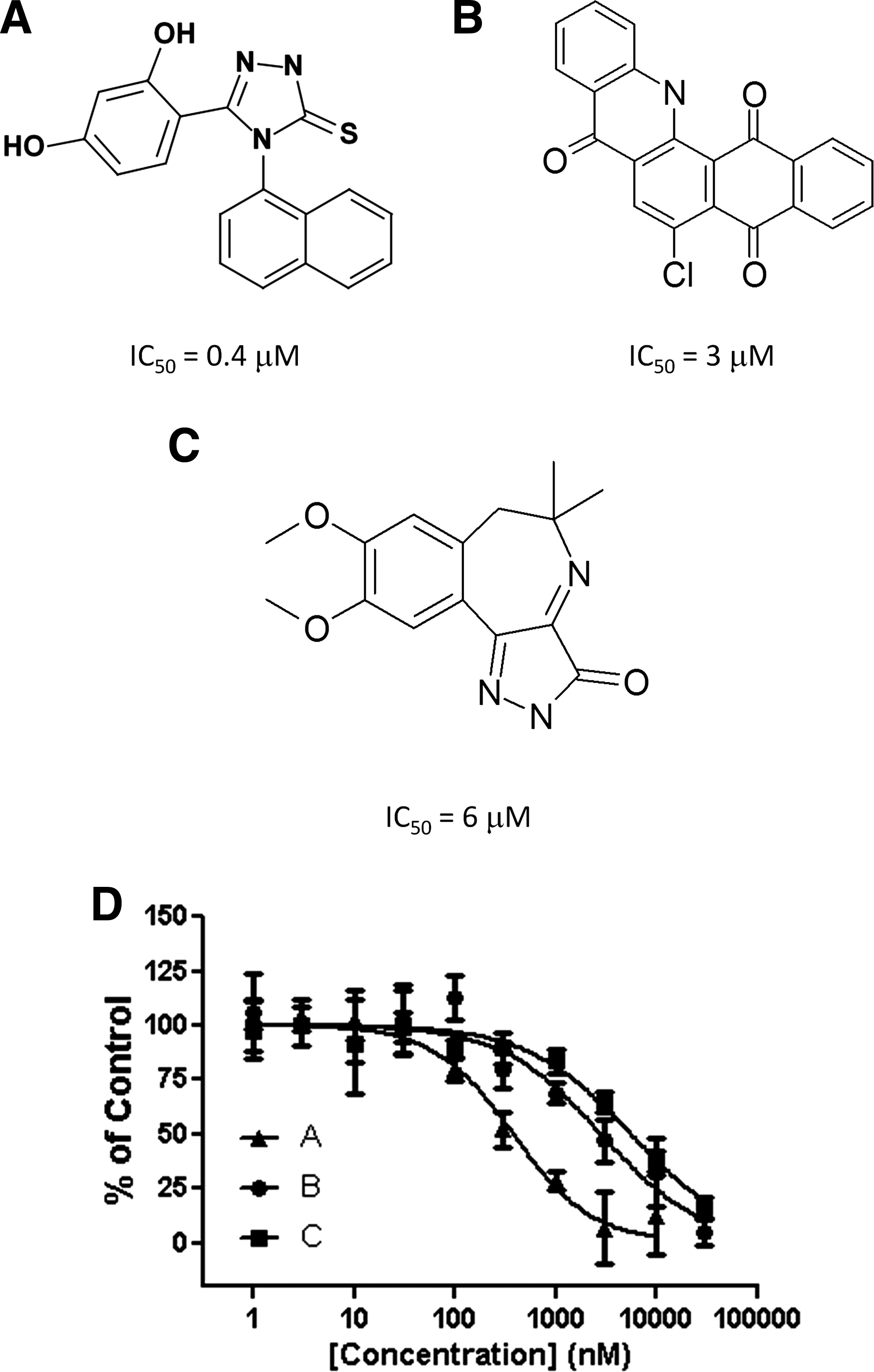
Structures of select positives from the red BODIPY-labeled GM HSP90α FP assay. The structures of three representative compounds
In summary, an HTS campaign utilizing a competitive green BODIPY-labeled GM FP assay was seamlessly performed. Unfortunately, the positives identified from this screen were mostly autofluorescent, prompting the need for a more reliable assay detection probe. Synthesis of a red BODIPY-labeled GM for use in an orthogonal confirmatory secondary screen proved to be a viable approach in reducing the number of false results. From this effort, a number of compounds emerged as “true positives” and may provide promising starting points for the development of HSP90α-directed therapeutic agents.
Footnotes
Disclosure Statement
No competing financial interests exist.