
Abstract
Cardiovascular toxicity is a leading contributor to drug withdrawal and late-stage attrition. Earlier and broader screening is a validated approach to build-in cardiovascular safety as demonstrated with human Ether-à-go-go-related gene (hERG) screening to reduce drug-induced arrhythmia. There is an urgent need for novel in vitro assays to address other mechanistic aspects of cardiovascular function, including contractility, heart rate, toxicity, hypertrophy, and non-hERG arrhythmia. Recent advances in label-free cellular impedance technology now enable tracking of spontaneous, synchronized beating of cultured cardiomyocytes. Analysis of beating allows integrated detection that is downstream of electrical and mechanical aspects of contraction. Here, we evaluate impedance-based cardiomyocyte responses against criteria required for drug screening. The throughput and sensitivity allowed for rapid assay development. Critical variables for rat neonatal cardiomyocyte assays included cell density and serum levels. Once optimized, consistent, stable beating for at least 3 days was straight-forward to achieve. In tests of compounds spanning a breadth of target classes, the potency values showed excellent precision, wide dynamic range, and consistency across multiple experiments. Cardiomyocyte impedance assays can extract multiple beat-related parameters. In these experiments, rate, amplitude, and rise slope were examined and each yielded acceptable precision. Potency values calculated by beat rate and amplitude were highly correlated for most compounds although selected compounds displayed unique profiles indicative of different mechanisms. Tests with known cardiovascular active drugs revealed concordance with clinical findings. Thus, impedance assays combine novel features including sensitivity to contractile activity, versatile data analysis, and robust/translatable data in a format with sufficient throughput to become a valuable addition to the cardiovascular in vitro screening arsenal.
Introduction
Cellular impedance assays have emerged as a highly versatile label-free approach capable of addressing a variety of applications in drug discovery. 5 –10 The technology is based on culturing cells on thin-film, interdigitated electrodes embedded in the bottom of each well of a microtiter plate. When a small alternating electrical current is applied, the cell layer impedes current flow to varying degrees depending on morphology, adhesion, and cell–cell contacts. A hallmark of impedance platforms is exquisite sensitivity that, in some configurations, enables resolution of cellular micromotion at 1 nm or one-third the thickness of the cell membrane. 11 Recent advances in technology have increased the data sampling rate to 12 ms, a fraction of the beat rate of cardiomyocyte cultures, thereby enabling label-free detection of cardiomyocyte beating characteristics. In principle, an impedance-based cardiomyocyte beat detection assay should offer features of a broad early drug discovery screen for cardiovascular toxicity, including (i) robust data, (ii) high throughput, (iii) label-free monitoring versatility, and (iv) detection downstream of both mechanical and electrical elements of contraction.
The goal of the present study was to evaluate the feasibility of developing a cardiomyocyte impedance assay for broad application in cardiac biology, pharmacology, and toxicity investigations using primary cardiomyocyte cultures. Experiments were performed focusing on criteria required for routine screening and pharmacology assessment. The ease of developing and optimizing impedance-based assays was challenged by using primary cultures of rat neonatal cardiomyocytes. The robustness of pharmacological data was explored with ligands spanning a range of potencies and pharmacological mechanisms. Intra-assay precision and inter-assay reproducibility were assessed using beat rate and amplitude data. Drugs with established cardiovascular effects in humans were tested to evaluate concordance with clinical data. The results indicate that cardiomyocyte impedance assays are robust and versatile and would be a valuable addition to the cardiovascular in vitro screening arsenal.
Materials and Methods
Myocyte Isolation and Culture
Cardiomyocytes were isolated from 3-day-old Han Wistar (Harlan, Indianapolis, IN) rats using Neonatal Cardiomyocyte Isolation System (Worthington Biochemical Co., Lakewood, NJ) following the manufacturer's instructions (Table 1). Briefly, isolated cell suspensions were preplated in a T150 flask. After 1 h, media and unattached cells were collected by centrifugation at 50 g for 5 min. The pellet was resuspended, and cells were counted using a Vi-CELL (Beckman Coulter, Brea, CA) with a 12 μm cutoff to avoid counting red blood cells. Cells were plated in 96-well xCELLigence Cardio E-plates at 30,000 cells per well. Media were replaced daily. For initial assay development different combinations of media (Dulbecco's modified Eagle's medium [DMEM], Ham's F-12, and CorAT; Mediatech, Manassas, VA), serum types (fetal bovine serum [FBS], dialyzed FBS, and horse serum; ThermoFisher, Waltham, MA), serum levels (0%, 2.5%, 5%, 10%, 15%, and 20%), and additives (insulin-transferrin-selenium and BrdU; Mediatech) were compared. Coating of E-plates with fibronectin or gelatin (Sigma, St. Louis, MO) was also evaluated. Optimal coating conditions were 1:100 dilution of fibronectin (Sigma F-1141) with a 3-h incubation at 37°C. Optimal media composition for compound screening was DMEM cell culture media supplemented with 20% heat-inactivated, dialyzed, FBS (ThermoFisher), 100 μg/mL penicillin, and 100 μg/mL streptomycin (Mediatech).
Protocol for Screening Compound Effects on Beating in Rat Neonatal Cardiomyocytes
1. Collect in ice-cold buffer, rinse twice, and discard buffer.
2. In a petri dish on ice, partially mechanically dissociated each heart by repeated nicking with a scalpel for 10 s at 2–3 Hz.
3. Hearts submerged in trypsin solution and incubated overnight.
4. Fibronectin in phosphate-buffered saline, 50 μL/well.
5. With 50 mL pipette, transfer hearts and solution to 50 mL conical tube containing 2 mg trypsin inhibitor. Cap and invert to mix. Incubate in water bath.
6. Tube closed with breathable cap and gently rotate (4 rpm).
7. Prewet cell strainer with 2 mL media, filter cells, rinse strain with 5 mL media.
8. Resuspend cell pellet in cell culture media.
9. Preplating removes rapidly adhering fibroblasts.
10. Gently collect nonadherent cells.
11. Count cells and dilute stock to plate 3 ×10⁴/well.
12, 13. Exchange media.
14. Compound stock solutions made in DMSO.
15. Compound stock solutions diluted in prewarmed media (3.3% DMSO).
16. Media–compound stocks added to E-plates (0.33% DMSO).
17. Impedance measurements collected as 30 s sweeps at flexible intervals. All data parameters available for postexperiment analysis.
DMSO, dimethyl sulfoxide.
Drug Source and Addition
Compounds were obtained from Sigma with the exception of furosemide, lapatinib, and parvastatin that were purchased from Tocris Bioscience (Bristol, United Kingdom). Compound stock solutions and dilutions were prepared fresh prior to each experiment. Compound dilutions were made in dimethyl sulfoxide (DMSO) and 6 μL was dispensed into a plate containing 174 μL of media. This mixture (20 μL) was added to cell culture plates containing 180 μL of media. Since temperature decreases reduce myocyte beat rates, media were prewarmed to 37°C prior to compound dilution and liquid handling steps were automated on a BioMek FX so that the total time cell plates were out of the incubator was reduced to fewer than 90 s.
Data Collection and Analysis
Data were collected with the xCELLigence Cardio System instrument using a single continuous 60 s sweep (for experiments aimed at optimizing culture conditions) or the mean of three consecutive 20 s reads separated by 2 s intervals (for compound testing). Beat rate, amplitude, and rise slope values were calculated using built-in xCELLigence software. The layout of the xCELLigence data was transformed in Excel and exported to Prism (GraphPad Software, La Jolla, CA) for graphing, statistical analysis, and calculation of potency values. Data in text are mean±standard deviation. Data in figures are plotted as mean±standard error of the mean (SEM). Significance was judged as P≤0.05 determined by unpaired t-test in Prism.
Results
Cardiomyocytes were isolated from neonatal rats according to established methods. 12 Using the xCELLigence Cardio instrument, synchronous beating was detected after the first media change at 24 h postplating on the E-plates. Consistency in the beat rate data was used to optimize culturing conditions. Different media types (DMEM, Ham's F-12, and CorAT), media supplements (ITS and BrdU), plate coatings (gelatin and fibronectin), serum sources (horse, fetal bovine, and dialyzed fetal bovine), and serum concentration (0%, 2.5%, 5%, 10%, 15%, and 20%) were evaluated based on experience and literature precedent. 13 –17 Among these variables, serum type and concentration had a prominent impact on beat rate variability and stability (examples in Fig. 1A, B). Under all conditions, beat rate fluctuated transiently after each complete media replacement. Stable rates (Fig. 1B) and profiles (Fig. 1C) were observed after 48 h in culture and were maintained through at least day 4. Changes in cell index (CI) were not a focus of this study but the increase in basal CI and relative decrease in beat CI are consistent with growth of mixed cultures of myocytes and fibroblasts (Fig. 1C).
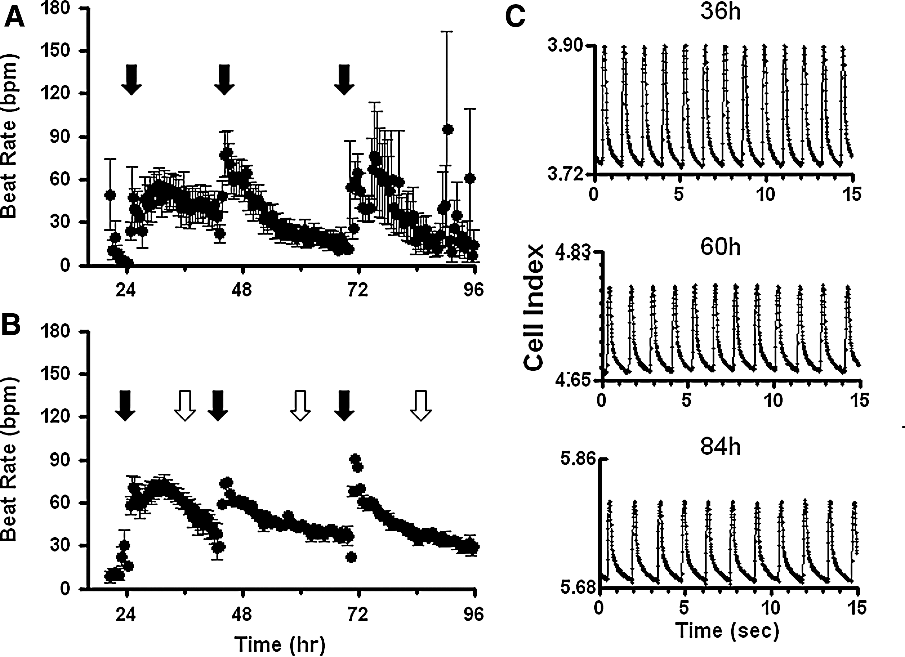
Comparing culture conditions on beat rate properties of rat neonatal cardiomyocytes—an example of how one variable (serum) affected the beat rate. Representative examples of impedance data from cardiomyocytes cultured in DMEM plus,
Cell seeding density was also observed to be a critical variable. Plating more cells decreased the beat rate, but increased amplitude (Fig. 2). Intermediate densities, between 24K and 32K, provided excellent signal and minimized variance for three leading parameters—beat rate, amplitude, and beat rise slope. Conditions were optimized to plate cells at 30,000 per well in 20% serum with media changes at ∼24 and ∼48 h. Compounds were tested in a 4-h window at least 2 h after media change (between 50 and 54 h postplating) (Table 1).
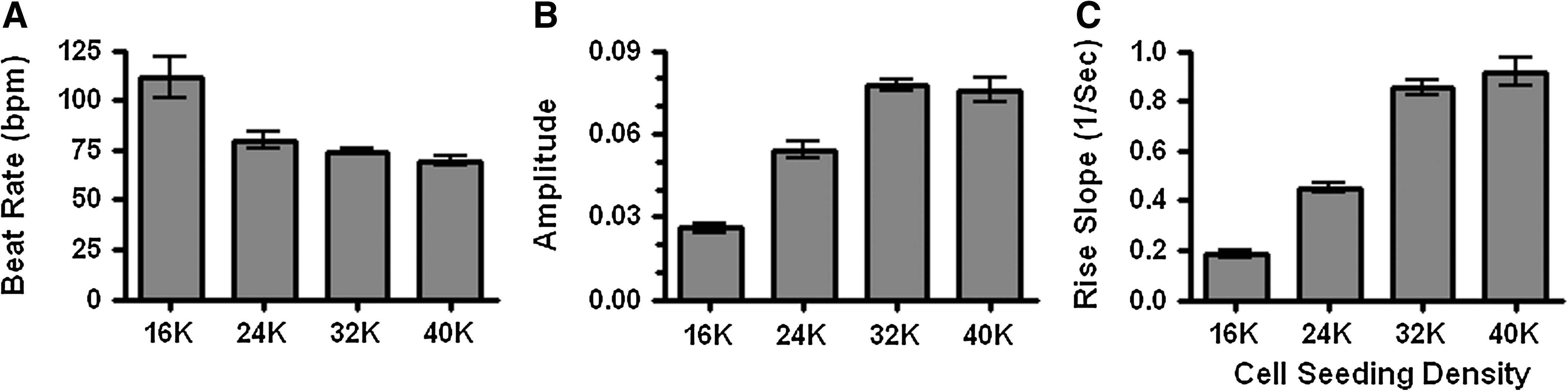
Effects of cell seeding density on cardiomyocyte beat parameters. Beat rate
For compound screening, assay tolerance to DMSO and serum-free conditions are important considerations. At the initial cell plating stage, serum was required with successively higher levels increasing beat rate and stability (data not shown). Attempts to remove serum after cultures were established (48 h) resulted in dramatically reduced beat rate and increased variance (Fig. 3A) consistent with previous findings. 12 DMSO sensitivity was also examined after both short- and long-term exposures. DMSO concentrations at or below 0.65% had minimal effects after a 20-min treatment (Fig. 3B). By contrast, after longer exposure (18 h), effects of DMSO concentrations as low as 0.11% were detectable (Fig. 3B). Thus, for responses longer than 20 min, DMSO-alone controls were particularly important.
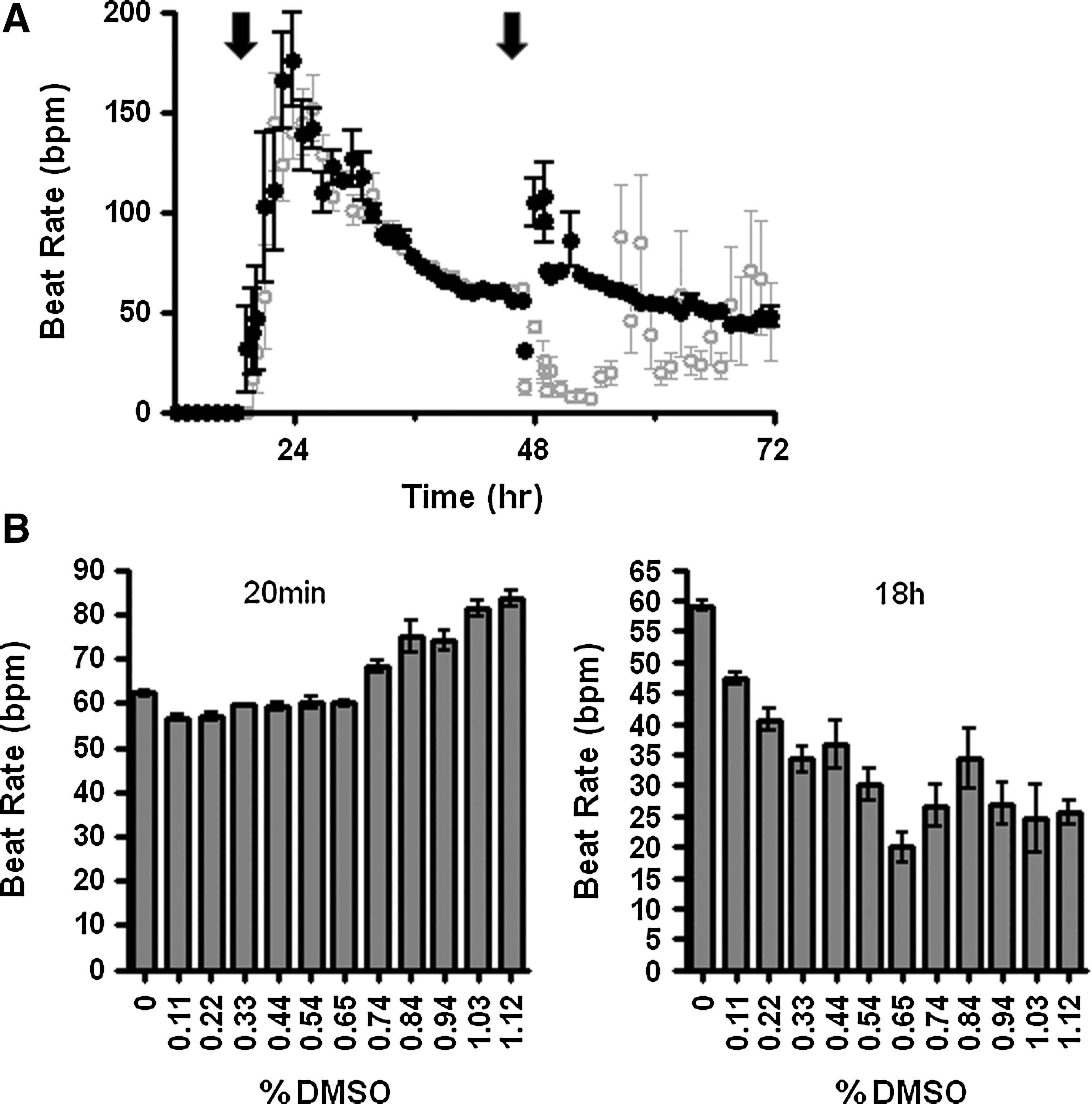
Evaluation of serum and dimethyl sulfoxide (DMSO) effects on beat rate. Stably beating cultures were established in media containing 20% dialyzed FBS.
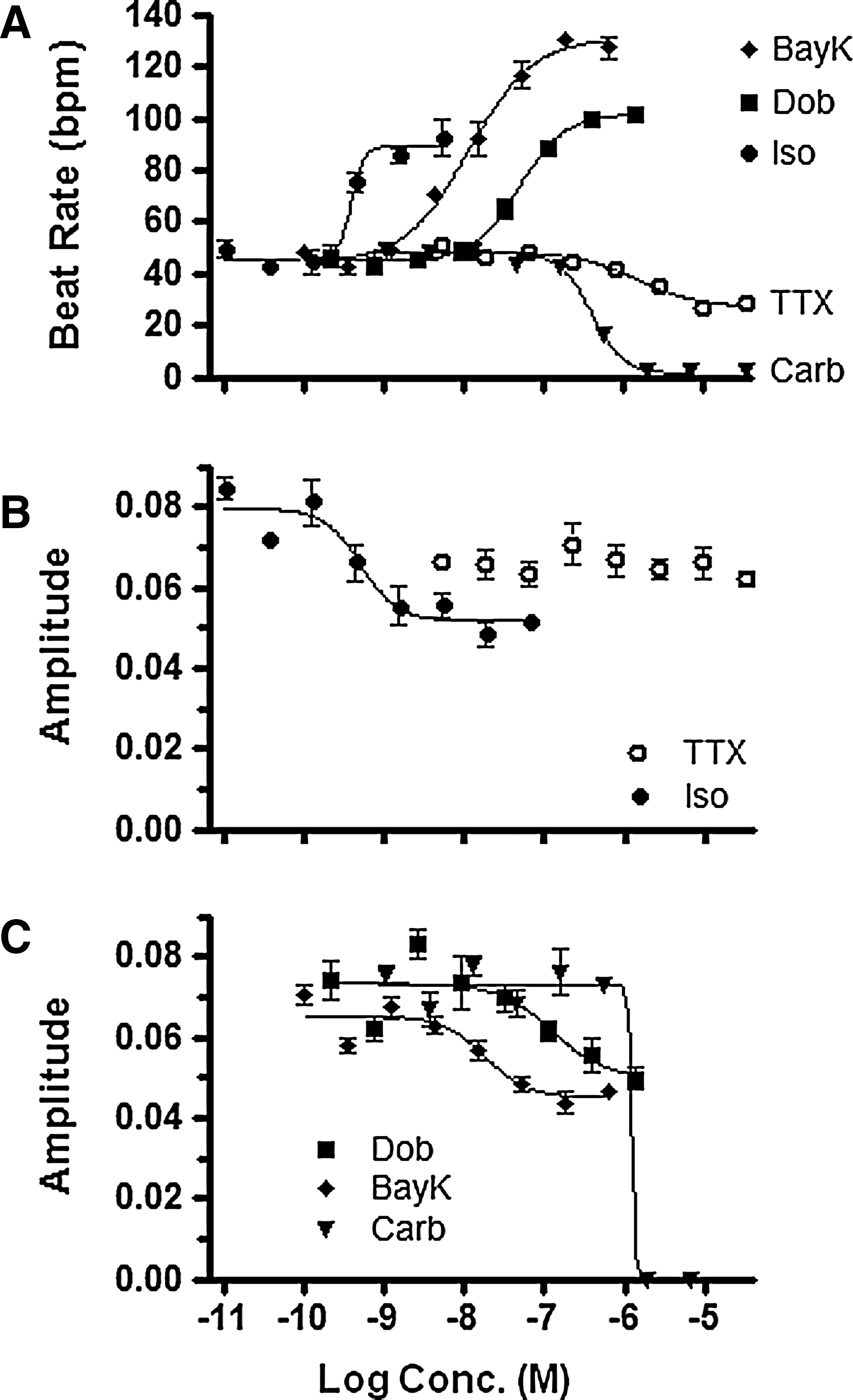
To evaluate the precision and dynamic range of impedance data, five reference compounds were selected representing diverse mechanisms of action, a range of potencies, and covering both positive and negative chronotropic/inotropic actions. Each compound was analyzed for effects on beat rate and amplitude. The data variance within an individual experiment was low for both measurements (Fig. 4). The three positive ionotropic compounds, BayK8644, dobutamine, and isoproterenol, increased beat rate and decreased beat amplitude, with the effects on beat rate being prominent. In contrast, the negative ionotropic agent, carbachol, decreased both beat rate and amplitude. Thus, a reduced beat amplitude can occur with compounds that increase or decrease beat frequency. At higher carbachol concentrations, cessation of beating was observed, but this was not a consequence of toxicity as judged by a lack of change in cellular adenosine-5′-triphosphate content (data not shown). As a third category, tetrodotoxin (TTX) decreased beat rate (EC50=0.8±0.3 μM) without altering amplitude. These particular results agree with findings by Guo et al. 18 using human iPS-derived cardiomyocytes where TTX (3 μM) reduced beat rate in impedance and micro-electrode array assays (49% and 23% of control, respectively), whereas beat amplitude was not significantly changed (105% and 113% of control, respectively). Thus, a comparison of beat and amplitude data can reveal important distinctions that may prove valuable for screening compounds with different mechanisms of action. Consistent with this application, the potency values generated by both beat rate and amplitude analysis showed good precision and reproducibility for this group of compounds (Fig. 5).
Sensitivity and precision in concentration-response mode for compounds spanning a range of potencies with diverse mechanisms of action. Beat rate
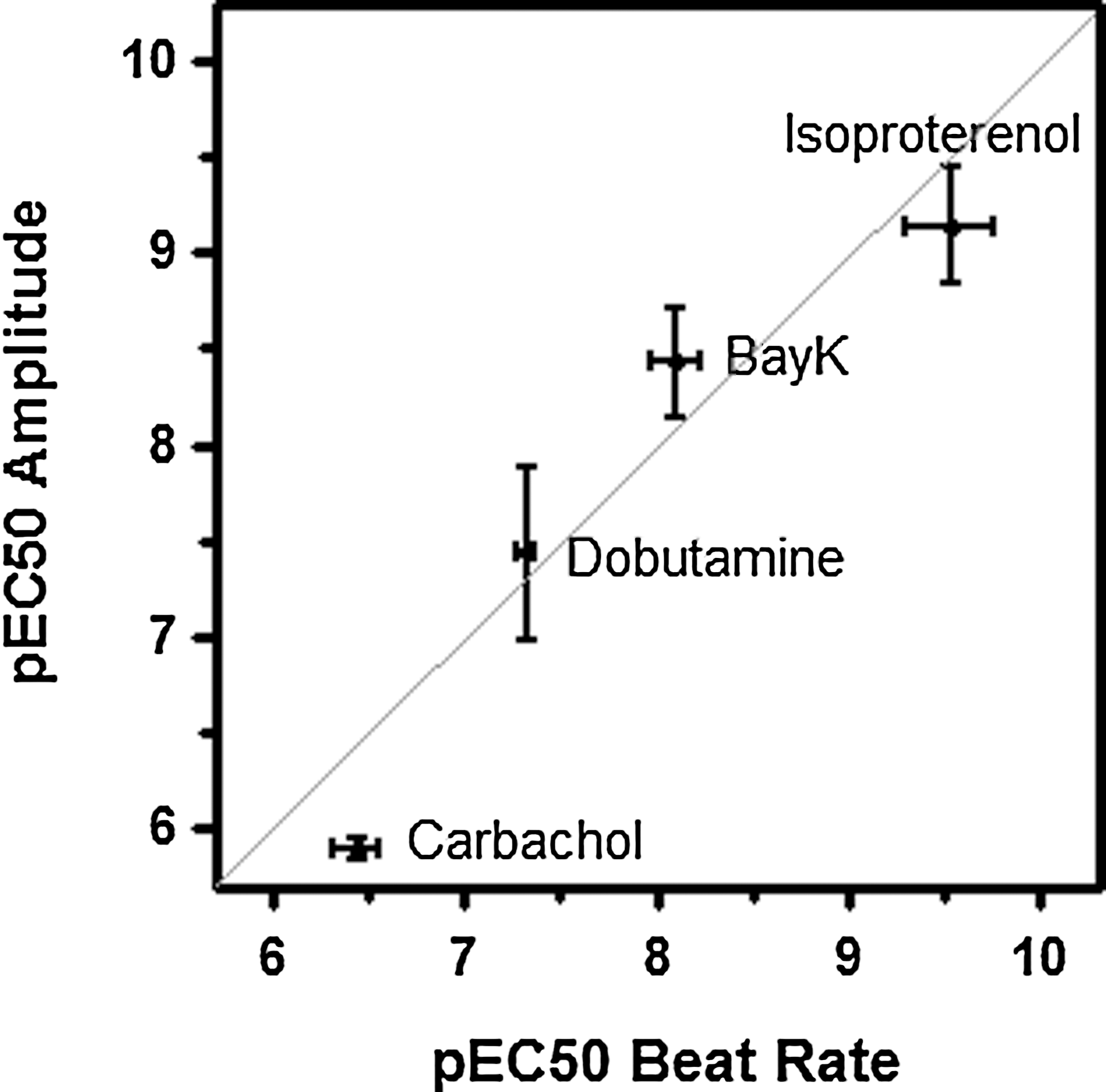
Reproducibility of potency values. Beat rate and amplitude EC50 values were determined for four reference compounds. Results from three experiments were plotted relative to the unity diagonal line. Data are average plus SEM. The correlation was judged to be significant (P<0.05) with a Pearson R 2=0.92.
To compare impedance data with clinical effects, tests were conducted with drugs having established effects on human cardiac contractility. Drugs with positive, negative, and neutral effects were selected (Table 2). Effects on beat rate were evaluated at 20 min after drug addition. The impedance data were concordant with clinical data for 20 of 21 compounds, including 6 of 6 inactives, 5 of 5 positive ionotropes, and 9 of 10 negative ionotropes. The exception, atenolol, is a β1-adrenergic antagonist. The lack of observed activity in cultured cardiomyocytes likely reflects the absence of adrenergic tone in the in vitro assay relative to in vivo.
Evaluation of Drugs with Established Effects on Contractility
Impedance-derived beat rate was evaluated 20 min after drug addition. For values greater than zero, P values were calculated by nonparametric t-test.
Discussion
The application of impedance-based label-free technology to cardiomyocyte biology appears poised to address a range of gaps in cardiovascular toxicity screening. These opportunities are made possible by advances in the technology that push data collection rates (∼0.01 s) to a fraction of the beat frequency of cardiomyocytes thereby generating high-resolution data. Initial evaluations of impedance cardiomyocyte assays focused on the beat profiles and the potential for predicting arrhythmia risks. 14,18 –20 Indeed, impedance data from others clearly distinguished drugs that induce clinical arrhythmia based on beat patterns in human and mouse stem-cell-derived cardiomyocytes. Further, compared with hERG channel and QT prolongation data, impedance data offered improved correlation with clinical torsades de pointes. 18 This suggests that impedance assays in cardiomyocytes from human or appropriate hERG-expressing species may be a valuable supplement to help predict clinical torsades de pointes. 3
Beyond arrhythmia predication, cardiomyocyte impedance assays have the potential for broad application in cardiac biology, pharmacology, and toxicology. In general, the ability to detect spontaneous synchronized contractions provides sensitivity to both mechanical and electrical events, and thus is well suited as a broad, initial screen for contractility and toxicities. To begin testing the scope of this potential, the primary focus of the present study was to investigate screening fundamentals, including assay development, robustness of potency determinations, versatility for extracting different beat-related parameters, and validation with known cardiovascular active drugs with non-hERG mechanisms of action.
Rat neonatal cardiomyocyte cultures were selected as a model system since primary cultures had not previously been tested on the xCELLigence Cardio instrument and would provide a rigorous challenge for evaluating assay development and data consistency. In addition, these cardiomyocyte cultures can be produced in sufficient amounts (i.e., six 96-well plates per rat litter) to enable moderate screening throughput and at a cost for cells (∼$50 per plate) that is substantially less than current commercial sources of stem-cell- and iPS-derived cardiomyocytes ($1,000–$3,000 per plate). Thus, for studies that do not require high hERG expression, rat neonatal cardiomyocyte cultures provide an operational advantage such that, if their biological properties prove suitable, then they could enable more widespread use of label-free methods for early phase drug discovery/safety applications.
For assay development, diverse media types and supplements were selected from published rat neonatal cardiomyocyte studies as well as emerging stem cell protocols. 13 –16 Different cell culture media, plate coatings, and cell seeding densities were explored in a matrix of combinations. Across these various parameter combinations, serum source and concentration proved critical. Serum was required to maintain substantial and stable beating with higher concentrations increasing beat rate (Fig. 3A and Refs. 12,21 ). Dialyzed FBS was included based on previous experience demonstrating improved consistency in impedance experiments 17 and literature findings that variations in serum hormones affect assay quality. 13 Cell seeding density also significantly influenced the data. Plating more cells significantly decreased beat rate, but increased beat amplitude. Although the signal magnitude changed with different cell densities, the data variance was similar at each of the cell densities and across rate, amplitude, and rise slope (Fig. 2). This ability to optimize the data based on cell density has not previously been reported and offers an unexpected opportunity to optimize assay conditions to meet experimental needs. In this case, we chose an intermediate seeding density to balance signal for both beat rate and amplitude data. Overall, we found that impedance assays provided low well-to-well variability in a 96-well format that allowed for evaluation of many assay conditions leading to straight-forward assay development.
For cardiomyocyte impedance assays to have significant impact in drug discovery, the pharmacological data must meet high quality standards required for SAR-driving assays. Ideally, the responses should correlate with clinical data and the potency values should have sufficient precision and reproducibility to discriminate a half-log difference in potency values. To begin assessing these characteristics, reference compounds were selected to span a range of potencies and different mechanisms of action. Within individual experiments, the precision was excellent across a wide dynamic range (Fig. 4). Resulting potency values were highly reproducible across multiple experiments (Fig. 5). Drugs with known positive and negative chronotropic/inotropic effects yielded impedance data concordant with clinical findings for most drugs (20 out of 21; Table 2). The exception, atenolol, is a β1-adrenergic antagonist that reduces contractility in humans, but was inactive in the impedance assay, presumably due to a lack of tonic adrenergic activity in vitro. Together, the data demonstrate high concordance with clinical data with the caveats common to in vitro screens.
A hallmark of existing whole-cell label-free platforms has been versatility. 5 –10 Multiple assay parameters, a range of kinetic options supported by continuous monitoring, and compatibility with diverse cell types all combine to allow tailoring experiments for increased relevance to biology and disease. 22 This flexibility is captured in the cardiomyocyte platform where assay parameters include beat rate, normalized beat rate, amplitude, normalized amplitude, rise time, rise slope, fall time, fall slope, half-max beat duration, beat pattern similarity, beat rhythm irregularity, and CI. In this report, we focused on beat rate and amplitude of spontaneously beating, neonatal rat cardiomyocyte cultures. Surprisingly, the option to normalize responses to the baseline for each individual well did not reduce data variance in the present experiments and so raw data were evaluated. A comparison of potency values derived from beat rate versus amplitude data revealed similar EC50 values and variance for most compounds (4 out of 5). The exception, TTX, had effects on beat rate, but not on amplitude, which suggests that these parameters may be useful for distinguishing mechanism of action. In addition to the diversity of parameters, impedance assays also offer options for assessing differences in response kinetics based on continuous, nondestructive monitoring. In the present study, end-point reads at 20 min postdrug addition were sufficient to detect positive and negative chronotropic/inotropic acting compounds (Table 2). Finally, the system is highly flexible for detection of endogenous receptor responses in primary cultures or stem cells thereby allowing comparison of different species and genetic manipulations. This combination of diverse and robust parameters in a label-free format provides the flexibility to accumulate data that may be relevant to specific disease states.
A challenge common to each of the whole-cell label-free platforms is extracting the maximal benefit from the rich data sets that are generated. 23 For example, in other impedance platforms, tracking the kinetics of receptor responses offers novel ability to decipher pleiotropic signaling and potentially biased agonism. 24 However, the notion of shifting from single end-point analysis to dynamic response profiles carries a daunting increase in complexity as demonstrated by Abassi and colleagues. 25 This is further magnified by the array of parameters available in cardiomyocyte impedance assays. Future versions of the analysis package could be improved with built-in multiparametric analysis or by developing links to analysis packages similar to those used for gene expression profiling. These modifications in the analysis software are needed to efficiently extract more information from these rich data sets without detracting from data delivery.
An opportunity to expand future applications exists with the hardware as well. All studies to date have quantified the beating profiles of spontaneously beating cardiomyocyte cultures (present study and Refs. 14,18 –20 ). Future embodiments of this technology could incorporate field potential stimulation to pace spontaneously beating cultures or initiate beating in adult cardiomyocyte cultures. Adult cardiomyocytes may bring additional drug discovery translational value compared with isolated neonatal cardiomyocytes and iPS cells differentiated to yield a cardiomyocyte phenotype in culture.
Overall, the data presented here indicate that cardiomyocyte impedance assays yield exceptionally robust data in a versatile assay format. Primary cultures of cardiomyocytes that form spontaneous and synchronous beating monolayers are readily adapted to this higher throughput platform. Thus, cardiomyocyte impedance assays are likely to substantially expand the capacity for in vitro cardiovascular screening in drug discovery.
Footnotes
Acknowledgments
The authors thank Christine Fernandes for dissecting neonatal rat hearts and Lois Ann Lazor for comments on the article.
Disclosure Statement
The authors are employees of AstraZeneca Pharmaceuticals.