
Abstract
Multidrug-resistant bacterial infections are an increasing source of healthcare problems, and the research for new antibiotics is currently unable to respond to this challenge. In this work, we present a screening strategy that integrates cell-based high-throughput screening (HTS) with in silico analogue search for antimicrobial small-molecule drug discovery. We performed an HTS on a diverse chemical library by using an assay based on a bioluminescent Escherichia coli K-12 (pTetLux1) strain. The HTS yielded eight hit compounds with >50% inhibition. These hits were then used for structural similarity-based virtual screening, and of the 29 analogues selected for in vitro testing, four compounds displayed potential activity in the pTetLux1 assay. The 11 most active compounds from combined HTS and analogue search were further assessed for antimicrobial activity against clinically important strains of E. coli and Staphylococcus aureus and for in vitro cytotoxicity against human cells. Three of the compounds displayed antibacterial activity and low human cell cytotoxicity. Additionally, two compounds of the set fully inhibited S. aureus growth after 24 h, but also exhibited human cell cytotoxicity in vitro.
Introduction
The development of resistance toward antibiotics is a topic of growing concern. The situation is constantly getting worse due to the emergence of multidrug-resistant bacterial strains. Among gram-negative Enterobacteriaceae, the extended-spectrum beta-lactamase -producing Escherichia coli strains are prone to multidrug resistance. Of gram-positive bacteria, hospital- and community-acquired methicillin-resistant Staphylococcus aureus is also a major concern. 1 Nevertheless, the antibacterial drug discovery efforts are still lagging behind. 2
High-throughput screening (HTS) is one of the initial methods for hit finding in small-molecule drug discovery. 3 It is based on testing a large amount of compounds, using either biochemical or cell-based biological assays relevant to the investigated therapeutic target. The size of the compound libraries screened can vary from thousands to as large as millions of compounds. 4,5 HTS assays are often run in singlicate, yielding hit rates between 0.01% and 1%. 6 HTS campaigns can thus become very expensive and time-consuming to run on extensive libraries.
There has been a growing interest in computational drug discovery methods over the past decades. Drug discovery has evolved to using HTS and virtual screening techniques side by side and, as a combination, they complement each other in the quest for novel active molecules. 7 Computational methods are often used to lower the costs of experimental approaches, for example, by reducing the size of the library to be screened by computationally selecting a subset of diverse molecules. 6 –8 Integrated in silico screening, which is used to expand the hit list for compounds with similar structures and to explore additional compound libraries, 6 has also become a successful concept in recent years. 9,10 Virtual, or in silico screening, can be divided into either structure-based or ligand-based methods. Structure-based methods are carried out on targets with known structures, while in ligand-based virtual screening, similarity searching of analogues based on the structures of bioactive compounds is employed. 11,12
A large number of HTS hits do not, however, always translate into successful lead compounds for drug development. A shift toward using cell-based and phenotypic approaches at the initial screening phase is a current trend in drug discovery strategies, aiming for an improved success rate. 13 Choosing a predictive assay can increase the probability of an HTS to produce clinically significant hits. Indeed, phenotypic approaches have a better success rate than target-based strategies in discovering first-in-class medicines according to a recent analysis. 14 Cell-based assays do not rely on a single specific molecular target and thus can yield diverse hits acting on different targets. In addition, cell-based assays take into account factors, such as membrane permeability, that may affect later drug developmental stages. 15
We have previously set up an antimicrobial HTS assay based on a bioluminescent E. coli strain K-12 (pTetLux1) in a 384-well format and validated the assay by screening a library containing known antibacterials and other bioactive compounds. 16 Briefly, the recombinant bacterial strain carries a plasmid with an inducible luciferase operon (luxCDABE) containing the genes necessary for light production. Antibacterial compounds, primarily affecting transcriptional or translational events, can be detected by an inhibition of light production. In this work, we used the developed assay to screen a diverse library containing 10,240 compounds with unknown biological activity. We then followed up the HTS results with in silico screening as a strategy to enhance the discovery of antibacterial compounds.
Materials and Methods
HTS Compound Library and Chemicals
A chemically diverse library containing 10,240 compounds was acquired from the Institute for Molecular Medicine Finland (FIMM) through the Drug Discovery and Chemical Biology Network. The compounds were preplated at FIMM in dimethyl sulfoxide (DMSO) at 30 nL volumes into 384-well plates using an acoustic liquid handling system (Echo®; Labcyte, Inc., Sunnyvale, CA). Seven HTS hits as well as additional 29 compounds selected by in silico screening were purchased from the original supplier (ChemBridge Corporation, San Diego, CA) and dissolved in DMSO to obtain 30 mM stock solutions. Antibiotics, erythromycin and rifampicin, used as positive controls in the assay were acquired from Sigma-Aldrich (St. Louis, MO). Ampicillin sodium salt was obtained from AppliChem (Darmstadt, Germany) and tetracycline hydrochloride from CalBiochem (La Jolla, CA).
Maintenance and Subculture of Bacteria
The recombinant E. coli K-12 strain was stored and lyophilized in vials. For preparation of liquid cultures, sterile milli-Q (MQ) water was added to the vial (1 mL). After 30 min, 50 μL of the suspension was used to inoculate a 5 mL culture in LB medium (Lennox; BD, Difco™, Sparks, MD) containing ampicillin as the selective agent (100 μg/mL). The culture was incubated overnight at 37°C, 100 rpm and used for preparing a bacterial dilution for the screening assay. Wild-type strains of E. coli (ATCC 25922) and S. aureus (ATCC 25923) were obtained from Microbiologics, Inc., (St. Cloud, MN) and used in the broth microdilution assay. Weekly subcultured slant cultures were grown at 37°C, from which an inoculum was used for starting suspension culture in either LB medium (E. coli) or MHB (S. aureus). Suspension cultures were incubated for 16–24 h at 37°C, 100 rpm.
Antimicrobial Assays
High-throughput screening
An assay protocol with preplated compounds and automated liquid handling using a Biomek FX station (Beckman Coulter, Fullerton CA) was used in the primary HTS of the compound library. Compounds were screened in singlicate in 384-well plates (320 compounds/plate) at a final concentration of 10 μM (0.1% DMSO). Each assay plate contained maximum signal wells (n=16, DMSO with induced bacteria) and background signal wells (n=16, DMSO with noninduced bacteria) for assay performance monitoring. The plates also included the positive control antibiotics, erythromycin (50 and 25 μg/mL) and rifampicin (5 and 2.5 μg/mL). The screening was performed using the same miniaturized protocol (30 μL well volume), plate setup, and liquid handling as previously reported—described in detail in Table 1. 16
High-Throughput Screening Protocol
1. Transfer by Echo into 384-well plates (CulturePlate-384, white; PerkinElmer, Inc., Waltham, MA). Plate layout: columns 1–2 (min wells and positive control erythromycin, columns 3–22: test compounds, columns 23–24: max wells and control rifampicin)
2–5. Performed using the Biomek FX station. Tet-HCl final well concentration 100 ng/mL.
6. Transfer plate to an external incubator, careful minimal shaking can be applied.
CFU, colony forming unit; DMSO, dimethyl sulfoxide.
Hit confirmation, primary testing of analogues, and dose–response experiments with E. coli (pTetlux1) assay
Compounds chosen for follow-up evaluation (including in silico selected analogues) were first retested manually with the same bioluminescent assay in 384-well plates at the same concentration as in the primary screen (10 μM, 0.33% DMSO, n=8). Compound stocks were diluted in DMSO (3 mM) and further diluted 1:100 in LB medium. Compound dilution in LB medium was plated (10 μL), followed by a dilution (10 μL) of E. coli (pTetLux1), at 5×10⁴ CFU per well. Bioluminescence was induced by tetracycline-HCl (10 μL, 100 ng/mL). Second, concentration–response assessment for the compounds in the bioluminescent assay was also carried out with the same setup. Inhibition percentages and the assay quality parameters were calculated as previously described. 16 Dose–response results were plotted in Origin software (OriginLab Corp., Northampton, MA), and IC50 (50% inhibitory concentration) values were calculated by fitting the data into sigmoidal dose–response curves using a logistic model where weight was given to error bars.
Absorbance-based growth inhibitory assay
From a suspension culture, a dilution in LB medium (MHB used for S. aureus) was prepared for the assay. Absorbance-based broth microdilution assays using the wild-type strains were performed in sterile, clear 96-well plates (Nunc A/S; Thermo Fischer Scientific, Hvidovre, Denmark). Erythromycin was used as positive controls in the assays at the following minimum inhibitory concentrations: 64 μg/mL (E. coli) and 0.5 μg/mL (S. aureus). Compounds were tested at three final concentrations (100, 50, and 10 μM) with a final DMSO concentration of 0.5%. Each concentration was tested in triplicates. Samples were first prepared into the assay medium and then plated at 100 μL per well. Thereafter, bacterial dilution in LB medium (100 μL) was added to the wells. The final bacterial inoculum was 5×10⁴ CFU per well (2.5×10⁵ CFU/mL). Plates were incubated at 37°C, at 250 rpm, and the absorbance at 620 nm was measured with Victor 2 (Wallac; PerkinElmer, Turku, Finland) at time points 0, 4, 7 h (S. aureus), and 24 h. Due to the slower growth rate of S. aureus, the measurement time point used was 7 h in the logarithmic phase. The percent inhibition of growth was calculated with the following formula: (1−Abstest/Absmax)×100, where Abstest is the absorbance of a test well and Absmax is the absorbance of the maximum growth control. The absorbance values at each time point were subtracted with background values at time 0 h.
Solubility Assay
The solubility in LB medium was assessed and analyzed using a protocol modified from Yestrepsky et al. 17 Each compound was diluted into suitable concentrations in DMSO and then further diluted 1:100 into LB medium correspondingly to the antimicrobial assays. The dilutions were plated (200 μL/well) in triplicates into a clear 96-well plate. The plate was incubated at 37°C for 5 min and thereafter absorbance was measured with the Multiskan GO plate reader (Thermo Fisher Scientific, Vantaa, Finland) at 620 nm. Compounds were tested at eight concentrations, ranging from 10 to 200 μM. The absorbance value for each compound sample was subtracted by the background value of the LB medium. The solubility values were determined by taking the mean of the first concentration that showed ≥0.005 difference from the background and of the next lower concentration. The error was expressed as the concentration difference×0.5.
Cytotoxicity Against Human Cells
Cell culture
Huh-7 cells (derived from human hepatocellular carcinoma) were a kind gift from Prof. Ralf Bartenschlager (University of Heidelberg, Germany). Cells were maintained in Dulbecco's modified Eagle's medium, supplemented with 10% fetal bovine serum (FBS; Gibco, Grand Island, NY), 100 μM nonessential amino acids, 2 mM
Cytotoxicity assay
The effect of most active compounds on the metabolic activity of human hepatocyte cells (Huh-7) was assessed by measuring the intracellular ATP content after compound exposure with Promega's CellTiter-Glo Cell Viability Assay (Madison, WI). In brief, cells were seeded at 2×10⁴ cells per well (200 μL) on white-walled 96-well microplates (ViewPlate; PerkinElmer, Inc., Waltham, MA), incubated at 37°C, 5% CO2 and 95% humidity overnight, and then exposed for 24 h to the compounds diluted into assay media (5% FBS). Following the exposure, cells were washed with 100 μL of phosphate-buffered saline (PBS), and 50 μL of fresh assay media as well as 50 μL of CellTiter-Glo reagent were added to each well. After 2 min of shaking, followed by 10 min of incubation at room temperature, the luminometric signal was measured by using the Varioskan Flash plate reader (Thermo Fisher Scientific). Polymyxin-B sulfate was used as a positive control (15,000 IU/mL, average cytotoxicity 86%). Compounds were initially screened at 10 and 100 μM (n=3) concentrations, and the most cytotoxic ones were further subjected to dose–response experiments (concentration ranging between 0.25 and 200 μM, depending on the potency of the compound). IC50 values were calculated by fitting the data into sigmoidal dose–response curves using a logistic model in Origin software (OriginLab Corp.).
Computational Methods
Library preparation
A virtual compound library (119,027 compounds) from the Institute for Molecular Medicine Finland (FIMM library version 2013) was chosen for virtual screening; compounds from this library are easily accessible to us for experimental testing. All compounds in the database were first converted from 2D (sdf format) to 3D using the Concord module in Sybyl-X, and one representative 3D conformation (lowest energy) was selected. Then, for all of these representative conformations, Gasteiger–Huckel charges were assigned and the compounds' energy minimized using the standard Tripos force field (Powell method and 0.05 kcal/[mol·Å] energy gradient convergence criteria). In addition, the ZINC database was also used for virtual screening. ZINC is a free database of commercially available compounds for virtual screening, which contains over 35 million purchasable compounds in ready to be used 3D formats.
Ligand-Based Virtual Screening
The maximum common substructure (MCS) shared by seven of the eight most active compounds (with % inhibition higher than 50% in primary screening) was first derived (Fig. 1). This MCS identification was done using Distill implemented in Sybyl-X v1.2 software (Tripos International, St. Louis, MO). Distill classifies compounds by hierarchical clustering based on their common substructure, where each node represents a substructure and the set of compounds containing that substructure. The set of active compounds was also used to develop two 3D pharmacophore models that describe the chemical features most important for activity using common feature pharmacophore generation from the Discovery Studio 4.0 platform (Accelrys, Inc., San Diego, CA). 12 Pharmacophore 1 (Fig. 2A) is constituted by hydrophobic, H-bond donor, and aromatic features, whereas pharmacophore 2 (Fig. 2B) contains hydrophobic, aromatic, and H-bond acceptor features.
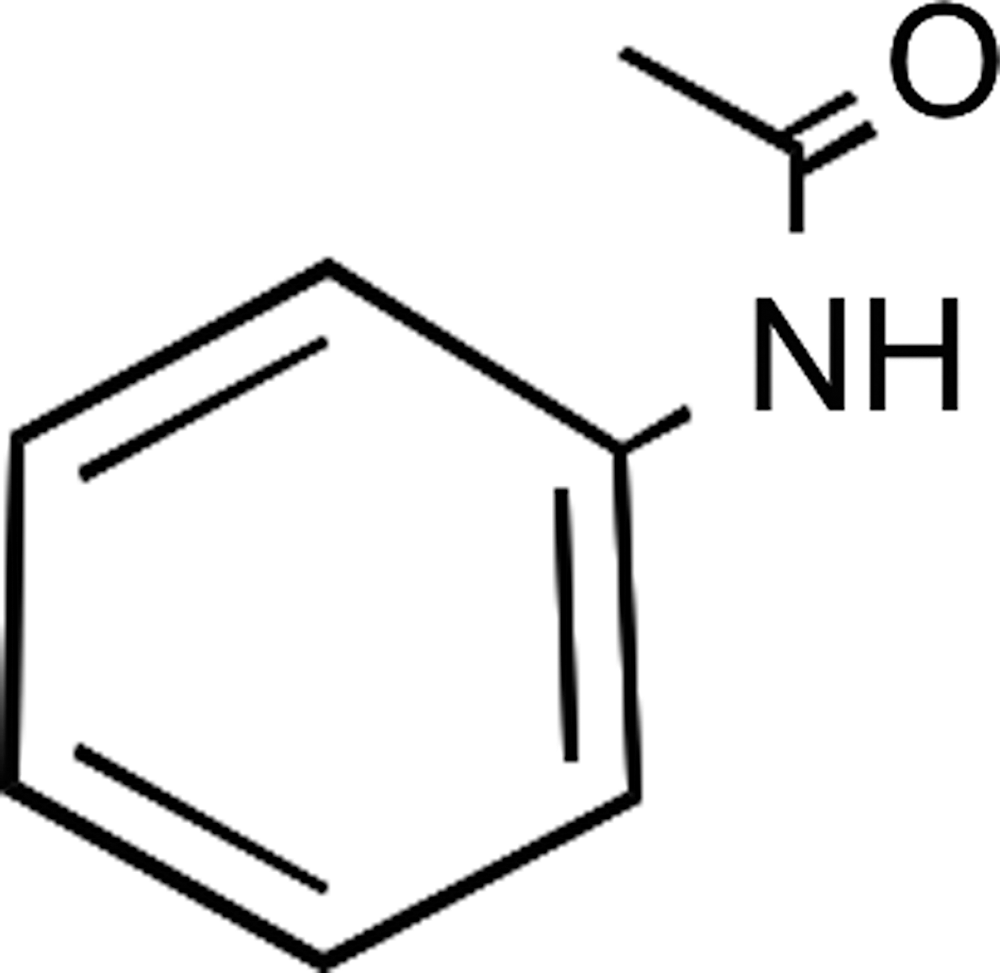
Maximum common structure shared by seven of the eight most active compounds from primary high-throughput screening (HTS).
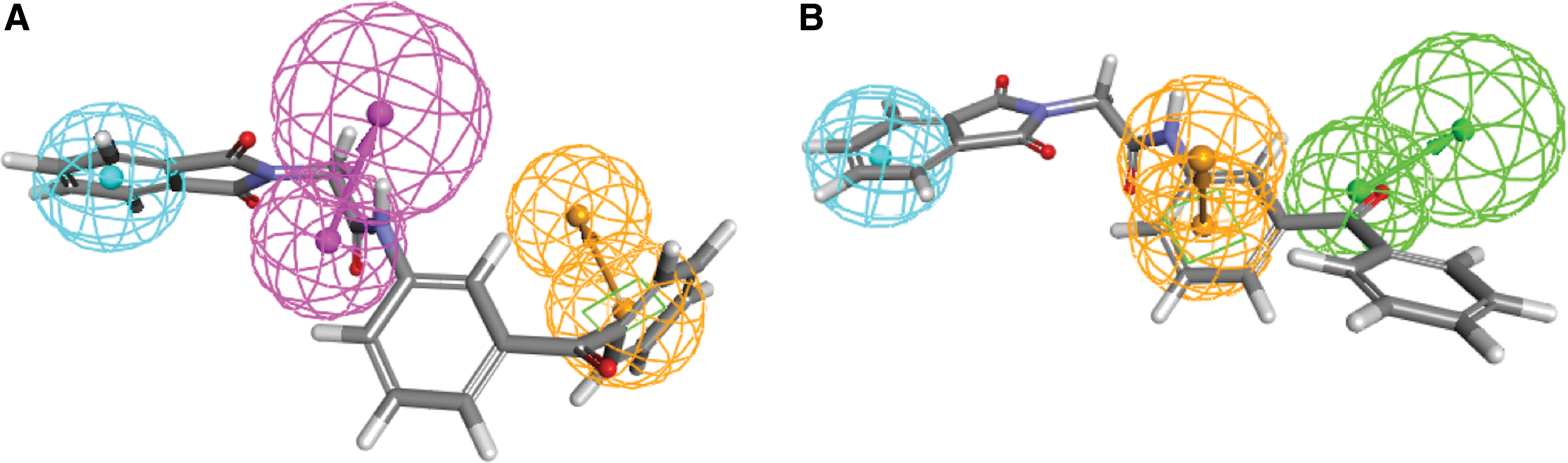
The two pharmacophore models used for virtual screening, pharmacophore 1
Database screening and compound selection by using, in parallel, MCS and 3D pharmacophore models were carried out on a Linux Pentium (2 CPUs). For each of the seven most active compounds containing the MCS, five compounds with the highest Tanimoto coefficients were selected from the FIMM library. Second, pharmacophore models were used as 3D queries. The results were ranked according to the fitting values of each compound to the pharmacophore. For pharmacophore 1, 25 compounds were retrieved, 20 from the ZINC database and 5 from the FIMM library; for pharmacophore 2, 5 compounds were selected from the FIMM library. Thus, a list of 65 prospective molecules was set up based on these two approaches. Of these, based on visual analysis by a medicinal chemist and on availability, a subset of 29 compounds was selected (Supplementary Table S2; Supplementary Data are available online at
Results and Discussion
HTS Assay, Virtual Screening, and Hit Confirmations
HTS was first carried out at 10 μM on a set of 10,240 compounds from the ChemBridge DIVERset library using the bioluminescent E. coli K-12 (pTetLux1) assay. The performance of the screen was very good; the average and standard error of the mean values for assay quality factors from 32 plates were: Z′=0.7±0.03, S/B=121.5±2.0, and S/N=14.1±2.1. The assay quality parameters using this assay in the larger scale screen are well in line with the values obtained from the previous assay validation screen with known bioactives. 16
With an HTS hit cutoff >50% inhibition, eight compounds were identified as active, resulting in a hit rate of 0.08% (compounds
After the initial HTS, a structural similarity and 3D pharmacophore search was performed based on the eight most active compounds from the primary experimental screen, aiming to identify new potential hits and to expand the set of compounds to be tested in the next round (see Materials and Methods section). The hit confirmation and expansion phase consisted of (i) confirming the activity of the repurchased seven HTS actives and (ii) primary in vitro testing of the 29 in silico selected structural analogues using the same assay and concentration as in the primary HTS (results for all 36 compounds tested are provided in Supplementary Table S2).
Six of the seven original HTS actives showed inhibitions that were either higher than or relatively close to the HTS values (Fig. 3). Only compound

Comparison of antibacterial HTS versus hit confirmation results (compounds
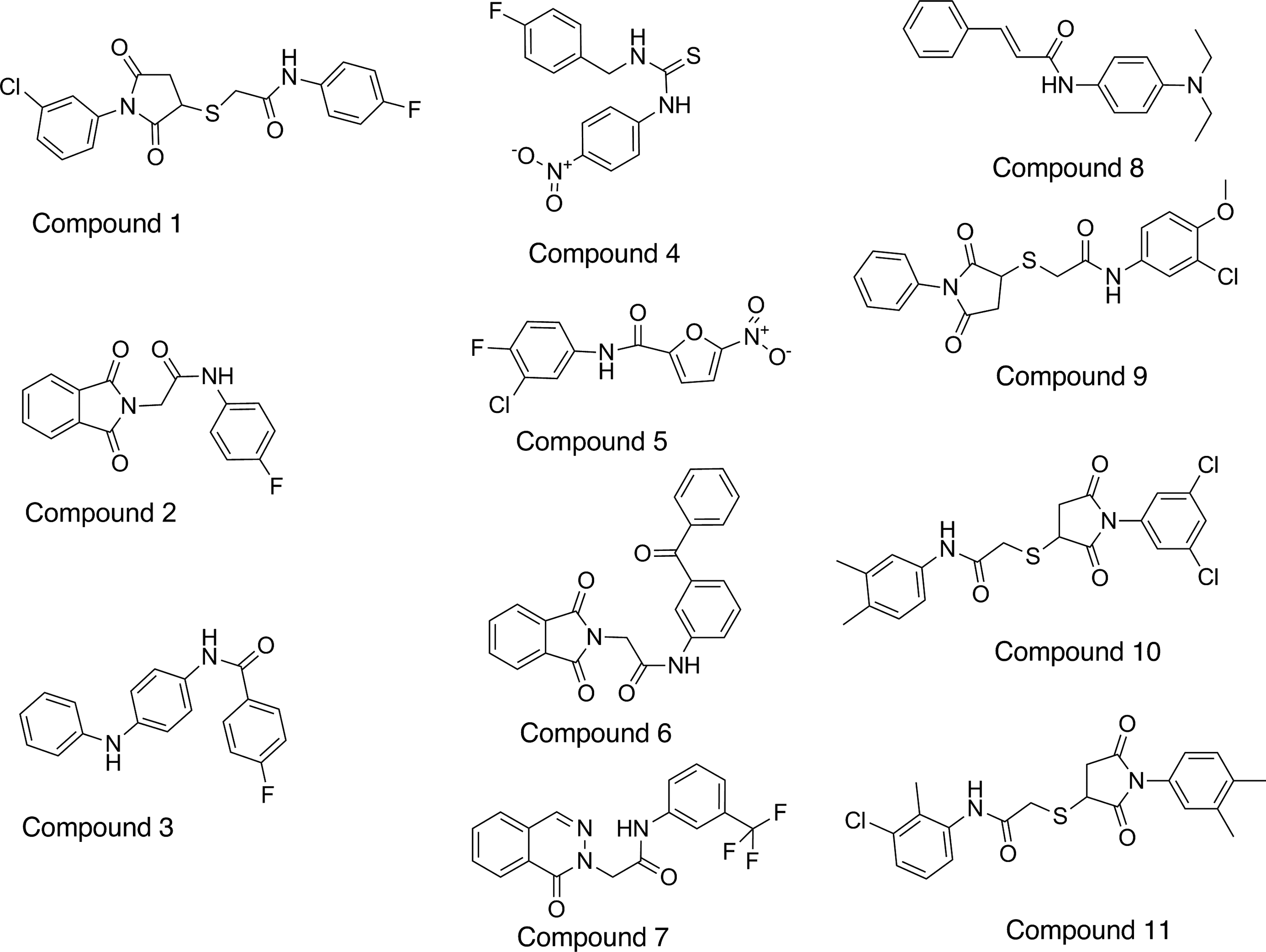
Chemical structures of 11 compounds selected for follow-up studies by using the E. coli (pTetLux1) bioluminescence assay. Compounds
Summary of Escherichia coli (pTetLux1) IC50 Values, Solubility, and Mammalian Cell Cytotoxicity In Vitro
IC50 (50% inhibitory concentration) calculated based on data from antibacterial bioluminescent assay in a 384-well format.
Compound solubility in assay medium (LB).
Cytotoxicity toward Huh-7 cells (%±SD (n=3) at 100 μM).
SD, standard deviation.
Activity on Standard Bacterial Strains
Furthermore, the inhibitory activity of the 11 compounds on bacterial growth was determined with an absorbance-based broth microdilution assay using ATCC strains of E. coli and S. aureus. These strains are routinely used in clinical antimicrobial testing. The results for antibacterial growth inhibitory activity against the standard strains are shown in Figure 5.
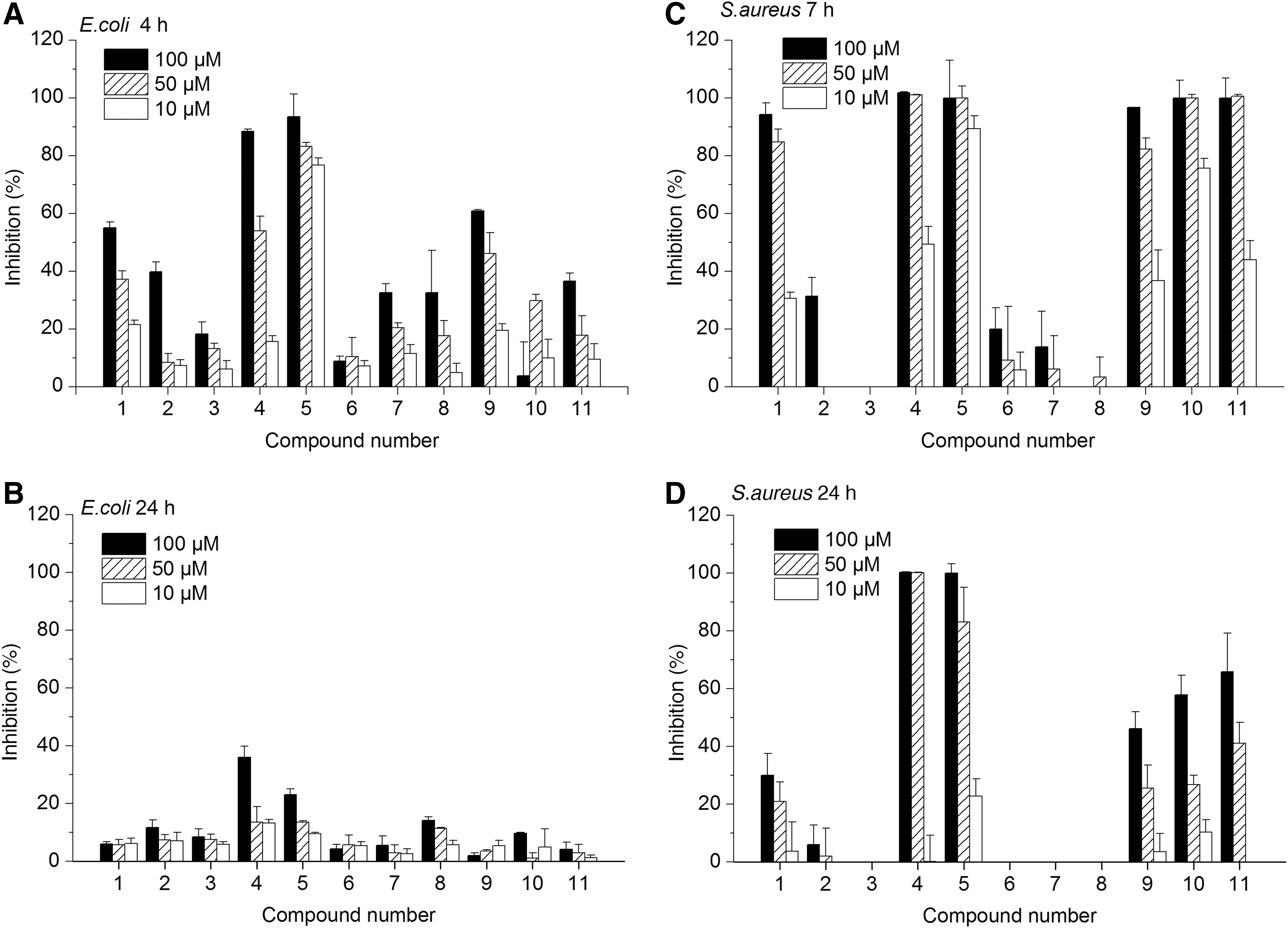
Antibacterial growth inhibitory activity on wild-type bacterial strains.
Four compounds inhibited E. coli growth over 50% in the logarithmic growth phase (at 4 h time point) at 100 μM (Fig. 5A). However, only compounds
Overall, the compounds did not show as high growth inhibitory activity against wild-type bacterial strains as was expected based on their low IC50 values from the E. coli K-12 (pTetLux1) assay (Table 2). For example, compounds
Follow-Up Characterization of HTS and In Silico Selected Hits
While preparing compound dilutions for the growth inhibitory assay, visual observation of the samples revealed that limited solubility of some of the compounds could be a reason behind their inactivity. Therefore, an assessment of compound solubility in assay medium was performed; the results are shown in Table 2. As reported earlier, compound solubility is best performed in the biological assay media rather than in pure aqueous solution because the solubility of a compound is affected by its environment, for example, by the composition of the buffer, temperature, and DMSO content. 18
Only compounds
Furthermore, it is well known that luciferase-based assays may identify false positives due to inhibition of the reporter protein (luciferase enzyme) and this is a possible caveat in our assay as well. This highlights the need to confirm the HTS actives as early as possible by using orthogonal assays, such as the broth microdilution assays used in our approach. Eukaryotic luciferases, firefly luciferase in particular, have been shown to be prone to interference, and chemotypes associated with inhibition of firefly luciferase have been defined. 19 However, this information cannot be applied for evaluating inhibition of other luciferases. For example, the substrates of firefly and prokaryotic luciferases are structurally very different. Only scarce information is available on inhibitors of prokaryotic luciferases, such as the luciferase originating from Photorhabdus luminescens, which is used in our assay. In addition to an orthogonal assay, a biochemical assay that would specifically evaluate reporter interference would be a valuable tool for the follow-up characterization. As we could not find a commercial source for luciferase from P. luminescens, we were not able to evaluate the direct reporter interference.
Differences in the activity between the growth inhibition and the pTetLux1 screening assay may also be due to the fact that the compounds are evaluated in the assays at very different bacterial growth phases (early logarithmic vs. stationary). Due to the specific characteristics of the pTetlux1 strain, the HTS assay identifies mostly compounds affecting transcriptional and translational events, which are most active in the logarithmic growth phase (assay endpoint 90 min). In broth microdilution assays, the first reliable measurement points are at 4–7 h due to the insensitivity of the absorbance-based detection and, typically, the endpoint is set at 24 h. As seen in Figure 5, with both wild-type strains, the growth inhibition by some of the compounds was observed in the logarithmic phase, but not anymore at the stationary phase endpoint 24 h. Thus, the effect seems to be bacteriostatic, affecting a target that slows down the growth of the bacteria, but does not kill them completely. Several studies suggest that the effect of some antibiotics is dependent on the microbial growth rate. 20 It has also been shown that at later phases of E. coli growth, nondividing cells in culture develop into persister cells, which are more resistant to antibiotics. 21,22 On the other hand, bacteria are known to express several efflux pumps, which enable them to transport xenobiotics out from the cell before they reach their targets. Gram-negative bacteria are particularly notorious in this respect; AcrAB-TolC, the main efflux pump of E. coli has been shown to extrude a large variety of unrelated compounds with very different structures (including antibiotics, dyes, and detergents). 23 The broad substrate specificity of AcrAB-TolC together with the effectiveness of the outer membrane as an entrance barrier makes the discovery of new antibacterials against gram-negative bacteria extremely difficult. It is possible that the inactivity of our HTS hits in broth microdilution assays at 24 h was caused by efflux pump activity.
Cytotoxicity experiments on human cells were also carried out to evaluate whether the compounds' activities are selective toward bacterial cells. Initially, the cytotoxicity of the actives was tested at the highest concentration used in the growth inhibitory assay (100 μM). By considering the bacterial growth inhibitory activity (Fig. 5), the preliminary solubility, and human cell cytotoxicity results (Table 2), compounds showing potentially selective antimicrobial activity are compounds
Novelty of Discovered Compounds
We searched the compound structures identified in this work both by 2D similarity and the literature. Two-dimensional similarity searches were carried for the six most interesting compounds (
Promiscuity, that is, nonspecificity of the compound's activity, may also be assessed by calculating a so-called promiscuity index (PCIdx) based on its PubChem activity profile.
27
For 8 of the 11 hits, the PubChem included only 0–21 bioassay results, and calculating the PCIdx was not considered reasonable. For compounds
A compound similar to number
Compound
Conclusions
In this study, an integrated approach utilizing a cell-based bioluminescent HTS assay in combination with virtual screening was employed to improve the efficiency of the antimicrobial screening process. The compounds identified in this work, HTS hits and in silico selected compounds, were evaluated in a comprehensive manner with a set of follow-up studies, such as activity assays on clinically important bacterial strains and a cytotoxicity assay on a human cell line. Two of the HTS hit compounds (compounds
Footnotes
Acknowledgments
This work was supported by the research funds of University of Helsinki (S.N., P.T.) and the Academy of Finland (P.T.). S.N. and M.K. are also grateful for personal grants from the Finnish Cultural Foundation (Suomen Kulttuurirahasto) and the Swedish Cultural Foundation in Finland (Svenska Kulturfonden). D.N. and L.G. gratefully acknowledge the support of the Drug Discovery and Chemical Biology Network of Finland and Centre of Drug Research of University of Helsinki. The authors are also grateful to Krister Wennerberg, Laura Turunen, and Ida Lindenschmidt at the Institute for Molecular Medicine Finland for plating the compounds for the initial HTS (supported by Drug Discovery and Chemical Biology Network, Biocenter Finland) and for the help with compound lists. The CSC− IT Center for Science Ltd., is thanked for organizing computational resources.
Disclosure Statement
No competing financial interests exist.