




Abstract
Immortalized cells, generated from two-dimensional cell culture techniques, are widely used in compound screening, lead optimization, and drug candidate selection. However, such cells lack many characteristics of cells in vivo. This could account for the high failure rates of lead candidates in clinical evaluation. New approaches from cell biology, materials science, and bioengineering are increasing the utility of three-dimensional (3D) culture. These approaches have become more compatible with automation and, thus, provide more physiologically relevant cells for high-throughput/high-content screening, notably in oncology drug discovery. Techniques range from simple 3D spheroids, comprising one or more cell types, to complex multitissue organoids cultured in extracellular matrix gels or microfabricated chips. Furthermore, each approach can be applied to stem cells, such as induced pluripotent stem cells, thereby providing additional phenotypic relevance and the exciting potential to enable screening in disease-specific cell types.
Introduction
In drug discovery, the use of cellular assays is routine, with strong evidence showing that compound hits and leads identified from such assays translate into better candidates for clinical evaluation. Over the past decade, cell-based screening technology in this area has rapidly developed in three major directions: (1) novel analytical technologies used to analyze cell responsiveness in high-throughput screening (HTS) or high-content screening (HCS) formats, (2) new approaches to generate and genetically manipulate the assay cells using, for example, CRISPR-Cas-9 (clustered, regularly interspaced, short palindromic repeats–CRISPR-associated protein) genome engineering, 1 and (3) increasing the relevance of the cell phenotype used in the assay to the in vivo situation. 2 Indeed, the use of tumor-derived cells has evolved to include broader use of human primary cells—particularly those derived from stem cell progenitors. 2
Much of cell culture is undertaken in two dimensions (2D). Has the importance of three-dimensional (3D) cell culture now been recognized in drug discovery? 3 The answer is certainly ‘yes’, given that the limitations of 2D cell culture may contribute to poor translation of preclinical assays to the clinic. 4 The field of 3D culture has been the subject of several excellent reviews. 3 –10 In this study, we briefly discuss approaches to culturing cells in 3D for use in HTS/HCS, lead optimization, and candidate selection.
3D Culture
Cells adopt 2D or 3D conformations based on the orientation of integrin-mediated adhesions to the extracellular matrix (ECM). Most cell culture, whether in medical research or drug discovery, is conducted using 2D systems. 5 These consist of cells grown in a monolayer that proliferate on flat polystyrene or glass surfaces. However, early work with cultured cells was undertaken with 3D tissue explants, where it was recognized that the cultures retained many of their in vivo-like phenotypes. 8,9 3D cell models have a minimum depth of 50 μm and possess both stroma and structure; two features absent in 2D cell culture. This preservation of tissue or organoid structures (notably in tumors) and consequently their optimal physiological function (such as cell proliferation rates, interaction with stromal elements, tissue organization, and drug sensitivity) are important in providing scientists with better models for conducting drug discovery. 8 –10
Cell tumor models are based on multicellular layer models (cell layers grown on porous membranes), matrix-embedded culture (cells embedded into 3D scaffolds), or multicellular spheroids. 5,11 While there are several 3D cell culture formats, the use of natural-based ECMs and cell aggregates, such as spheroids, predominates. Many cells, however, spontaneously aggregate in spheroids ex vivo, establishing specific microenvironments and displaying apical and basolateral polarities similar to those found in vivo. Cellular spheroids can be generated from many cells, including embryoid bodies, mammospheres, tumor spheroids, hepatospheres, and neurospheres.
As several authors 8 –10 have argued, drug discovery using 3D cell culture better reflects compound interaction with cells and tissues in vivo. There are several published reports comparing the pharmacology of compounds screened at cells grown under 2D versus 3D conditions, and a summary of these can be found in Edmondson et al. 5 and citations therein. A particularly relevant example has been reported 12 in studies with cancer cells overexpressing human epidermal growth factor receptor (HER2). In this study, cells grown in spheroids were compared to 2D culture. In the latter, it was seen that HER2 and HER3 formed heterodimers, whereas in 3D spheroids, homodimerization of HER2 occurred. This latter association resulted in an enhanced antiproliferative potency of trastuzumab (Herceptin)—a monoclonal antibody specifically targeting HER2. 3D culture, therefore, facilitated HER2 homodimerization, leading to enhanced activation of HER2 and induced a signaling pathway switch from phosphoinositide 3-kinase (PI3K) to mitogen-activated protein kinase (MAPK). The authors of this study concluded that human multicellular tumor spheroids allow identification of novel targets for treatment of HER2-positive breast cancer patients. 12
Advanced 3D culture technologies, coupled with improved detection technologies (notably confocal optical imaging), allow drug screening in protocols that are not feasible with 2D cultures. 6 –8 SLAS/SBS standard microtiter plates (96 or 384 well) are widely used in conjunction with 3D culture models. This is most relevant when screening for novel antitumor agents using cell spheroids in multiwell plate-based assays in HTS/HCS protocols. 5 Such cell assemblies are formed using hanging drop methods, rotating wall vessels, or surfaces modified to reduce cell attachment (Fig. 1). 13 Of these, hanging drop and low-attachment microtiter plates have found broad use due to their high compatibility with automated screening instrumentation and detection systems. 3D Spheroids are also widely used for functional assays that exploit key features of tumor physiology. These include coculture assays using stromal and immune cells to modulate tumor growth, migration, or invasion. Spheroids can also be cocultured with embryoid bodies containing differentiated endothelial cells, providing a means to screen for tumor angiogenesis inhibitors. 13 It is probable that compound screening of patient-derived cells will be a major application of 3D culture. As an example, circulating tumor cells derived from patients with breast cancer proliferate in 3D culture as tumor spheres, providing an approach to screen compounds on cells from individual patients. 14
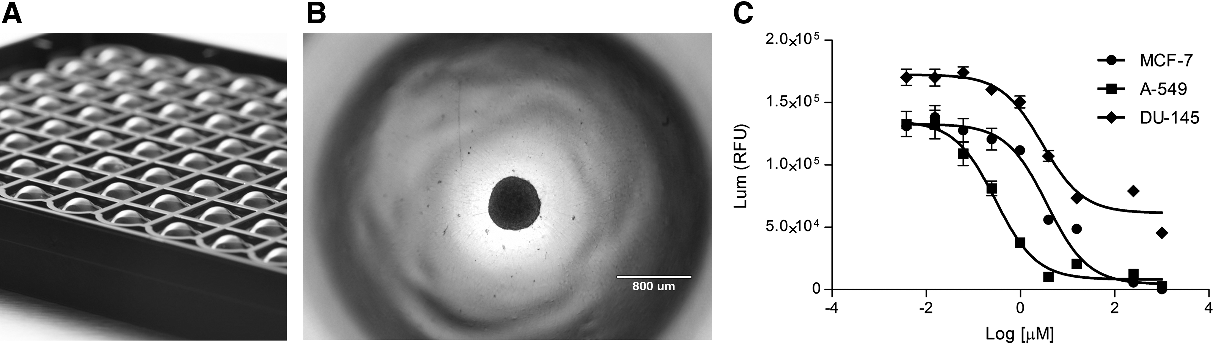
Multiwell spheroid microplates
3D Culture, Surfaces, Scaffolds, and Stem Cells
Cells used in 3D culture are acquired from many sources, including autologous or allogeneic cells, human- or animal-derived primary cells, genetically engineered cells and stem cells. Pluripotent stem cells (PSCs), in particular, can be directed to differentiate into specific somatic cell lineages by inductive signals that mimic changes occurring during embryogenesis. Stem cells self-organize, self-pattern and undergo self-morphogenesis, all of which depend on the spatial location of the cell. Therefore, it is not surprising that many current applications of 3D cell culture specifically involve stem cells. 15
The majority of 3D cell culture involves the use of hydrogel-based matrices or solid scaffolds. 16 Emerging techniques include bioprinting, which allows for precision stem cell patterning and differentiation. Naturally derived hydrogels for 3D culture comprised proteins and ECM components, such as collagen, laminin, and fibrin. Matrigel (Corning® Matrigel® Matrix), notably, has been widely used since it contains many of the common ECM components found in basement membranes. 3D culture of stem cells in ECM gels allows formation of tissue organoids (organotypic culture) that recapitulate key features of organ function. Accordingly, tissue organoids representative of intestinal, retinal, pancreatic, mammary, colonic, and cerebral tissues have now been generated. Historically, these approaches were widely used to study tissue-specific developmental processes. However, they also offer novel approaches for compound screening in a more physiological manner and allow screening of drug candidates in target tissues. As has been stated “…self-organizing tissues with high functionality may be useful for drug screening…particularly with patient-specific induced PSCs.” 14
However, a limitation of natural hydrogels lies in their isolation from animal sources, which may cause cell batch-to-batch variations as well as the potential for pathogen contamination. Consequently, there is a growing interest in synthetic hydrogels, particularly those utilizing polyethylene glycols (PEGs). Today, chemically defined synthetic hydrogels are available, which are both biocompatible and devoid of animal-derived materials or pathogens. 16,17 The control of stem cell differentiation involves spatial and mechanical cues, as well as biochemical signals. Some cues can be imparted by solid surface biomaterials designed to direct growth and differentiation. Consequently, a diverse range of synthetic hydrogel structures, including 3D matrices, scaffolds, and microfluidic formats, are now being adapted for use in microtiter plate formats. 3D matrices, notably, can be derived from several PEG-based hydrogels with different porosities, pore sizes, permeabilities, and mechanical characteristics, each of which reflects a specific tissue ECM. 18 These materials can be further optimized to present oligopeptidic sequences tailored for recognition by specific cellular adhesion molecules. Finally, they can also be applied to microcarrier culture, which utilizes beads derived from several porous polymers as 3D support for anchorage-dependent cells. 19,20
3D culture is, therefore, gaining acceptance into drug discovery, particularly as primary cells or PSC-differentiated cells are becoming available in the large reproducible amounts required for screening. 3D culture is already having an impact in the area of preclinical lead optimization, particularly in screening compounds for metabolic liability or cellular toxicity. This is perhaps unsurprising, since many 3D coculture methods have been refined using liver cells for compound evaluation.
3D Cell Culture Limitations
One major limitation of using 3D culture in drug screening lies in the technical aspects that relate to assay protocols. 5,6 These include the need to optimize and standardize procedures for cell harvesting, cell lysis, production scale-up, as well as control of pH and temperature to reduce well-to-well and lot-to-lot variation. Since 3D culture can be more heterogeneous than 2D culture, interpretation of data is more challenging. In addition, the potential for compound nonspecificity may be increased due to the more complex culture conditions used, as well as physicochemical issues, including compound access to the cells within the 3D complex. Nonetheless, as occurred in the adoption of 2D cell culture procedures in HTS/HCS, defined protocols—and novel instrumentation—are now being developed for 3D culture that could circumvent these limitations.
A second, more general, limitation of 3D culture is the lack of organoid vascularization, consequent lack of oxygenation, and removal of metabolic side products. In this respect, current 3D culture is inferior to 2D cell culture. However, the hypoxic interior of the spheroid may actually better mimic the hypoxic core of a tumor, as well as access of the novel compound to cells in the interior. 3D cultured cells exhibit lower sensitivity than those grown in 2D culture, although other factors may be involved here, such as cell surface receptor expression, cancer gene expression, and uniformity of cell differentiation stages—all of which differ between cells from 2D or 3D cultures. 5
More fundamental is the development of 3D cell culture models for growing cells in a matrix that mimics the in vivo ECM dynamically. Many 2D or 3D environments are static and lack exposure to mechanical forces that influence tissue and organ morphogenesis (including shear forces provided by blood flow). Potentially, the next wave of 3D cell cultures lies in models that better mimic the microstructure of living organs, particularly those using microfabrication technologies. In the most advanced concepts, organ-on-a-chip systems dynamically link multiple miniaturized organs through channels lined by endothelial cells. 21 It remains to be seen if such systems can be utilized in HTS or whether they will play a more limited role in preclinical lead ADME/toxicology evaluation.
Conclusion
The availability of techniques to generate large reproducible batches of human primary cells is now impacting drug discovery, notably in compound screening, but also lead optimization. It is unclear if the use of 3D culture will reduce the compound attrition rate, although the cellular responses to drug treatments in 3D are similar to what are seen in vivo. 5 The adoption of 3D culture in many drug discovery organizations indicates that these technologies have advantages, particularly in oncology, but increasingly in neuroscience and regenerative cell therapy. 22 As 3D culture technologies, protocols, and automation technologies continue to improve, and the available range of human primary cells becomes ever broader, drug discovery is indeed going 3D. It remains to be determined if it is time to say “goodbye to flat HTS”!
Footnotes
Disclosure Statement
No competing financial interests exist.