
Abstract
Historically, two-dimensional (2D) cell culture has been the preferred method of producing disease models in vitro. Recently, there has been a move away from 2D culture in favor of generating three-dimensional (3D) multicellular structures, which are thought to be more representative of the in vivo environment. This transition has brought with it an influx of technologies capable of producing these structures in various ways. However, it is becoming evident that many of these technologies do not perform well in automated in vitro drug discovery units. We believe that this is a result of their incompatibility with high-throughput screening (HTS). In this study, we review a number of technologies, which are currently available for producing in vitro 3D disease models. We assess their amenability with high-content screening and HTS and highlight our own work in attempting to address many of the practical problems that are hampering the successful deployment of 3D cell systems in mainstream research.
Introduction
Over the last century, there has been a steady progression in the diversity and complexity of tissue culture methodologies used in biomedical research, the earliest reported examples of in vitro cell culture being that conducted by Ross G. Harrison who observed neuronal sprouting from frog embryo spinal cords on a microscope slide in 1907. 1 At present, cell-based research is often performed on a variety of planar surfaces that have been modified to promote the growth of two-dimensional (2D) cellular monolayers. These monolayers are utilized for majority of in vitro evaluations in research and have proven very effective. However, it is evident that while these approaches provide a convenient means of treating and analyzing cells, they do not reliably permit the formation of multicellular structures, which in turn form microenvironments similar to that found in vivo. 2 –8 Hence the interest in generating more biologically relevant in vitro models, such as three-dimensional (3D) culture systems.
Limitations associated with 2D models have been identified; such as the loss of tissue-specific architecture, mechanical and biochemical cues, and cell-to-cell interactions. 9 –12 Conversely, the microenvironment generated by 3D cell culture appears more representative of that observed in vivo, resulting in relevant cell-to-cell and cell-to-extracellular matrix (ECM) signalling. 13 –16 Such signalling cascades are deemed essential for a multitude of cellular processes, including differentiation and proliferation. 9,17,18
In contrast to conventional 2D methods, cells cultured in a 3D format may exhibit unique biochemical and morphological features similar to their corresponding tissues in vivo 19 (summarized in Table 1). It should be noted that the cell type, as well as the 3D culture method, impacts on cell organization and formation of the 3D structure. However, the concentric arrangement of heterogeneous cell populations in 3D cultures, as well as their growth pattern, mimics the initial (i) avascular stages of solid tumors in vivo, (ii) not-yet-vascularized micrometastatic foci, (iii) intercapillary tumor microregions with a high proliferative activity close to the capillaries, (iv) quiescent cells as intermediates, and (v) necrotic areas at larger distances. 20,21
The Advantages and Disadvantages of 2D and 3D Cell Culture
2D, two dimensional; 3D, three dimensional; ECM, extracellular matrix.
Two-dimensional cell-based assays are well established in the drug discovery process, particularly in cancer. 22 However, their value in predicting clinical responses to new agents is limited. This unpredictability is attributable to the fact that such systems do not accurately mimic the response of cells in the 3D microenvironment present in vivo. 23
Billions are spent every year on developing targets identified from in vitro systems through to Phase III trials in patients. The vast majority of these compounds fail due to either unacceptable toxicities or limited efficacy in humans. 13 This in itself demonstrates that the more traditional 2D cell systems are ineffective in predicating clinical responses. Indeed, 3D models tend to have better drug predicative value compared with 2D. 13,24,25 Furthermore, issues are associated with the ECM component in 2D culture, which appears to be overcome in a number of 3D systems. 25 –29
Incorporating 3D cell culture with in vitro screening processes such as high-throughput screening (HTS) and high-content screening (HCS) is necessary to identify clinically relevant compounds. Drug discovery is heavily reliant on HTS; the process of identifying hits by testing a large number of diverse chemical structures against disease targets and is characterized by its simplicity, efficacy, low cost per assay, and high efficiency. 30 In addition, HCS-facilitated phenotypic screens yield more complex biologically relevant information and increased data generation relative to conventional in vitro assays, such as protein enzyme assays, binding assays, and endpoint assays. 31,32 Highly sensitive fluorescence-based HCS assays are important for and complement HTS, therefore contributing to the industry-wide initiative to simplify, miniaturize, and speed up assays. 30 Active compound hits, identified by HTS and HCS screens, act as templates for further drug development. 31 The features of both HTS and HCS, and the potential impact of combining these technologies, are summarized below (Fig. 1).
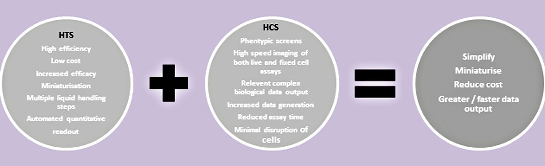
The combination of HTS and HCS strategies will enhance drug development. HCS, high-content screening; HTS, high-throughput screening.
HTS and HCS technologies are, for the most part, conducted and optimized with cells cultured in 2D monolayers. However, creating the means to facilitate screening of 3D models using the same technologies is essential. Unfortunately, not many 3D cell culture technologies are compliant with both HTS and HCS. We believe that this is perhaps a consequence of technological development being promoted above design principles. 33 Very few commercially available products are readily available technologies designed to improve the accuracy of in vitro 3D cell culture analyses in a routine and cost-effective manner. 34 In this study, we discuss the merits of 3D cell culture models and the technologies that are currently attempting to mainstream their utilization in HTS. The need to identify the optimal method to facilitate their generation and use in translational research in the most effective and efficient way possible, is essential.
3d Cell Culture of in Vitro Models
To create an environment that mimics more closely that found in tumors in vivo, 3D systems must simulate a pathophysiological cellular microenvironment in a tumor, reconstruct a tissue-like cytoarchitecture with cell-to-cell and cell-to-ECM interactions, and exhibit growth, differentiation, and therapy responses similar to those observed in vivo. Multicellular structures appear to be the best described 3D tumor model system, which meets all of these criteria. 23,35 –38
As mentioned previously, 2D models are not a reflection of the in vivo state as only a portion of the cell membrane engages with neighboring cells, while the remainder of the cell is exposed to the bulk culture medium. 39 It is therefore a crucial aspect of cancer research that tumor and normal cells are cultured in a manner that most closely resembles the in vivo environment. An important factor in the 2D culture system is the absence of a true ECM. ECM is a critical cellular factor important in normal and deregulated cellular homeostasis 26,40 –43 as it affects cellular properties such as structure, adhesive potential, mechanotransduction, and outcomes of exposure to soluble effectors. 44 Indeed, it has been noted that cells in 2D lose their ability to differentiate, while this property can be recovered in 3D. 44,45 Consequently, ECM-related cellular morphology results in modifications in cellular functions in 2D versus 3D, with 3D demonstrating physiological relevance. 44 –47
The geometry of and pathophysiological gradients exhibited by multicellular 3D models are comparable to in vivo tumors. Beyond a critical size (≥100 μM), most 3D models from cell lines develop a necrotic core surrounded by a viable rim of cells (>100 μM). The periphery of the 3D model is composed of proliferating cells, which resemble actively cycling tumor cells adjacent to capillaries in vivo. In contrast, the innermost cells become quiescent and eventually die by apoptosis or necrosis, forming the necrotic core (Fig. 2). This cell death is a consequence of limited inward and outward diffusion of positive and negative regulators, resulting in decreased oxygen (hypoxia) and/or nutrients, accumulation of waste products, and low pH. 48,49 Such heterogeneity in cellular physiology and tumor milieu are linked to alterations in sensitivity to many antitumor therapies. 20,21,23,48,50 –52
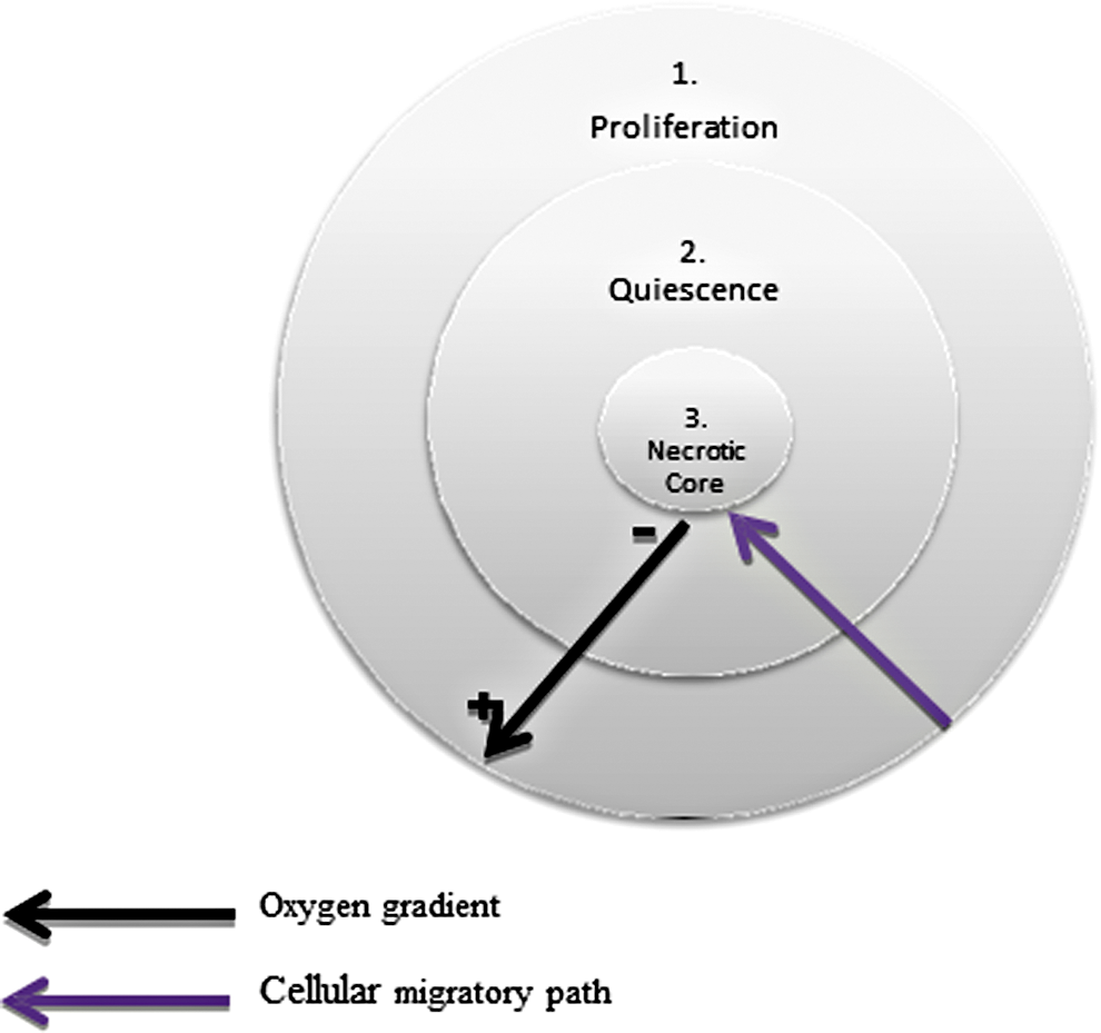
Cellular gradients in 3D cell culture models. Cells originate on the outer proliferative layer (1) and ultimately undergo apoptosis/necrosis in the core (3). This migration is initiated by cell-to-cell and cell-to-extracellular matrix interactions, which regulate the movement of cells from the proliferative (1) to the quiescent (2) zones. From here, cells generally enter the necrotic core either as a direct result of necrotic cell death or through accumulative apoptosis followed by secondary necrosis. Hypoxia is one of several factors that contributes to the development and maintenance of the necrotic core. 3D, three dimensional. 39
One of the biggest challenges in visualizing 3D structures is overcoming the inherent practical limitations imposed by their geometry. With regard to HCS and imaging approaches to 3D cellular assays, many have implemented the use of confocal analyzing modalities to obtain detailed information regarding cellular behavior in 3D. However, even with these advanced automated imaging systems, physical limitations are imposed such as light scattering and absorption. 32 We have found that these limitations are often manifested as poor light penetration of the sample, which in many cases can lead to incomplete imaging of the multicellular structures, and long acquisition times. These issues can be further exacerbated if cells are grown on scaffold materials such as gels or biocompatible polymers, which may further reduce optical clarity. There has, however, been some exciting recent developments that may improve the capabilities of light microscopy platforms, in particular light sheet microscopy. 53 Although this approach offers improved image quality and resolution, it has not yet been routinely adopted by laboratories focused on HTS. In addition, much emphasis has been placed on methods for improving the optical characteristics of the specimen, for example, assay systems where highly pigmented cells can be clarified by incorporating tissue clearing methods in the assay preparation work flow. 54,55
As alluded to previously, one of the main issues with 2D culture is the gap between in vitro and in vivo cellular modeling and drug efficacy studies. A number of techniques now exist to culture patient tumor tissue in either a tissue culture type system such as organoids 56 –58 or using patient-derived xenograft (PDX) murine models. 59,60 This allows for the gene and protein characterization of the patient's cancer and/or normal tissue, in addition to the determination of the patient's cells response to a number of drugs in either system. 56,61 Again, 3D systems would appear to be superior at evaluating drug efficacy, with breast cancer as one such example. 62 Importantly a number of these systems are fully compatible with HTS and HCS and will lead to improved personalized medicine strategies for patients. Some groups have used multiple cell types in 3D model systems such as the lung 63 and breast. 64 These models consisted of epithelial cells, fibroblasts, and other stromal components resulting in physiologically relevant microenvironments. 63,64 Model systems using multicellular types provide enhanced 3D models as more unicellular 3D spheroids will vary in size and lack a vascular system. 13 ADMET (absorption, distribution, metabolism, elimination, toxicity) studies also aid in the drug screening process and help to ensure that only the most efficacious therapeutic targets are investigated leading to a greater number of successful compounds. These types of studies help to characterize the drugs properties and also identify possible drug–drug interactions. 65,66
These models could be modified to use a wide variety of cells and/or patient tissues cultured in 3D systems, 67,68 including those cultured in our own colloidal suspension medium (CSM), which will be discussed later. The latter is currently being utilized for the optimization of assays involving patient samples in a range of cancer and normal cells.
Although traditional 3D culture tends to promote the culture of single cell types, work is ongoing to develop this to encompass multiple cell types, which could further mimic the ECM and stromal compartment within tumors. These advances will also lead to superior 3D models of normal cells such as the kidney and liver, therefore enhancing the capability of toxicity testing in vitro. This would also benefit ADMET and PDX models as normal models could be produced from corresponding normal tissues derived from the patient, therefore allowing an increased scope to investigate drug effects on seriously ill patients. Support in the literature for 3D cell culture highlights discrepancies observed between 2D and 3D culture and in the majority of cases promotes 3D as the preferred method. 69 –73 Some examples are described in Table 2. The idea is that by simulating an in vitro environment, which encourages cells to proliferate, aggregate, and differentiate as they do in vivo, we may yield more biologically and clinically relevant data. The creation of a more natural cellular environment may influence intracellular interactions and downstream signalling pathways and possibly challenge current hypotheses generated by results obtained from 2D cell-based assays. 74 –76
Pharmacological and Genomic Comparisons Between 2D and 3D Cell Culture
3d Cell Culture Technology for Hts and Hcs
There are many options in today's market for those wishing to generate 3D models in vitro, however, there are still many concerns regarding these technologies. There have been numerous publications which highlight these issues, 34,77 –80 for example, in 2010, HTStec conducted a survey which attempted to understand the drive behind the transition from 2D monolayers to 3D. 81 They found that there was no complete system, no one relatively simple method that could transform single cells into robust 3D structures, which could be easily manipulated, treated, imaged, analyzed, and upscaled. The survey highlighted issues such as (i) poor reproducibility, (ii) labor intensiveness, (iii) overcomplicated systems, (iv) inability to upscale, (v) difficulty in recovering cultures, (vi) incompatibility with automated systems, (vii) lack of flexibility, (viii) poor imaging quality, and (ix) long-term stability problems. 77 Industrial drug development requires a universal 3D culture system. However, many emerging technologies focus on specific applications and fail to provide standardized 3D models compatible with automation. Routine 3D cell culture relies on technologies that have considered cost, ease of use, application, and reproducibility. These factors are especially crucial for the field of drug discovery. 34,79,80,82
The methods chosen for discussion in the later sections of this review are purely examples of the type of technology in question and are by no means an exhaustive list and include anchorage dependent and independent methods (Fig. 3). We will also briefly describe a novel technology, currently under development, that we have engineered specifically for 3D culture in HTS and HCS settings.

Chart detailing the principle methods involved in 3D cell culture model production.
Anchorage-Dependant Technologies
Anchorage-dependent scaffolds represent the space available for a tissue to develop. The cross talk between the scaffold and the cells is controlled by the scaffold characteristics and the properties of the material from which it is made. Scaffolds offer physical support for cell growth and accommodate cell adhesion, proliferation and ECM production. 83
Hydrogels
Hydrogels are cross-linked networks composed largely of water, 84 engineered to represent basement membranes. They are primed with ECM proteins such as laminin, collagen IV, and entactin. 85 Many hydrogels also contain growth factors, which promote cellular interactions and 3D model formation. Further applications of hydrogels include co-culture, regenerative medicine and drug screening. Their ability to mimic essential features of the ECM allow them to be bioengineered on a molecular level, offering scientists the possibility to adapt 3D models to specific cell types and to investigate different aspects of cell–matrix interactions. 77 For the most part hydrogels are available in a liquid form. Cells are suspended in the hydrogel and the user is required to carefully coat tissue culture plates with the cell/hydrogel mixture while ensuring gelation does not occur before the plate is fully prepared. An alternative overlay method can also be employed which involves precoating plastics with a layer of the hydrogel and allowing gelation to occur before adding the second hydrogel/cell layer. 86 While both methods are sufficient for 3D culture, the latter is generally favored. Hydrogels are effective in generating 3D models, however certain issues exist which may make them unsuitable for large scale data generation. Hydrogels often need to be dispensed at specific temperatures to prevent premature gelation. This factor may disqualify them from use with some liquid handling systems associated with high throughput screens. This coating of the well surface may render some hydrogels noncompliant with HCS imaging systems. To culture 3D models in a hydrogel and analyze them using a HCS, 3D structures may need to be recovered from the matrix and transferred to a fresh vessel. As well as adding extra steps to an already relatively labor intensive workflow, if not handled with care transferring 3D models between vessels could potentially compromise their integrity. 87
Inert matrices
Another anchorage-dependent method of generating 3D cell culture models is the utilization of inert matrices. An inert matrix is composed of a rigid, porous scaffold and engineered into thin membranes that fit into conventional cell culture plastic ware, ranging from Petri dishes to 384-well plates. 88 The aim of these matrices is to provide cells with the space and freedom to proliferate, interact and differentiate as they would in vivo. The inert nature of the scaffold removes the issue of contamination arising from animal sources, while cells are never further than 100 μm from the nutrient medium owing to a membrane thickness of 200 μm and a pore size of 36–40 μm. While originally designed for generic 3D culture of cells, specific applications have now been developed for these scaffolds. These include in vitro cosmetic and drug testing as well as toxicity and cell invasion assays. 77 As efficient as this system is at culturing cells in 3D format, it is quite labor intensive. The plates may require rehydrating and wash steps before cell seeding and, in a similar manner to hydrogels, the structure of the scaffold itself makes this system unsuitable for HCS. In fact, cells contained in this matrix cannot be viewed under a light microscope without the addition of a visible dye. In our laboratory, we have also witnessed auto-fluorescence in these plates. Certain lipophilic dyes, Nile red, for example, also have a tendency to bind to the scaffold, which may further hinder imaging. Recovering the cultures generated is generally not an option. To do so, they would require treating with trypsin followed by agitation, 89 both of which could potentially compromise their integrity. Analysis is required in situ, generally by absorbance assays, such as MTT [3-(4,5-dimethylthiazol-2-yl)-2,5-diphenyltetrazolium bromide] and MTS [3-(4,5-dimethylthiazol-2-yl)-5-(3-carboxymethoxyphenyl)-2-(4-sulfophenyl)-2H-tetrazolium]. These scaffolds have been utilized in conjunction with a variety of cell types, including various cancer lines such as HepG2 (human hepatic carcinoma) 90 and HT-29 (human colorectal adenocarcinoma), 91 and have successfully produced 3D models suitable for downstream processing. However, the labor intensive factor associated with this technology, with regard to setup, imaging, and cell recovery, cannot be overlooked. While inert scaffolds may be suitable for small scale experimentation, there are technologies available which are more compliant with HCS systems as discussed in later sections.
Anchorage-Independent Technologies
Anchorage-independent (scaffold-free) 3D culture methods promote self aggregation of cells in the medium by providing more freedom to move as opposed to the spatial restrictions imposed by scaffolds. Opting to go scaffold free means a product will be selected from one of the following categories: low adhesion plates; micropatterned surfaces; or hanging drop plates (HDPs).
Low adhesion plates
Low adhesion plates consist of a polystyrene surface often treated with hydrophobic or hydrophilic coatings, which renders them biologically inert therefore greatly reduces the binding of attachment proteins or cells. This encourages the natural migration of cells toward each other and the formation of an ECM. 92 A drawback of these 3D culture systems is the issue of reproducibility. Low adhesion plates possess the capability to produce large numbers of 3D models. However, we have discovered in the course of our work that to generate cultures of a uniform size, much optimization with regard to cell seeding density is required and even after optimization uniformity is not guaranteed.
Low adhesion surface technology is applied to a range of tissue culture vessels, including both 96- and 384-well plates. In addition to the standard flat bottom version, these plates are also available in the form of U-bottom and V-bottom wells, the latter promoting miniaturization by requiring less culture medium. This feature encourages the movement of cells toward each other and promotes the formation of 3D models in culture medium, potentially in a shorter period of time than if left to aggregate naturally. 93 By utilizing this method, the incidence of generating single cultures of uniform size in the troughs of these wells is increased, which is ultimately compatible with HTS. In both examples, the vessels arrive precoated, which reduces the labor involved, therefore all that is required by the end user is to seed their cells as they see fit.
The potential production of reproducible 3D models in a short space of time using these methods make them ideal candidates for HTS technologies. 93 They are designed in such a way as to reduce cross talk and background noise in luminescence and some fluorescence assays, 78 however they do not lend themselves well to HCS. High-content imaging requires the use of high image grade quality plates to obtain sufficient image resolution to perform the downstream image analysis. Treatments on the low adhesion plates can interfere with the imaging surface and the plates are required to be flat bottomed to capture the highest quality images. Certain U-bottom plates do offer a relatively high grade imaging surface and images can be obtained from the very center of these wells, where the well profile tends to promote the accumulation of 3D structures. 94,95 In many of these cases, similar to the hydrogels, 3D cultures may need to be liberated from the plates and transferred to an appropriate surface to facilitate analysis using a HCS. This process adds to the labor intensiveness of the procedure as well as the possibility of damaging the cultures.
Micropatterned surfaces
Other modified surface technology is available in the form of micropatterns. The surface of these plates is imprinted with microstructure patterning to control cell adhesion and promote 3D cell culture. These plates are specifically designed for 3D culture and have the advantage of possessing a transparent bottom film to enable microscopic observation, as well as availability in a selection of patterns and adhesive properties to cater for a variety of cell types. It is recommended that cells are optimized using all plate types to discern the optimum culture conditions. Cells are seeded on the plates; however, they cannot adhere strongly and migrate randomly along the patterned surface. This migration encourages the formation of cell-to-cell interactions and the subsequent generation of 3D models in vitro. Cells use the micropatterned surface as a scaffold which supports the 3D models as they grow, and being loosely attached to the surface, they can be harvested easily by simply removing with a pipette. 96 These plates have many features, which make them compliant with HTS. For example, micropatterned plates have little well-to-well and plate-to-plate variation; therefore it should be possible to generate 3D models of uniform size using this technology. Optimization utilizing the different patterns and adhesion properties infers control over 3D model location and geometry. The plates come ready to use in, so far, as they do not require any other treatment or extra equipment. Caution is advised, however, as bubbles can easily form in the media, which may disturb the culture. Damage can also occur to the micropatterned surface itself if care is not exercised when pipetting. One must also be careful when refreshing the culture medium. 96 While many micropatterned surfaces are compliant with HCS, as with many anchorage-independent technologies, their use is restricted only to the microplate formats currently available.
Hanging drop
Hanging drop technology is a well-known 3D cell culture system. HDPs are available in both 96- and 384-well formats and are composed of an assembly stack, which allows culture of cells, media exchange, and harvesting. Hanging drops are created by carefully pipetting up to 50 μL of cell suspension into wells from the top of the plate. Plate geometry allows the drops to form and suspend from the bottom of the wells through surface tension. The cells are confined within this small droplet and are thus encouraged to interact and aggregate (Fig. 4).

Diagram illustrating a HDP with cell suspensions. HDP, hanging drop plate.
Three-dimensional cell culture using this method allows the user to exert control over the size of the cultures generated, aiding reproducibility and promoting compatibility with many HTS instruments. To image the 3D models produced by the hanging drop method, they must be harvested and transferred to an appropriate plate. This can be achieved either by centrifugation or by the addition of excess media to the hanging drops. These transfer methods require careful optimization and caution as they both put the integrity of the cultures at risk. Care must be taken to reduce evaporation of the hanging drops; hence some plates are equipped with peripheral reservoirs designed to buffer against and combat this issue. Care must be taken to not contaminate the wells with the chosen buffer solution. Even though this method of 3D model formation is generally compatible with HTS, a significant level of technical expertise may be required to gain maximum impact from these products. 97 –99 While the standard HDP systems are in a 96-well plate format, 384-well HDPs have recently become available at similar pricing to the 96-well products and therefore represent a significant saving per assay. 100 However, in our experience, 96-well HDP systems are complex in terms of liquid handling, and the recent advance to 384-well format may be beyond the liquid handling capability of some laboratories.
Collodial suspension medium
After examining and trialing a number of technologies available on the market, we sought to engineer our own. We aimed to design a one-step system that is capable of accommodating cells from initial seeding, through 3D model formation to final analysis in the same culture vessel, while also being compatible with HTS and HCS. We felt that an anchorage-independent CSM would best achieve this. Our optimized cell suspension medium is of low viscosity; a feature which has been uniquely formulated to comply with liquid handling systems, HTS, HCS, and flow cytometry. It allows cells the freedom to naturally migrate, self aggregate, proliferate, and differentiate as they would in vivo without restrictions imposed by artificial matrices. As its principle component is standard culture medium, our CSM can be formulated to meet the specific nutritional requirements of individual cell lines. We have cultured a range of cell lines using our technology, including tumor-forming and nontumor-forming stem and primary cells, and we are currently testing a number of protocols to coculture a wide range of cell types in addition to the development of normal 3D models to examine the effects of new therapeutic approaches.
Our CSM is simple to use, requiring little technical expertise, and 3D cultures can be transferred between culture vessels, fed, and sampled with ease. When the desired stage of 3D model formation has been achieved, the culture can be treated with an inactivation solution. This solution dissociates the CSM and allows sedimentation of the 3D structures, permitting their recovery from the medium and allowing, for example, media exchange, fixation, drug addition, and imaging, without impacting on their geometry or composition. Data suggests that our system is capable of generating and maintaining large volumes of 3D culture models in a variety of cell culture vessels. Characterization of these structures is ongoing. 101 –103 ,* These traits combined with its capacity for use in miniaturization experiments make this technology compatible with both HTS and HCS platforms, for which it has been specifically engineered.
Discussion
There are many options available with regard to 3D cell culture systems. The method chosen will depend upon the desired experimental output. Unfortunately, in many cases, the input vastly exceeds the output. A 3D cell culture technology that can accommodate both the quantitative aspect of HTS and the qualitative aspect of HCS could hold major potential in relation to drug screening, target identification, and validation, in addition to elucidating mechanisms of drug resistance. The ability to facilitate this while remaining economically viable would be a significant advancement in the field of drug discovery and have many positive implications for the bench-to-bedside approach. We are hopeful that our CSM will provide means to do just that.
Footnotes
Acknowledgments
S.-L.R. is supported by a Supervisor Scholarship from QUT. K.O.B. is supported by a Senior Clinical Research Fellow (SCRF) award Queensland Government, Office of Health and Medical Research.
Disclosure Statement
Dr. Anthony Davies is the inventor of the Colloidal Suspension Media (CSM) and is also a major shareholder and a director of the company that currently commercializes this technology. For all other authors, no competing financial interests exist.