
Abstract
Angiogenesis has emerged as an important therapeutic target in several major diseases, including cancer and age-related macular degeneration. The zebrafish offer the potential for high-throughput drug discovery in a whole vertebrate system. In this study, we have taken advantage of the transgenic Tg (fli1a:EGFP) zebrafish line to screen the U.S. Drug Collection Library and identified 11 old drugs with antiangiogenic activity, including Closantel, an FDA-approved broad-spectrum salicylanilide antiparasitic drug for a variety of types of animals. Closantel was confirmed to have antiangiogenic activity in zebrafish with a half-inhibitory concentration (IC50) at 1.69 μM on the intersegmental vessels and 1.45 μM on the subintestinal vessels. Closantel also markedly suppressed cancer growth in zebrafish xenotransplanted with human lymphoma, cervical cancer, pancreatic cancer, and liver cancer cells, generally in a dose-dependent manner. These data reveal that Closantel has antiangiogenesis and anticancer effects and could be a potential drug candidate for animal and human cancer treatments. Further study is needed to clarify the mechanisms involved in the antiangiogenesis and anticancer effects of Closantel.
Introduction
Angiogenesis has become an attractive target for drug therapy because of its key role in many diseases, such as cancer, age-related macular degeneration, rheumatoid arthritis, and endometriosis. 1 –4 An endothelial cell model of migration, proliferation, apoptosis, and tube formation is currently a popular angiogenesis assay used for antiangiogenic drug screening, but this assay lacks the biological complexity of in vivo systems and has a poor predictive power. 5 Animal models, including the chick chorioallantoic membrane assay, corneal neovascularization assay, and Matrigel plug assay, preserve biological complexity but are costly and of low efficiency. 6 Recently, the zebrafish (Danio rerio) has emerged as a valuable model organism in drug discovery, including target identification, disease modeling, lead discovery, and compound toxicology. 7 Zebrafish provide a system for drug screening that combines the biological complexity of in vivo models with the suitability for high-throughput screening. 7 –12
The processes of vasculogenesis and angiogenesis in zebrafish are similar to those in other vertebrates. During early embryogenesis, zebrafish angiogenic vessel patterning is simple, primarily occurring in the head, intersegmental vessels (ISVs), and subintestinal vessels (SIVs). The ISVs sprout from the aorta and form between each pair of somites, connecting to the dorsal longitudinal anastomotic vessel. 13 Blood vessel development continues during the subsequent days by angiogenesis processes, and in particular, at 48 hpf (hours postfertilization), SIVs originate from the duct of Cuvier. At the molecular level, vasculogenesis and angiogenesis in zebrafish are also similar to other vertebrates. Several important genes, including vascular endothelial growth factor (VEGF), Flk-1/KDR, Fli-1, Flt-1, Tie-1, and Tie-2, have been cloned in zebrafish and shown to express patterns similar to those in mammals. 14,15 Studies have shown that treatment of zebrafish with antiangiogenic drugs inhibits formation and growth of blood vessels, and several antiangiogenic compounds identified from zebrafish angiogenesis model are now in preclinical or clinical trial stages. 16 –19
Despite the recent regulatory approval of recombinant antibodies and small molecules targeting the VEGF pathway, the clinical efficacy of these therapies for various cancers is limited. 20 Many antiangiogenesis small molecules currently under clinical trials have failed due to absorption, distribution, metabolism, and excretion (ADME) issues; off-target effects; target-associated toxicity; or low potency. 21,22 FDA-approved drugs usually have no ADME problems and have approved drug safety. Drug reprofiling could potentially find existing drugs with previously undiscovered antiangiogenic activity and unanticipated mechanisms of action, which may allow more effective combination treatments and more efficient targeting of tumor neoangiogenesis and thus improve clinical efficacy. 23 In this study, we have taken advantage of the Tg (fli1a: EGFP) zebrafish line, 24 in which the vascular system is visible through endothelium-specific enhanced green fluorescent protein (EGFP) expression, to screen old drugs for antiangiogenic activity from the U.S. Drug Collection Library. We identified 11 positive hits, one of the most potent being Closantel, which is a veterinary drug used as an anthelmintic for cattle, sheep, and goats, 25 and has not been previously reported to display antiangiogenic activity in a dose-dependent manner. Furthermore, Closantel demonstrated in vivo anticancer potential in zebrafish xenotransplanted with a variety of human cancer cells.
Materials and Methods
Zebrafish Care and Maintenance
The Tg (fli1a: EGFP) y1 zebrafish line was obtained from Dr. Brant Weinstein at the National Institute of Child Health and Human Development. Adult Tg (fli1a: EGFP) zebrafish were housed and maintained in accordance with standard procedures. Embryos were generated by natural pairwise mating according to The Zebrafish Book. 26 Four to five pairs of adult zebrafish were set up for each mating, and on average, 200–300 embryos per pair were generated. Embryos were maintained at 28°C in fish water (0.2% Instant Ocean salt in deionized water, pH 6.9–7.2, conductivity 480–510 μS/cm, and hardness 53.7–71.6 mg/L CaCO3). Embryos were cleaned and staged at 6 and 24 hpf. 27 The University Animal Care and Use Committee approved the animal procedures described here. These procedures are consistent with the American Veterinary Medical Association Panel on Euthanasia.
Library Screen
The U.S. Drug Collection Library (MicroSource Discovery Systems), containing 1,280 FDA-approved drugs, was used in this screen. All drugs in the library were provided in dimethyl sulfoxide (DMSO) solution at a concentration of 10 mM. Tg (fli1a: EGFP) embryos were arrayed into 96-well microplates (Nest; NEST Biotech) with five embryos per well in 200 μL fish water before the 24 hpf stage of development, the stage when ISVs begin to sprout from the dorsal artery (DA) 13 The library drugs were added to each well at an initial concentration of 10 μM (1:1,000 dilution). The positive control drug for this assay was 10 μM lovastatin, a VEGFR antagonist, 28,29 and the negative control was 0.1% DMSO. After 24 h of exposure to the drug, the embryos were visually inspected and photographed for the number of ISVs formed and embryonic viability under a stereo fluorescence microscope (Nikon AZ100 fluorescence microscope). Hits were defined as drugs with the ability to significantly inhibit angiogenesis and reduce the number of mature ISVs, which normally connect the DA to the dorsal longitudinal anastomotic vessel (DLAV) in viable embryos.
Dose–Response Assay of Antiangiogenesis
Subsequent dose–response study was carried out to confirm the angiogenesis suppression of the positive hits identified from the primary screen and determine the half-inhibitory concentration (IC50) of the hits on angiogenesis. Closantel was repurchased from Sigma-Aldrich (St. Louis, MO). Stock solutions at concentrations ranging from 0.1 to 50 mM were prepared in 100% DMSO, and serial dilutions were made before experiments. Twenty-four hours postfertilization (for ISV assay) or 48 hpf (for SIV assay) embryos were distributed into six-well microplates (Nest; NEST Biotech), 30 embryos per well in 3 mL fish water for a treatment period of 24 h. After treatment, embryos were anesthetized with 0.016% MS-222 (tricaine methanesulfonate; Sigma-Aldrich); then, the number (N) of ISVs was counted, and the area (S) of SIVs was quantified with NIS-Elements D3.1 software. Drug effect was calculated following the formulas (a) and (b).
Closantel Dose Determination for Anticancer Efficacy Assessment
To determine no observed adverse effect level (NOAEL) for anticancer efficacy assessment of Closantel, zebrafish at 3 dpf were distributed into six-well microplates, 30 zebrafish per well in 3 mL fish water. Closantel was added to the fish water, and zebrafish were exposed by semistatic immersion until 6 dpf. Dead zebrafish were counted and removed on a daily basis. Closantel concentrations ranging from 0.025 to 10 μM were tested to determine its NOAEL in zebrafish.
Cell Culture
Human Burkitt's lymphoma Ramos cells, human cervical cancer Hela cells, human pancreatic cancer PANC-1 cells, and human liver cancer HepG2 cells were purchased from the American Type Culture Collection (ATCC). Human Burkitt's lymphoma Ramos cells were maintained in RPMI 1640 essential medium, and the other 3 cell types were maintained in DMEM (Gibco). All media were supplemented with 10% heat-inactivated fetal bovine serum, 100 units/mL penicillin, 100 μg/mL streptomycin, and 2 mM
Anticancer Efficacy Assessment in Xenotransplanted Zebrafish Model
Cell labeling
Human cancer cells were collected by centrifugation and resuspended in phosphate-buffered saline (PBS). The cells were fluorescently labeled by incubating with 10 μg/mL CM-DiI (Invitrogen) containing 0.5% DMSO and adjusted to a density of 100 × 106 cells/mL (100 cells/nL) in Hank's Balanced Salt Solution. Labeled cells were injected within 2 h. Cells prepared for injection in this way routinely had fewer than 5% dead cells and were in a single cell suspension, yielding controllable and reproducible numbers of injectable cells that were highly and uniformly fluorescent. The dye was transferred from mother to daughter cell, and fluorescent single cells were clearly visible after several doublings on the periphery of late arising tumors. 30
Cell transplantation
The transplantation protocol was similar to that described by other investigators. 30,31 CM-DiI-labeled human cancer cells were loaded into a pulled glass micropipette (VWR blood capillaries #53508–400) that was drawn on an electrode puller and then trimmed to form a needle with a resulting internal diameter of ∼15 microns and outer diameter of ∼18 microns. The microneedle was attached to an air-driven Cell Tram (Eppendorf). The tip of the needle was inserted into the yolk of a 48 hpf zebrafish, and the pulse time was controlled to deliver ∼500 cells in 15 nL using positive pressure. The number of injected cells was standardized by fixing cell density and injection volume. After a 1-hour recovery period at 28°C, implanted zebrafish were examined under a fluorescence microscope (AZ100; Nikon) for the presence of xenotransplanted cells that reside only in the yolk and are then transferred to 35°C for the duration of the experiment.
Anticancer efficacy assessment
Xenotransplanted zebrafish were used to assess anticancer efficacy of Closantel. At 24 h postxenotransplantation, Closantel at three various concentrations (1/4 NOAEL, 1/2 NOAEL, and NOAEL) was added to the treatment solution (fish water) for a drug treatment period of 72 h. The xenotransplanted zebrafish treated with 0.1% DMSO was used as vehicle control. After treatment, xenotransplanted zebrafish were photographed under a fluorescence microscope. Nikon NIS-Elements D 3.10 software was used to quantify all the cancer cell fluorescence intensity (S) in xenotransplanted zebrafish. Drug effect was calculated using the following formula:
Acute Toxicity Assessment of Closantel
Zebrafish at 3 dpf were distributed into six-well microplates, 30 zebrafish per well in 3 mL fish water. Closantel at five various concentrations (0.025, 0.05, 0.1, 0.25, and 0.5 μM) was added to the treatment solution (fish water) for a drug treatment period of 48 h. Dead zebrafish were recorded and removed from treatment solution on a daily basis. After treatment, zebrafish were observed under a dissecting microscope (Olympus Co.) and photographed with a digital camera. The following organs and tissues were evaluated: body length, heart, circulation, liver, intestine, pancreas, kidney, head, eye, tail, pigmentation, otoliths, and muscle. To detect ocular apoptosis, zebrafish were stained with 2.5 μg/mL acridine orange for 30 min. After two brief washes in fish water, zebrafish eyes were visually inspected and photographed under a Nikon AZ100 stereo fluorescence microscope.
Statistical Analysis
All data are presented as mean ± SE. Statistical analysis and graphical representation of the data were performed using GraphPad Prism 5.0 (GraphPad Software). If a single concentration of a drug was assessed, the Student's t-test was used to identify drugs that exhibit a significant effect compared to the vehicle control group. If multiple concentrations of a drug were assessed, ANOVA was first used to assess whether there were any differences in the mean among the various concentrations of each compound; if a significant difference was determined (P < 0.05), Dunnett's test, which is appropriate for multiple pairwise comparisons against a control, was then performed.
Results
We used transgenic zebrafish with fluorescent blood vessels as a high-throughput assay to screen an FDA-approved drug library in a whole-organism system. A known antiangiogenic drug lovastatin 28,29 was used as a positive control that markedly inhibited ISV formation in zebrafish (Fig. 1A). A total of 11 hits were identified with antiangiogenic activity, including Oxibendazole, Closantel, Suloctidil, Gemfibrozil, Levaluterol, Chloroxine, Vinpocetine, PCI-32765, Phenytoin Sodium, Mena, and Phenazopyridine (Fig. 1B).

Chemical screen to identify antiangiogenesis compounds in the U.S. Drug Collection Library using Tg (fli1a: EGFP) zebrafish.
Of the 11 existing drugs identified as hits, we further assessed the dose–response of antiangiogenesis of Closantel on the ISVs and SIVs of zebrafish and found that Closantel suppressed angiogenesis in vivo in a dose-dependent manner. As shown in Figures 2A and 3A, the growth of angiogenic ISVs and SIVs was markedly inhibited in zebrafish treated with Closantel for 24 h. Some of the ISVs and SIVs were completely absent, while others were incompletely formed resulting in a significant reduction in the total number of mature ISVs and total area of SIVs compared with the control (Tables 1 and 2). These inhibitory effects on angiogenesis were dose dependent (Figs. 2B and 3B), and the IC50 on the ISVs and SIVs was 1.69 and 1.45 μM, respectively.
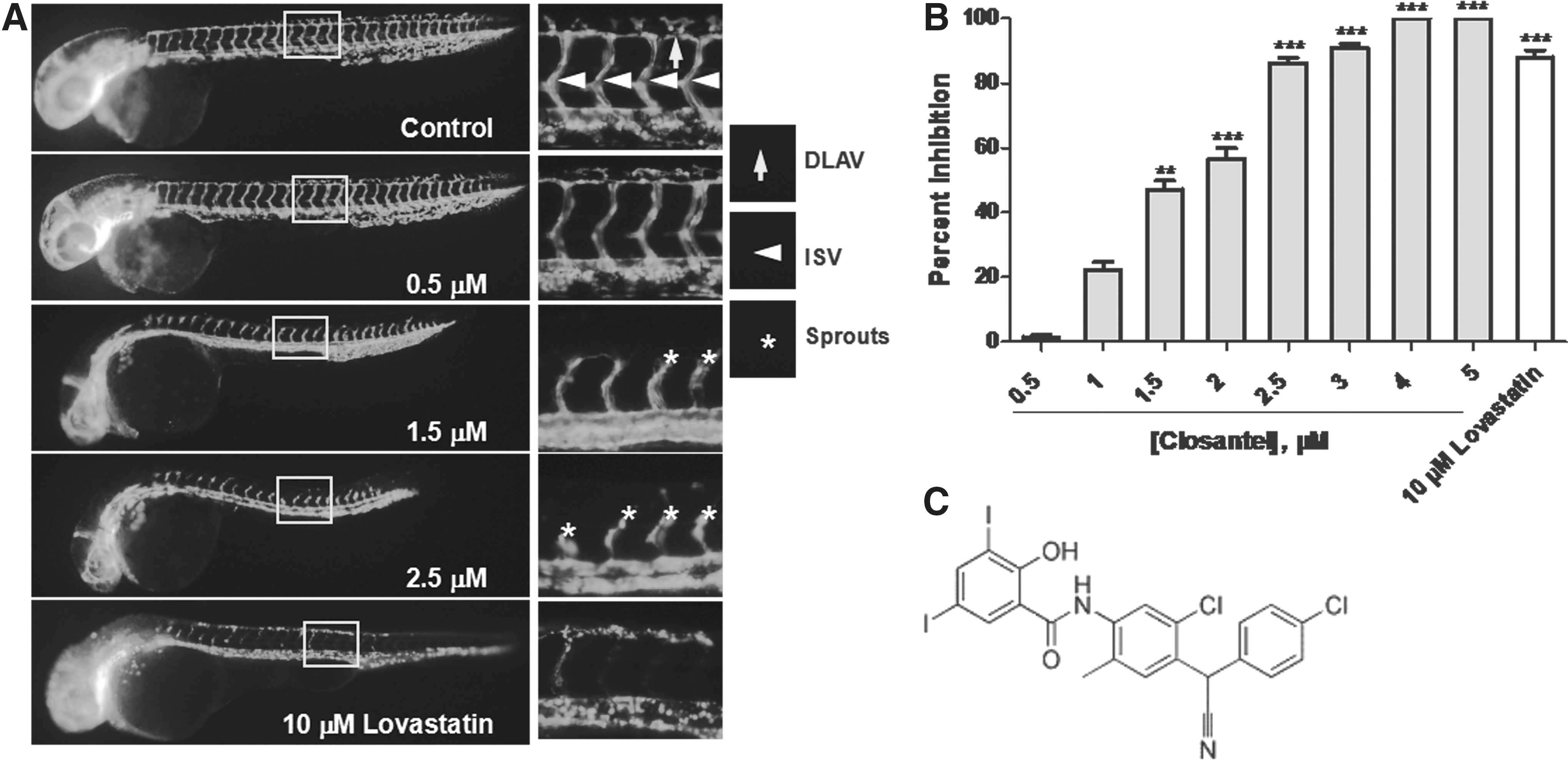
Closantel inhibits zebrafish ISV angiogenesis in a dose-dependent manner.
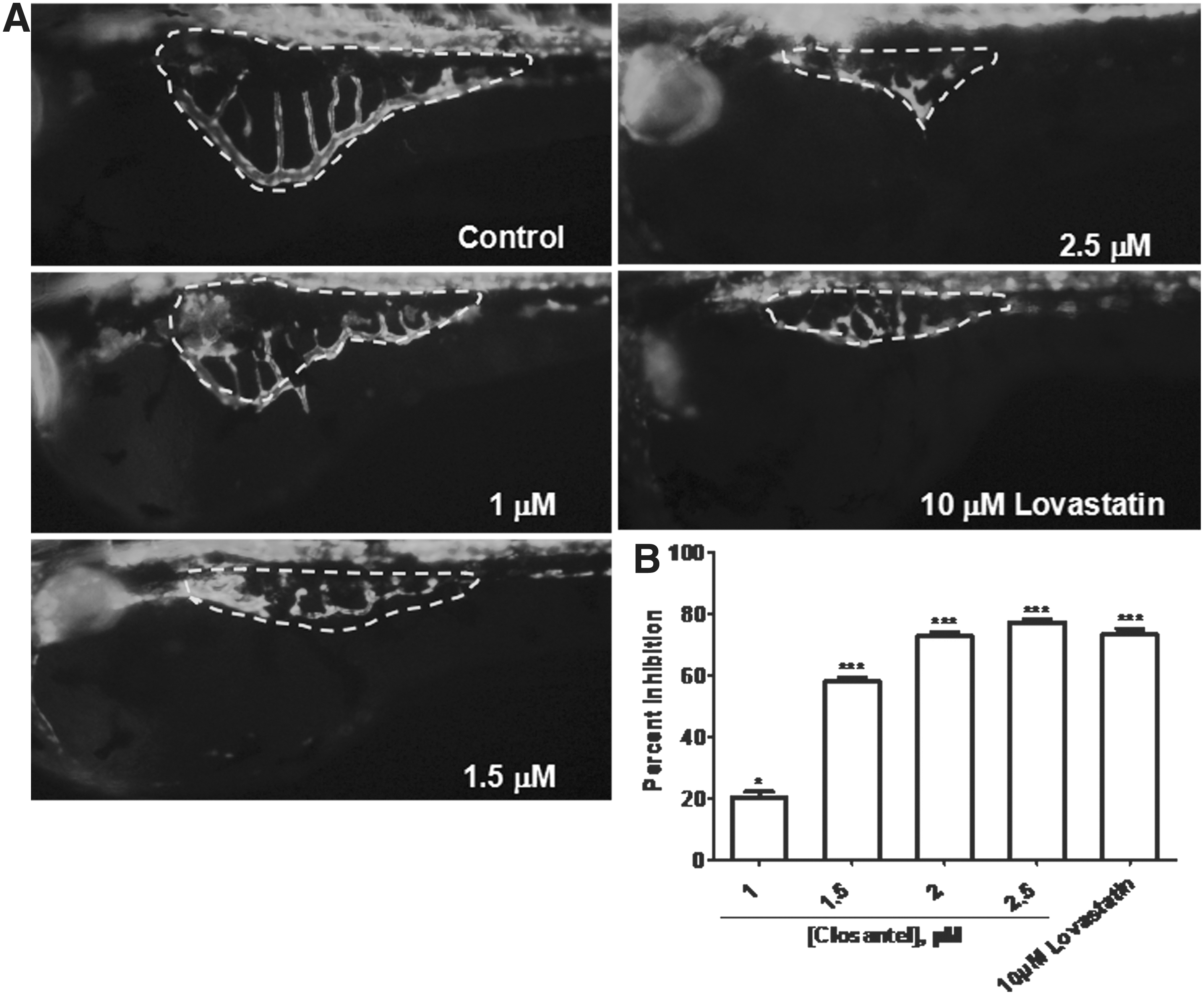
Closantel inhibits zebrafish SIV angiogenesis in a dose-dependent manner.
Percent Inhibition of Closantel on the ISVs in Zebrafish
n = 10.
P < 0.001 versus control.
ISVs, intersegmental vessels.
Percent Inhibition of Closantel on the SIVs in Zebrafish
n = 10.
P < 0.001 versus control.
SIV, subintestinal vessels.
To investigate the anticancer potential of Closantel through antiangiogenesis in the process of cancer growth and metastasis, the zebrafish xenografted with human Ramos, Hela, PANC-1, or HepG2 cancer cells were used. NOAEL concentration of Closantel was 0.1 μM in zebrafish, and thus, three concentrations at 0.025 μM (1/4 NOAEL), 0.05 μM (1/2 NOAEL), and 1 μM (NOAEL) were selected for assessing anticancer efficacy of Closantel in xenotransplanted zebrafish. Each DiI-stained human cancer cell was successfully xenotransplanted into the yolk sac of zebrafish, as indicated in Figure 4A-a. After 72 h of xenotransplantation, human cancer cells in the untreated control group migrated away from the primary site, displaying an irregular boundary of the tumor xenograft (Fig. 4A-b). Compared with the control group, the xenografts of Ramos, Hela, PANC-1, and HepG2 cells had smaller metastatic areas after treatment with Closantel (Fig. 4B). Moreover, all the xenografts of these cells treated with Closantel had an obviously reduced fluorescence intensity, indicating that Closantel could kill or suppress migration, proliferation, or metastasis of human Ramos, Hela, PANC-1, and HepG2 cancer cells in zebrafish xenografts. The cancer inhibition percentages of Closantel on these human cancer cells xenotransplanted into zebrafish were calculated according to the formula (c), and we observed a significant dose-dependent inhibition of tumor cell growth (Fig. 4C and Table 3).
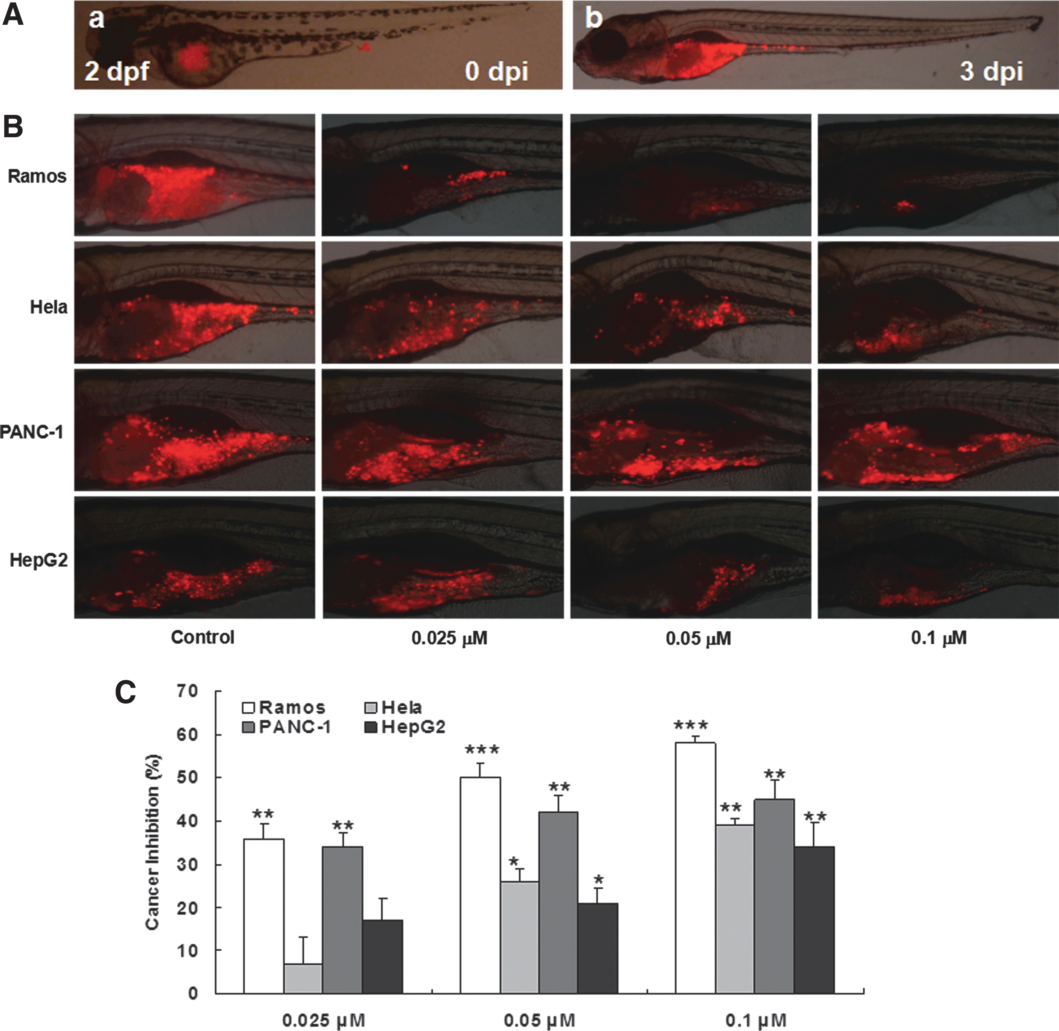
Inhibitory effect of Closantel on the growth of Ramos, Hela, PANC-1, and HepG2 cells in zebrafish xenografts. CM-DiI-labeled cancer cells were imaged under a fluorescence microscope at 2 dpi.
Percent Inhibition of Closantel on Human Cancer Cells Xenotransplanted into Zebrafish
n = 10.
P < 0.05, ** P < 0.01, *** P < 0.001 versus control.
We assessed acute toxicity of Closantel in zebrafish and found that Closantel at 0.1 μM and lower concentrations did not induce any observable side effect. All zebrafish treated with 0.5 μM Closantel were dead. Compared with untreated zebrafish, ∼12% heart rate reduction was observed in zebrafish treated with 0.25 μM Closantel. Ocular apoptosis was found in zebrafish treated with 0.25 μM Closantel, as indicated in Figure 5.
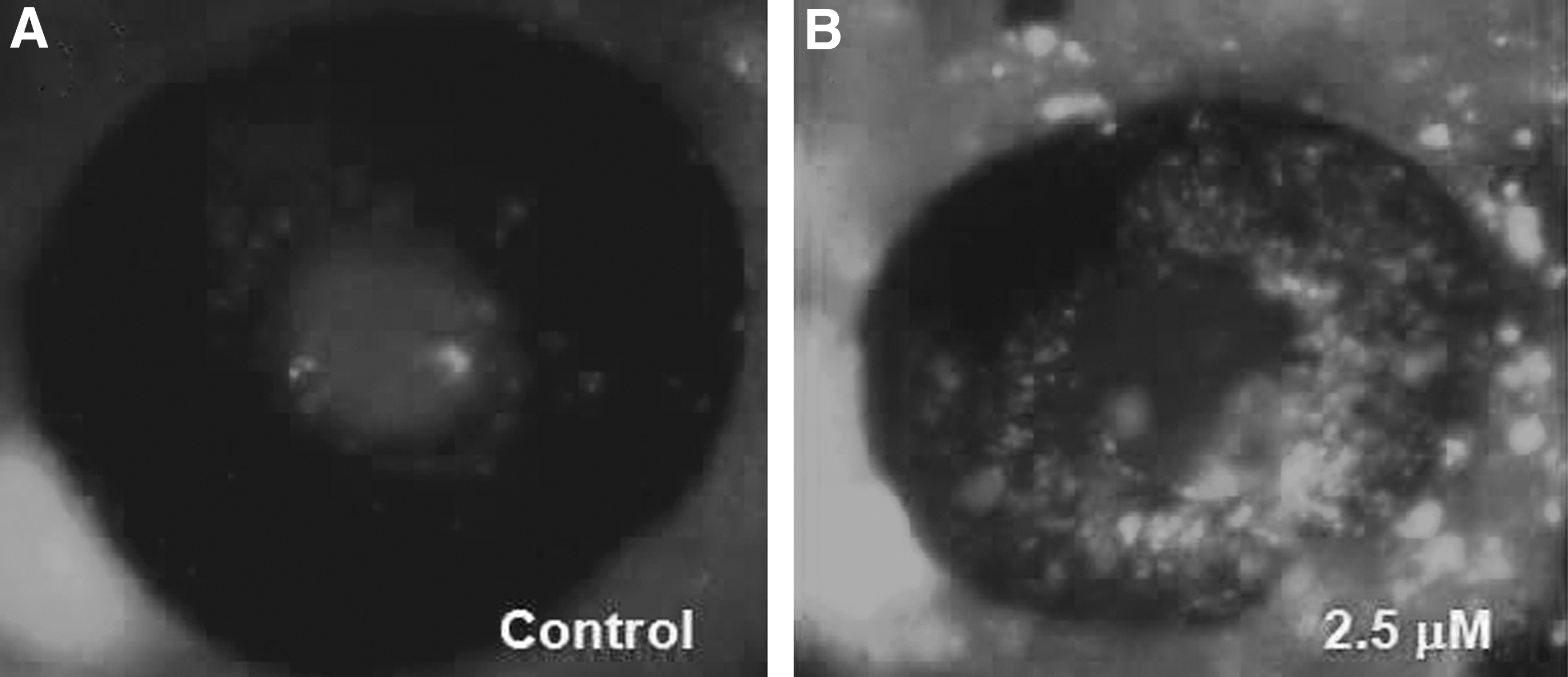
Closantel induced ocular apoptosis in zebrafish:
Discussion
Angiogenesis is essential for normal vascular development and malignant tumor growth. The modulation of angiogenesis is an attractive therapeutic strategy for the treatment of a wide variety of human diseases, and at least 11 antiangiogenesis drugs, including Thalidomide, 17 have been marketed worldwide to treat cancer patients.
In this study, we used an established transgenic zebrafish with fluorescent blood vessels to develop a powerful assay that enabled high-throughput screening of an FDA-approved drug library in a whole-organism setting. Zebrafish angiogenesis signaling and molecular pathways are highly conserved compared with mammalian systems, 32 and the fact that antiangiogenesis drugs in humans also suppress angiogenesis in zebrafish suggested that zebrafish could be used as a predictive animal model for in vivo antiangiogenic drug screening and efficacy assessment. 16,17 The bright consistent fluorescence of blood vessels in Tg (fli1a: EGFP) zebrafish embryos suggested that they could provide a convenient, cost-effective, and high-throughput assay for rapidly identifying antiangiogenic agents.
Using this Tg (fli1a: EGFP) zebrafish model, 11 old drugs were identified with antiangiogenic activities in zebrafish. Of these 11 drugs, Closantel was found to inhibit angiogenesis in zebrafish, with an IC50 at 1.69 μM on ISVs and 1.45 μM on SIVs. Closantel is an FDA-approved broad-spectrum salicylanilide antiparasitic drug active against several adult and developmental stages of trematodes, nematodes, and arthropods and mainly indicated to control fascioliasis (liver fluke) in sheep and cattle. 33 Closantel was confirmed as a potent and specific inhibitor of chitinase of Onchocerca volvulus (OvCHT1) and other filarial chitinases. 34 Other investigators reported that the anthelmintic mode of action of Closantel relied on its role as a proton ionophore. 35,36 Very recently, antibacterial action of Closantel for methicillin-resistant Staphylococcus aureus has been reported. 37
In addition to its antiangiogenesis effect on zebrafish vessel development, Closantel also markedly suppressed cancer growth in zebrafish xenotransplanted with human lymphoma, cervical cancer, pancreatic cancer, and liver cancer cells. Collectively, these results imply that Closantel possesses antiangiogenesis and antitumor activities and could be a potential anticancer drug for animals and humans.
There were several reports that Closantel overdosage could induce retinal degeneration in humans 38 and various animal species. 39 –41 Consistently, here we found that Closantel induced ocular apoptosis and reduced heart rate. A systematic toxicity and safety assessment for Closantel is needed in a future study.
Our findings of Closantel's in vivo antiangiogenic activity in the zebrafish model provide new insights into its clinical application and therapeutic potential for treating angiogenesis-dependent diseases. We are currently further investigating the mechanisms involved in the antiangiogenesis and anticancer effects observed following Closantel treatment as well as the relationship between antiangiogenesis and anticancer effects of Closantel.
Footnotes
Acknowledgment
This work was sponsored in part by the Zhejiang Provincial Major Research Program (2014C03009).
Disclosure Statement
No competing financial interests exist.