




Abstract
Atherosclerosis is a major cause for cardiovascular diseases. High-density lipoprotein (HDL) may reduce atherosclerosis through several different mechanisms. HDL is composed of lipids, cholesterol, cholesteryl esters, triglycerides, and phospholipids, mainly phosphatidylcholine plus specialized proteins called apolipoproteins (apos). In this study, we prepared vitamin E containing HDL (VE-HDL) that contains egg phosphatidylcholine, cholesterol, vitamin E, and two kinds of recombinant human apolipoproteins (rhapo)—rhapoA-I and rhapoE in vitro by the facilitation of cholate. After that, we studied the effects of VE-HDL on foam cell formation, cellular cholesterol efflux, oxidative low-density lipoprotein (oxLDL)-stimulated oxidative stress, and apoptosis of macrophages to evaluate the protective efficacy of VE-HDL on macrophages. As the results showed, we prepared a new type of reconstituent HDL with apolipoproteins and vitamin E for the first time. VE-HDL has protective efficacy on macrophages. It has the prospect of becoming a therapeutic agent on atherosclerosis in the future.
Introduction
Atherosclerosis is a major cause for cardiovascular diseases. A large amount of studies show plasma levels of high-density lipoprotein (HDL) cholesterol are strongly inversely correlated with the risk of atherosclerotic cardiovascular disease, which is the leading cause of death in the developed countries. 1 –3 HDL may reduce atherosclerosis through several different mechanisms, including reverse cholesterol transport, reducing formation of foam cell, protecting against endothelial dysfunction, antioxidant activity, and inhibiting modifications of low-density lipoprotein (LDL), and so on. 4
HDL is composed of lipids, cholesterol, cholesteryl esters (CEs), triglycerides, and phospholipids, mainly phosphatidylcholine plus specialized proteins called apolipoproteins (apos). 5 The predominant species of HDL are spherical microemulsion particles, which are composed of a core of triacylglycerol (TG) and neutral CE and a monolayer of free cholesterol (FC), phospholipid (PL), and apolipoproteins. 6 The exchangeable apolipoprotein family that is associated with HDL includes apolipoprotein A-I (ApoA-I), apolipoprotein A-II (ApoA-II), apolipoprotein A-IV (ApoA-IV), apolipoprotein C (ApoA-C), and apolipoprotein E (ApoE).
ApoA-I, the major protein component of HDL, is secreted into the bloodstream as a lipid-poor monomer and can stabilize all HDL subclasses. ApoA-I has antiatherosclerostic functions such as antioxidant, anti-inflammatory abilities, and cholesterol efflux capacity. 7,8 ApoE which is expressed by macrophages is a very important regulatory protein in lipoprotein metabolism. 9 Its functions include taking part in reverse cholesterol transport as an extracellular cholesterol acceptor. Meanwhile, ApoE is also known to be a target of the low-density lipoprotein receptor-related protein (LRP), 10 which is highly expressed in human atherosclerotic plaques.
Therefore, we planned to prepare a new kind of HDL nanoparticle, which contains ApoA-I and ApoE. This new HDL nanoparticle could make use of the specific binding of ApoE to upregulated LRP on atherosclerotic arteriosus surfaces as targeted part of HDL nanoparticle, and it could take advantage of antiatherosclerostic effects of ApoA-I and ApoE at the same time.
In natural HDL, human exchangeable apolipoproteins (ApoA, ApoC, and ApoE) combined with cholesterol ester by hydrophobic interface to form the core of HDL particles, meanwhile, the hydrophilic interface of apolipoproteins exposed on the surface of HDL particles. This structure could make HDL particles steadily disperse in plasma as well as the water phase. Based on the particular structure of apolipoproteins and HDL particles, we supposed that we could use hydrophobic vitamin E to replace cholesterol ester during the process of preparing reconstituent HDL to form the core of HDL particles. By this method, we could obtain a new type of vitamin E-containing HDL (VE-HDL), which could have stronger antioxidant ability and could be applied to effective targeted treatment.
In this study, we prepared vitamin E-containing HDL (VE-HDL) that contains egg phosphatidylcholine, cholesterol, vitamin E, and two kinds of recombinant human apolipoproteins (rhapo)—rhapoA-I and rhapoE in vitro by the facilitation of cholate. After that, we studied the effects of VE-HDL on foam cell formation, cellular cholesterol efflux, oxidative LDL (oxLDL)-stimulated oxidative stress, and apoptosis of macrophages to evaluate the protective efficacy of VE-HDL on macrophages.
Materials and Methods
Reagents
Vitamin E, egg phosphatidylcholine, cholesterol, and cholate were purchased from Sigma-Aldrich. RhapoA-I and rhapoE were prepared and preserved in our laboratory. oxLDL was purchased from Yiyuan biotechnology (Guangzhou, China). The β-actin polyclonal antibody was obtained from Abclonal. The rabbit polyclonal antibodies to Bcl-2, Bax, nuclear factor 2 (Nrf2), cleaved-caspase-3, and Caspase-8 were purchased from Abcam (United Kingdom). The macrophage RAW264.7 cells were obtained from the type culture collection of the Chinese Academy of Sciences (Shanghai, China).
Separation of Native HDL from Human Plasma
Native human HDL (density 1.063–1.21 g/mL) was separated by density gradient centrifugation as reported. 4 After filtration sterilization, the solution of HDL was reserved at 4°C for further studies.
Preparation of VE-HDL
Recombinant human apolipoproteins—rhapoA-I and rhapoE were prepared with same methods as mentioned previously. 11 In brief, the two kinds of recombinant human apolipoproteins (rhapoA-I and rhapoE) were respectively expressed in Picha pastoris and purified with cation exchange chromatography (SP Sepharose XL, Sweden) and reverse phase chromatography (Source 30, Sweden). They were identified by western-blot and amino-terminal sequence analysis. Based on our previous studies, 4 we prepared VE-HDL, which contains two kinds of recombinant human apolipoproteins (rhapoA-I and rhapoE) and vitamin E as shown in Table 1. An ethanol solution (1 mL) containing 10 mg vitamin E, 7.2 mg egg phosphatidylcholine, and 1.6 mg cholesterol was rapidly injected into 12 mL of phosphate buffer (pH 7.4) through a glass syringe with a 25-gauge needle. After mixing for 15 min under a stream of N2, 5.4 mg cholate, 10 mg rhapoA-I, and 5 mg rhapoE in phosphate buffer (1 mL) were added to the lipid mixture with stirring. The mixture was incubated for 30 min at room temperature, and then, it was incubated at 4°C for 12 h. The solution was dialyzed totally at 4°C (about 2 days) against phosphate buffer to remove ethanol and cholate. We analyzed and photographed morphological feature and grain diameters with transmission electron microscope.
Protocol Table for VE-HDL Preparation and Purification
1. 50 mL centrifuge tube, 12 mL pH 7.4 phosphate buffer.
2. An ethanol solution containing 10 mg vitamin E, 7.2 mg egg phosphatidylcholine, and 1.6 mg cholesterol, rapidly injected into phosphate buffer.
3. Mixing for 15 min under a stream of N2.
4. 5.4 mg cholate, 10 mg rhapoA-I, and 5 mg rhapoE in phosphate buffer (1 mL) and added to the lipid mixture with stirring.
5. Mixture was incubated for 30 min at room temperature.
6. Mixture was incubated at 4°C for 12 h.
7. Solution was dialyzed totally at 4°C (about 2 days) against phosphate buffer to remove ethanol and cholate.
rhapoA-I, recombinant human apolipoprotein A-I; rhapoE, recombinant human apolipoprotein E; VE-HDL, vitamin E-containing HDL.
Cell Culture
The mouse macrophage RAW264.7 cells were maintained in Dulbecco's modified Eagle's medium (DMEM) containing 10% fetal bovine serum and penicillin–streptomycin at 37°C, 5% CO2. Cell viability was determined by Cell Counting Kit-8 (CCK-8) assay. For experiments, the mouse macrophage RAW264.7 cells (seeded at 5 × 10⁵ cells/mL) were maintained in DMEM. After being 70%–80% confluence, the cells were treated with 80 mg/L oxLDL for 24 h as described previously. 12,13 In some experiments, cells were exposed to 25, 50, and 100 mg/L VE-HDL for 24 h, respectively, in the presence of 80 mg/L oxLDL. Cells treated with DMEM were used as controls.
Effects of VE-HDL on Foam Cell Formation
RAW264.7 cells were incubated with DMEM, or oxLDL (80 mg/L), or 80 mg/L oxLDL and different doses of VE-HDL (25, 50, and 100 mg/L, respectively) for 24 h. Concentrations of intracellular total cholesterol (TC), FC, and the protein concentration were determined by kits (Applygen, China) after the cell pellets were solubilized in lysis buffer. The CE was calculated as subtracting FC from TC. The results were expressed as a percentage of CE over TC.
Oil Red O Staining
To determine the effects of VE-HDL on oxLDL-induced foam cell formation, lipid staining was performed. Lipid staining was assessed histologically using Oil Red O staining. RAW264.7 cells were incubated with DMEM or oxLDL (80 mg/L) or 80 mg/L oxLDL and different doses of VE-HDL (25, 50, and 100 mg/L, respectively) for 24 h. The cells were then fixed with 4% w/v paraformaldehyde for 30 min in room temperature. Oil red O staining was followed by the instruction of Oil red O staining Kit (Genmed, China). In brief, after washed with PBS, cells were stained with 0.05% Oil red O in isopropanol and water (3:2 by volume). The staining was evaluated by microscopic examination (Olympus, Japan).
Cellular Cholesterol Efflux Assays In Vitro
Macrophages RAW264.7 were plated in DMEM medium and loaded with 3 μCi/mL [ 3 H] cholesterol for 24 h and equilibrated for 18 h. Then, macrophages were incubated with BSA or VE-HDL (25, 50, and 100 mg/L, respectively) for 24 h before being determined the radioactivity within the medium by liquid scintillation counting. Macrophages incubated with BSA were used as blanks to determine HDL-independent efflux. Efflux rate is given as the percentage of counts recovered from the medium in relationship to the total counts present on the plate.
Effects of VE-HDL on oxLDL-Stimulated Oxidative Stress of Macrophages
As there is vitamin E in our new VE-HDL, we supposed that it will have stronger antioxidant ability than native HDL. To compare the antioxidative activity between VE-HDL and native HDL, we detected both VE-HDL and native HDL on oxLDL-stimulated oxidative stress of macrophages. RAW264.7 cells were incubated with DMEM or oxLDL (80 mg/L) or 80 mg/L oxLDL accompanied with native HDL (100 mg/L), or 80 mg/L oxLDL accompanied with different doses of VE-HDL (25, 50, and 100 mg/L, respectively) for 24 h. Concentrations of malondialdehyde (MDA) and activity of superoxide dismutase (SOD) were determined by kits (Jiancheng, China).
Western Blot Analysis of Nuclear Nrf2 in Macrophages
RAW264.7 cells were incubated with DMEM or oxLDL (80 mg/L) or 80 mg/L oxLDL and different doses of VE-HDL (25, 50, and 100 mg/L, respectively) for 24 h. Then, the lysis buffer A (10 mM HEPES, 10 mM KCl, 0.1 mM EDTA, 0.1 mM EGTA, 1 mM DTT, and 1 mM PMSF) was added to the cells and then pellets were collected. The lysis buffer B (20 mM HEPES, 0.4 M NaCl, 1 mM EDTA, 1 mM EGTA, 1 mM DTT, and 1 mM PMSF) was added to the above pellets, and then, the supernatant was collected to perform western blot analysis. SDS-PAGE analysis was performed using a 12% gel. For western blot analysis, proteins in the gel were transferred to polyvinylidene fluoride membrane. The membrane was blocked with 5% nonfat milk powder, which was dissolved in Tris-buffered saline–Tween (TBST) for 1 h and then incubated with the rabbit polyclonal antibody to Nrf2 or β-actin for 12 h at 4°C. After being washed by TBST, the membrane was incubated with the goat anti-rabbit IgG conjugated to HRP (Abclonal), diluted to 1:2,000. The bound antibody was detected using EasyBlot ECL Kit (Beyotime, China).
Effects of VE-HDL on oxLDL-Stimulated Apoptosis of Macrophages
The viability of macrophages was monitored with using CCK-8 assay. In brief, RAW264.7 cells were incubated with DMEM or oxLDL (80 mg/L) or 80 mg/L oxLDL and different doses of VE-HDL (25, 50, and 100 mg/L, respectively) for 24 h. Then, the medium was replaced with 200 μL of fresh culture medium and 20 μL CCK-8 solution was added to each well. After being incubated for 2 h, the absorbance was detected at 450 nm using a microplate reader (Bio-Rad Instruments).
To detect the expression of apoptosis-related proteins, immunocytochemistry was performed. After being treated with different dosages of VE-HDL, RAW264.7 cells were fixed with freshly prepared paraformaldehyde (4% in PBS [pH 7.4]), rinsed with PBS, and incubated in permeabilization solution. Then, the cells were incubated with rabbit polyclonal antibodies to Bcl-2, Bax, cleaved-caspase-3, and caspase-8 (diluted to 1:1,000; Abcam), respectively, for 4 h at room temperature. Following several washes with PBS, the cells were incubated with goat anti-rabbit IgG conjugated to HRP (1:2,000 dilution; Abclonal) for 1 h again. The color reaction was developed as the direction of DAB Kit (Bioss, China). The macrophages were imaged under bright-field microscopy.
Results
Preparation of VE-HDL
Cholate can facilitate penetration of apolipoproteins between the phosphatidylcholine acyl chains. Taking use of cholate's functions, we synthesized large-particles, mature rhHDL in vitro with rhapoA-I, rhapoE, vitamin E, egg phosphatidylcholine, and cholesterol. Basing on morphological observation with transmission electron microscope, we confirmed that the micellar complexes of VE-HDL were prepared. After being dialyzed, the solution of VE-HDL was proven to consist of particles of different diameters (150–200 nm) (Fig. 1).
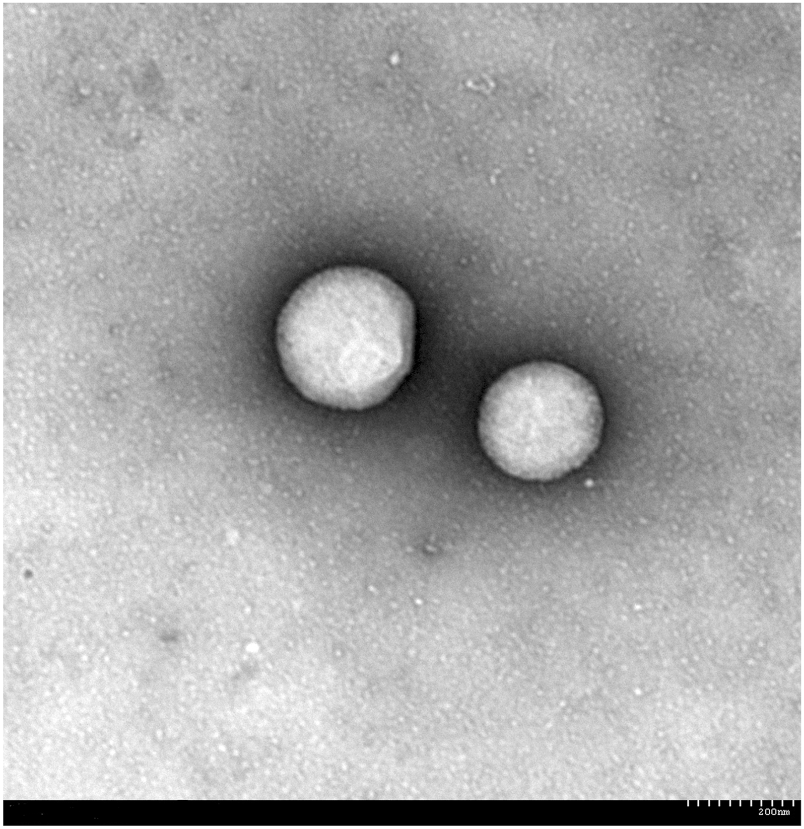
Morphologic observation of VE-HDL by transmission electron microscope ( × 10,000). VE-HDL, vitamin E-containing HDL.
Effects of VE-HDL on Foam Cell Formation
Macrophage foam cells, a defined pathologic feature of atherosclerotic lesions, are characterized by CE accumulation within intracellular lipid droplets. 14 During the period of foam cell formation, LDL plays a major role as a cholesterol donor. In our study, after incubation with oxLDL, the concentrations of intracellular TC and CE increased markedly. Meanwhile, the ratio of CE/TC was over 60%, which means that the macrophages were induced to form cells. VE-HDL could decrease TC, CE, and the ratio of CE/TC, so, it may prevent foam cell formation induced by oxLDL (Table 2).
Effects of VE-HDL on Foam Cell Formation (x
P < 0.001 versus normal control group; b P < 0.05, c P < 0.01 versus model control group.
CE, cholesteryl ester; TC, total cholesterol.
The uptake of oxLDL by macrophages induces foam cell formation and promotes the development of atherosclerosis. 15 After being treated with oxLDL and different dosages of VE-LDL, lipid staining was assessed histologically using Oil red O staining. Oil red O-positive lipid droplets were observed in model control group macrophages after being treated with oxLDL for 24 h. After being treated with different dosages of VE-HDL, the fat deposition in macrophages reduced. Higher concentrations of VE-HDL resulted in large decreases in the number of droplets (Fig. 2).

Oil Red O staining of macrophages ( × 200). NC group was incubated with DMEM, MC groups were treated with oxLDL (80 mg/L), and LD, MD, and HD groups were treated with oxLDL (80 mg/L) and VE-HDL (25, 50, and 100 mg/L, respectively). DMEM, Dulbecco's modified Eagle's medium; HD, high-dose; LD, low-dose; MC, model control; MD, middle-dose; NC, normal control; oxLDL, oxidized low-density lipoprotein.
Cellular Cholesterol Efflux Assays In Vitro
HDL plays a predominant role to prevent atherosclerosis by taking part in the process of reverse cholesterol transport. HDL could promote the efflux of excess cholesterol from peripheral tissues (including macrophages) and return it to the liver for biliary excretion. In our studies, after treated with VE-HDL at different dosages, the medium and macrophages were collected to perform cellular cholesterol efflux assays. As a result (Fig. 3), VE-HDL treatment caused a dose-dependent increase of cellular cholesterol efflux in macrophages at 24 h.
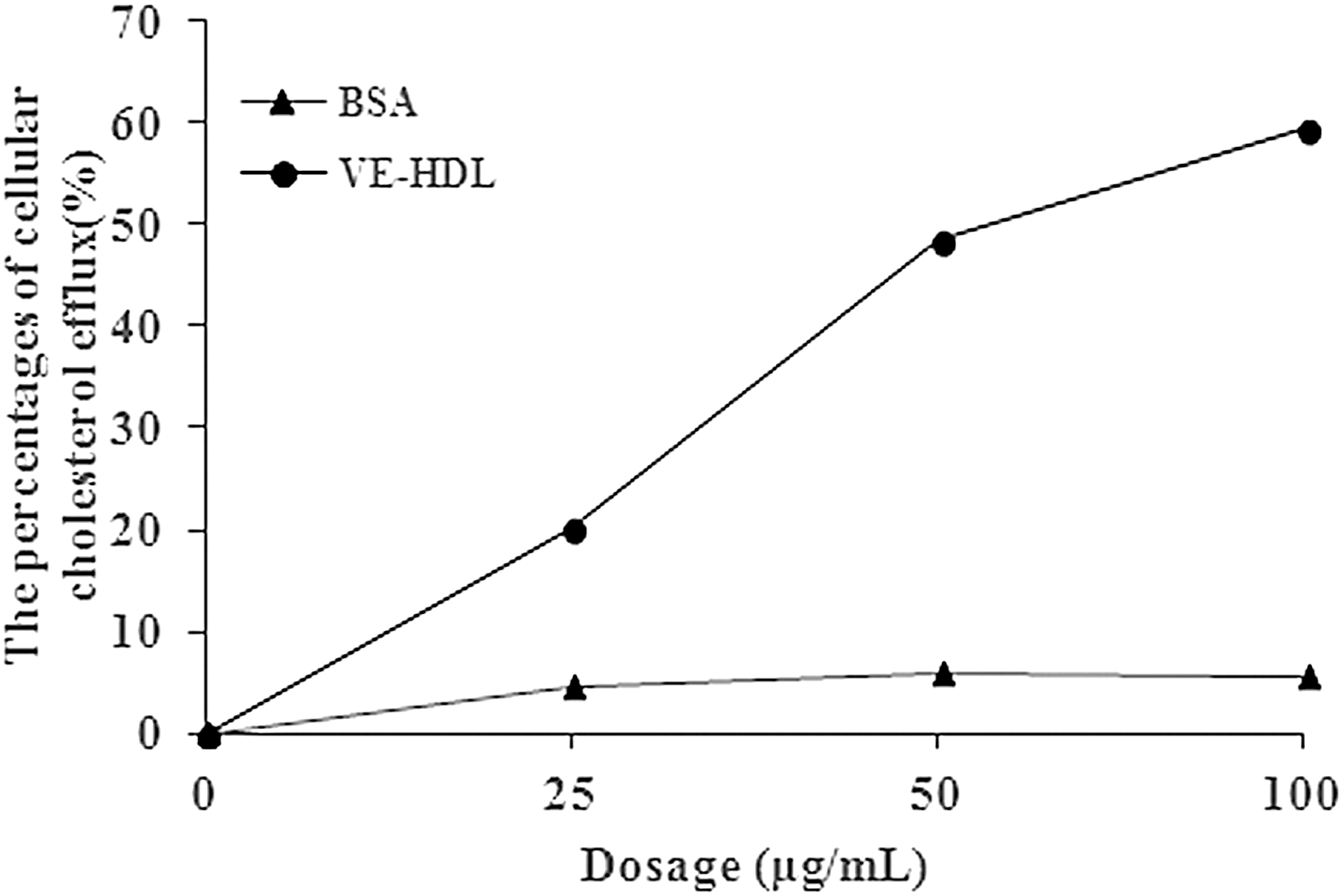
Cellular cholesterol efflux assays. Dose–effect curve of cellular cholesterol efflux facilitated by BSA (triangles), VE-HDL (circles).
Effects of VE-HDL on oxLDL-Stimulated Oxidative Stress of Macrophages
The enzyme SOD neutralizes superoxide radicals by changing it into molecular oxygen and hydrogen peroxide, thereby preventing the formation of highly aggressive compounds. It is proved to play a key role in cellular defenses against oxidative damage. In our study, after microphages were treated with oxLDL, the activity of SOD reduced markedly. Both VE-HDL and native HDL could increase the cellular SOD and enhance antioxidative ability of macrophages (Fig. 4A). MDA is an index of lipid peroxidation and a marker of oxidative stress. As a result, both VE-HDL and native HDL could reduce the concentration of MDA, which is induced by oxLDL in macrophages (Fig. 4B). The effect of VE-HDL is better than native HDL on oxLDL-stimulated oxidative stress of macrophages in the same dosage (100 mg/L). The results mean that VE-HDL has stronger antioxidant ability than native HDL.

Effects of VE-HDL on oxLDL-stimulated oxidative stress of macrophages. Effects of VE-HDL on SOD
Nrf2 is a crucial transcription factor mediating protection against oxidants. Antioxidant properties of Nrf2 are thought to be mainly exerted by stimulating transcription of antioxidant proteins. 16,17 In resting cells, Nrf2 is mainly located in cytoplasm, associated with Kelch-like ECH-associated protein 1 (Keap1), and degraded by proteasome. Upon oxidative stress and kinase-activating signaling pathways, Nrf2 escapes from the Keap1-Cul3 complex and translocates into the nucleus, then plays a part in antioxidation. In our studies, after the macrophages were incubated with VE-HDL, the concentration of Nrf2 in the nucleus increased (Fig. 5).
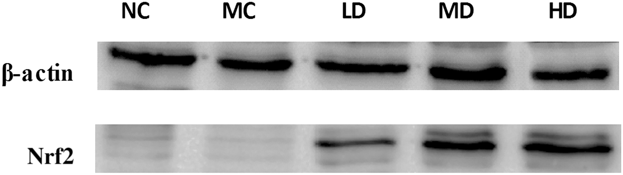
Western blot analysis of nuclear Nrf2 in macrophages. NC group was incubated with DMEM, MC groups were treated with oxLDL (80 mg/L), and LD, MD, and HD groups were treated with oxLDL (80 mg/L) and VE-HDL (25, 50, and 100 mg/L, respectively).
Effects of VE-HDL on oxLDL-Stimulated Apoptosis of Macrophages
As the result of CCK-8 assay, oxLDL treatment causes inhibition of growth on RAW267.4 macrophages. Meanwhile, cell debris could be observed in culture solution. VE-HDL could protect macrophages by inhibiting the function of oxLDL (Fig. 6). The result of immunocytochemistry showed that oxLDL and VE-HDL could affect the expression of apoptosis-related proteins. Being treated with oxLDL could increase the expression of Bax, cleaved-caspase-3, and caspase-8, meanwhile, could reduce the expression of Bcl-2. VE-HDL could protect macrophages by decreasing the apoptosis caused by oxLDL. After being treated with different dosages of VE-HDL, the expression of Bax, cleaved-caspase-3, and caspase-8 was reduced, and the concentration of Bcl-2 in macrophages was elevated (Fig. 7).

Effects of VE-HDL on oxLDL-Stimulated apoptosis of macrophages (CCK-8 Assay). NC group was incubated with DMEM, MC groups were treated with oxLDL (80 mg/L), and LD, MD, and HD groups were treated with oxLDL (80 mg/L) and VE-HDL (25, 50, and 100 mg/L, respectively). CCK-8, Cell Counting Kit-8. ***p < 0.001 versus normal control; ▴▴ p < 0.01 versus model control; ▴▴▴ p < 0.001 versus model control.
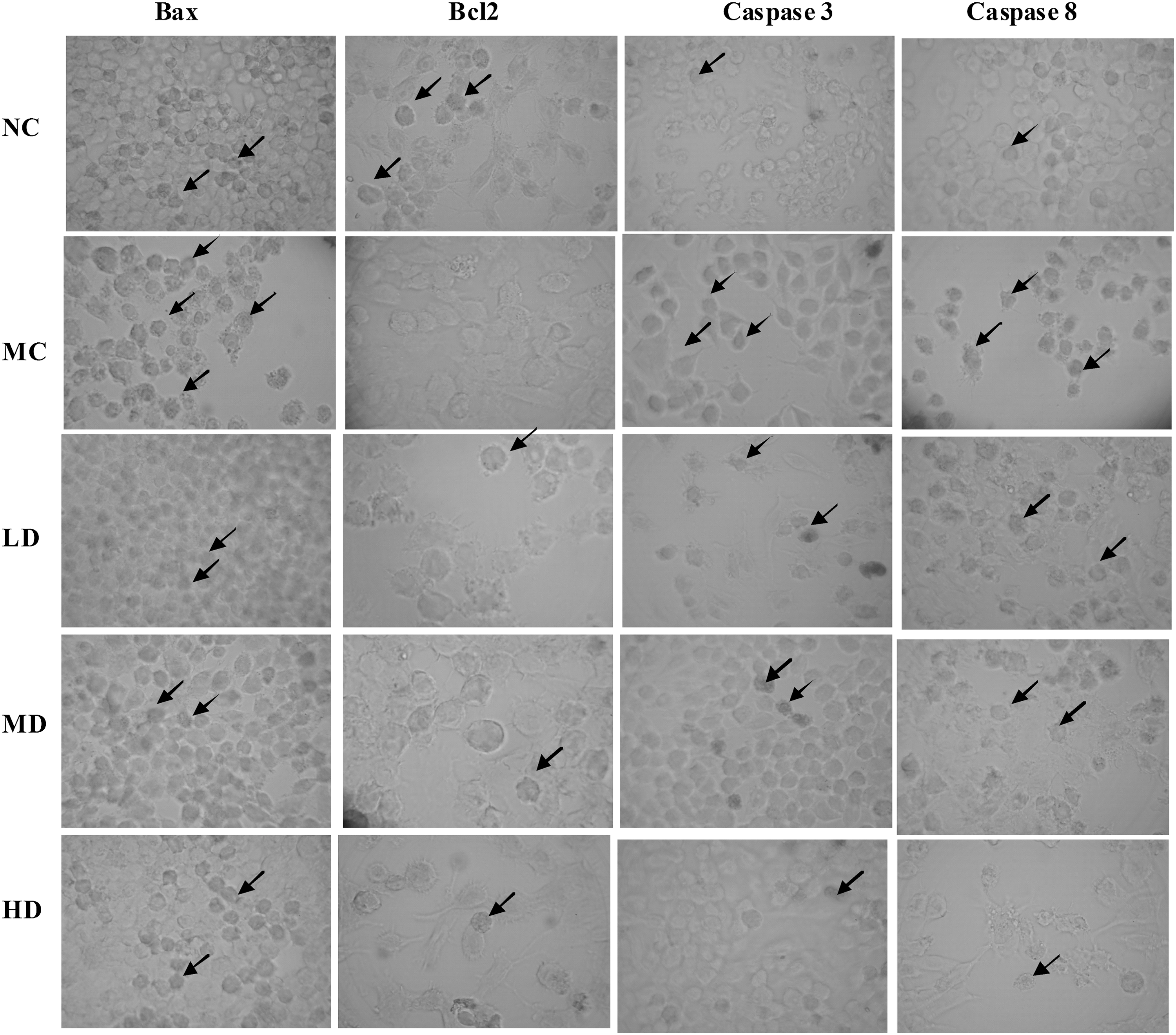
Effects of VE-HDL on apoptosis-related proteins in macrophages (immunocytochemistry, 200 × ). NC group was incubated with DMEM, MC groups were treated with oxLDL (80 mg/L), and LD, MD, and HD groups were treated with oxLDL (80 mg/L) and VE-HDL (25, 50, and 100 mg/L, respectively). The arrows point out the positive cells.
Discussion
To obtain a new type of reconstituent HDL, which could have stronger antioxidant activity, we used hydrophobic vitamin E and apolipoproteins (recombinant human ApoA-I and recombinant human ApoE) to prepare vitamin E-containing HDL. Cholate can facilitate penetration of apolipoproteins between the phosphatidylcholine acyl chains. Since cholate could disrupt the phosphatidylcholine lattice by intercalation and solubilization, it must be removed by dialysis after being incubated at 4°C. 4,18 In our studies, we obtained VE-HDL that contained particles of different diameters (150–200 nm). As the phosphatidylcholine to cholate ratio could influence the particles diameters of reconstituent HDL, we can get VE-HDL with different particles diameters to meet the experimental needs in the following studies.
Then, we studied the protective efficacy of VE-HDL on macrophages by a series of experiments. The results showed us that VE-HDL could induce cellular cholesterol efflux, reduce the fat deposition in macrophages, and inhibit foam cell formation. Meanwhile, we noticed that VE-HDL could increase the cellular SOD and reduce the level of MDA. It means that VE-HDL could reduce oxLDL-stimulated oxidative stress and enhance antioxidative ability of macrophages. To find the mechanisms of the antioxidative effect of VE-HDL, we tested the Nrf2 protein in nucleus. The result showed that the concentration of Nrf2 in the nucleus increased after the macrophages were treated with VE-HDL. This transformation means that Nrf2 escapes from the Keap1-Cul3 complex in cytoplasm, translocates into the nucleus, and then plays a part in antioxidation. So, Nrf2 pathway may be one of the mechanisms of antioxidation of VE-HDL. Furthermore, we measured the effects of VE-HDL on apoptosis of macrophages. VE-HDL could inhibit the apoptosis of macrophages, which was caused by oxLDL through regulating the expression of apoptosis-related proteins.
As mentioned above, we prepared a new type of reconstituent HDL with apolipoproteins and vitamin E for the first time. VE-HDL has protective efficacy on macrophages. It has the prospect of becoming a therapeutic agent on atherosclerosis in the future.
Footnotes
Acknowledgments
This work was supported by grants from the National Natural Science Foundation of China (81401221), China Postdoctoral Science Foundation (2016M591490), Jilin Science and Technology Funds (20140520047JH, 20150204007YY, 2014 G073, and 20140204022YY), Jilin Province Postdoctoral Program, Education Department of Jilin Province (2015500), Health Department of Jilin Province (2014Q023), and Norman Bethune Program of Jilin University (2015333).
Disclosure Statement
No competing financial interests exist.