
Abstract
The aim of the investigation was to search out the possible corrective effect of hydro-methanolic extract:: 3:2 of Musa balbisiana flower on streptozotocin (STZ)-induced diabetic male rat. In this concern, glycemic profile, oxidative stress profile, lipid profile, and toxicity profile were studied where a genomic approach has been taken to explain the mechanism of action of the said extract for such recovery. Such enzyme kinetics domain and genomic domain of concerned profile were not covered till now to explore the mechanism of diabetes management by this plant part. As hexokinase is one of the important enzymes of glycolysis and glycogenesis, and GLUT-4 an important transporter of glucose in skeletal muscle, liver and adipose tissue these genes have been selected here. For focusing the status of apoptosis in hepatic tissue, an important organ for carbohydrate metabolism, Bax and Bcl-2 gene expression were included, which are the novelty of this study. The hydro-methanolic extract was administered orally at the dose of 10 mg/100 g body weight for 28 days to diabetic rats. Abovementioned extract exhibited a significant recovery in parameters like fasting blood glucose; serum insulin; glycated hemoglobin; antioxidative enzymes in hepatic and renal tissue; and carbohydrate metabolic enzymes in hepatic and skeleto-muscular tissue along with proappoptotic gene Bax and antiappoptotic gene Bcl-2, glycemic genes like Hex-I, and GLUT-4 in hepatic tissue of STZ-induced diabetic rat. This investigation demonstrated the potentiality of hydro-methanolic extract (3:2) of M. balbisiana flower for correction of diabetes and diabetes-induced oxidative stress.
Introduction
Diabetes mellitus, a metabolic disease is associated with multiple etiologies and characterized by a bunch of symptoms. This pathophysiological condition exists with abnormal high blood glucose and deficiency of insulin or resistance to insulin or both. 1 Diabetes is the root for raising several metabolic complications such as nephropathy, retinopathy, and cardiomyopathy. 2 To avoid the risk of serious complications from diabetes such as heart and blood vessels diseases, controlling of only blood glucose level is not sufficient but regulation of blood lipid levels is also necessary. Hyperlipidemia is a characteristics feature of streptozotocin (STZ)-induced diabetic rats. 3 This noncommunicable global health disease has been spreading at an alarming rate around the world. Above 230 million people are suffering from diabetes worldwide and the number will be reach 350 million by 2025. 4
Since diabetes is a chronic multifactorial syndrome its treatment is complicated and needs multiple therapeutic strategies. Oxidative stress plays a pivotal role in the pathogenesis of diabetic complications. 5 So, an antidiabetic drug should be a combination of both hypoglycemic and antioxidative properties. Currently available synthetic drugs are not satisfactory effective therapy for treating this disease due to its many side effects. Since diabetes invites many diseases there is a need for strong treatment to keep away from other diseases. Though, there are various approaches to reduce the ill effects of diabetes and its secondary complications, herbal formulations are preferred due to lesser side effects and low cost.
The use of herbal medicines for the treatment of diabetes mellitus has gained importance throughout the world. Phytomedicine is capable of treating the disease with fewer side effects and less expense and will be of great help to diabetic patients especially due to the extended belief that natural treatments cause less harm to the organism than synthetic ones. Some of the plants have potent antidiabetic activities and having immense folk reputation. There are numerous traditional medicinal plants reported to have hypoglycemic properties such as Eeugenia jambolana, 6 Camellia sinensis (Green tea), 7 Trigonella foenum (Fenugreek), 8 and Momordica charantia (Bitter gourd). 9 Many of these are less effective in lowering glucose levels in severe diabetes. Musa balbisiana (colla) is a banana plant species, which belongs to Musaceae family and it has already been proven as having antihyperglycemic activity but that study has covered some limited and basic parameters to search out the antidiabetic efficacy of flower of M. balbisiana. 10,11
No study has been conducted on the phytoingredient present in this plant focusing on phytomolecule–gene interaction, the important domain of pharmacogenomics. For this purpose, gene expression of hexokinase gene has been considered as the enzyme closely involved in glucose metabolism through glycolysis and gluconeogenesis having Km value far less than glucokinase. 12 Moreover, GLUT-4 is involved in glucose transportation through membrane of skeletal muscle, adipocytes and hepatocytes. So, its gene expression in this experiment has been considered. 13 Hepatic tissue is the prime site of metabolism especially carbohydrate metabolism, So, apoptosis level of this tissue in diabetes and extract treated condition have been studied by focusing on one proapoptotic gene, that is, Bax and another antiapoptotic gene, that is, Bcl-2. This is a novel experiment, in respect to previous studies, covering this plant for management of diabetes. Moreover, we are in the dark about this plant's mechanism of action for diabetes management at genomic level.
This study was designed to find out the untouched area: carbohydrate metabolic enzymes activity and gene expression of relevant biosensors about the mechanism of diabetic rectification potentiality of this extract. For this purpose, we have studied the glycemic sensors, oxidative biomarkers, toxicity profiles, and gene expression of Bax, Bcl-2, Hex-I, and GLUT-4.
Materials and Methods
Plant Material Collection
The fresh M. balbisiana's flowers were collected from local market of Paschim Medinipur, West Bengal, India and were recognized by Prof. Ram Kumar Bhakat, taxonomist of Botany and Forestry department, Vidyasagar University, Midnapore. The voucher specimen Bio-Med/V.U/M.B/16/09 have been submitted in the Herbarium of the Botany Department.
Preparation of Flower Extract
The hydro-methanolic:: 3:2 extract of flower of M. balbisiana has been prepared as per our standard method reported earlier. 14 In brief, by using electric grinder the dry flower was crushed and then pulverized. Then 100 g flower dust was suspended in 500 mL of hydro-methanol:: 3:2 and kept in room temperature with intermittent stirring for first 2 h. After completion of 48 h at room temperature, the mixture was filtered through Whatman grade No. 3 filter paper and filtrate was evaporated under low pressure using rotary evaporator (HAHN-SHIN HS-2000NS, Korea) and then residue was preserved in refrigerator at 4°C for experiment. The extract was dissolved in distilled water and administered orally to experimental diabetic rats at a specific dose.
Selection of Animal and Animal Care
The present experiment was conducted by using matured 18 normoglycemic Wistar strain male albino rats having fasting blood glucose (FBG) level 80–90 mg/dL, 3 months of old, and weighing about 120 ± 10 g. Six rats were housed in each hygienic polypropylene made cage. Earlier to the experiment, rats were acclimated for 15 days in our laboratory condition. Standard atmosphere in the animal house was maintained with dark-light cycle (12:12 h) and room temperature at 25°C ± 2°C. Rats were fed with nutritionally adequate pellet diet and water ad libitum for specific duration. The principles of laboratory animal care and instruction given by our Institutional Ethical Committee (IEC) [IEC/9/4/18, dated February 2, 2018] were followed throughout the experiments.
Experimental Induction of Diabetes Mellitus
Diabetes mellitus was induced in vivo through a single intraperitoneal injection of STZ at the dose of 4 mg/0.1 mL of citrate buffer/100 g body weight into overnight fasted rats. After 48 h of STZ injection rats having FBG level more than 300 mg/dL were selected for this experiment. On seventh day of STZ injection, these hyperglycemic rats were used for this study. In this way, twelve rats were made diabetic.
Experimental Design
The animals were divided into three groups; six animals were included in each group.
Group I (Vehicle treated control group): Normoglycemic adult animals of this group were treated with distilled water orally (0.5 mL/100 g body weight/rat) for 28 days.
Group II (Vehicle treated diabetic group): In this group, diabetic rats received vehicle (0.5 mL distilled water/rat/day) orally for 28 days.
Group III (Hydro-methanolic (3:2) extract treated diabetic group): Diabetic rats of this group were treated with oral administration of the hydro-methanol::3:2 extract of flower of M. balbisiana at the dose of 10 mg/0.5 mL/100 g body weight/day for 28 days. Animal feed was supplied 2 h before and after extract/vehicle treatment. It has been adopted to minimize the drug–nutrient interaction if any.
Treatment was started from the seventh day of STZ injection to the rats. On 29th day of drug treatment, all the animals were sacrificed by decapitation using euthanasia and their body weights were recorded. Blood was collected from dorsal aorta by a syringe and the serum was separated by centrifugation at 4,000 rpm for 10 min for the estimation of different parameters. Different tissues like liver, kidney, pancreas, and skeletal muscular tissues were dissected out from each animal, washed properly in normal saline, soaked in blotting paper, and stored in deep freezer (−20°C) for the measurement of different relevant biomarkers and genomic sensors in this concern.
Measurement of FBG
FBG level was measured in all of the rats in each group as per our reported method by Single Touch Glucometer on every seventh day. 15
Assay of Serum Insulin Level and Glycated Hemoglobin Level
The level of serum insulin was measured by enzyme-linked immunosorbant assay using the kit (Ray Biotech, Norcross, GA). The intra-assay variation was 4.9%. The tissue samples were run at a time to ensure no inter-assay variation. The insulin level in serum was expressed in μIU/mL. 16 Glycated hemoglobin (HbA1C) level was measured according to standard method. 17
Assay of Hexokinase Activity in Hepatic Tissue and Skeletal Muscle
Hexokinase activity in hepatic tissue and skeletal muscles were determined spectrophotometrically. The assay mixture consists of 3.7 mM glucose, 7.5 mM MgCl2, 11 mM thioglycerol, and 45 mM HEPES buffer. All the samples were homogenized at 50 mg tissue/mL concentration by using ice-cold 0.1 M phosphate-buffered saline (pH 7.4). After that, 0.9 mL of assay mixture was added with 0.01 mL NADP and 0.03 mL of ATP in a spectrophotometer cuvette and mixed properly. Then, into this cuvette, 0.1 mL supernatant tissue was added and the absorbance was noted at 340 nm. One unit of this enzyme activity was expressed as unit/mg of tissue. 18
Assay of Glucose-6-Phosphate Dehydrogenase Activity in Hepatic Tissue and Skeletal Muscle
The activity of glucose-6-phosphate dehydrogenase in hepatic and skeletal muscles was measured spectrophotometrically according to the standard method. 19 Concentration of tissue was 50 mg/mL and it was homogenized in ice-cold 0.1 M phosphate-buffered saline (pH 7.4). Activities of glucose-6-phosphate dehydrogenase in hepatic and skeletal muscles were measured by substrate, as glucose-6-phosphate. The absorbance was recorded at 340 nm.
Biochemical Assay of Catalase Activity of Hepatic and Renal Tissues
Hepatic and renal catalase (CAT) activity was measured according to the standard protocol. Both that tissues were homogenized separately by using 0.05 M Tris-HCl buffer (pH 7.0) at a concentration of 50 mg/mL. Centrifugations of these homogenized samples were performed at 10,000 × g for 10 min at 4°C. In a spectrophotometer cuvette, 0.5 mL of 0.00035 M H2O2 and 2.5 mL of distilled water were added and mixed properly. Optical density was noted at 240 nm before the addition of supernatant. From the sample, 40 μL supernatant was added to the cuvette and at 30 s interval six readings were noted. 20
Biochemical Assay of Superoxide Dismutase Activity of Liver and Kidney Tissue
Superoxide dismutase (SOD) activity was estimated in hepatic and renal tissues by assessing the percentage of inhibition in auto-oxidation of pyrogallol by SOD according to the standard protocol. The buffer preparation was performed by 50 mM TRIS (pH 8.2). In a cuvette, 2.04 mL of TRIS buffer, 20 μL of sample, and 20 μL of pyrogallol were taken and mixed properly. The OD was taken at 420 nm for 3 min period. 21
Biochemical Estimation of Lipid Profile in Serum
The concentrations of total cholesterol (TC), 22 triglycerides (TG), 23 low-density lipoprotein cholesterol (LDL-C), 24 and high-density lipoprotein cholesterol (HDL-C) 25 in serum were estimated by using diagnostic kits (Span Diagnostic Ltd., Surat, India) on a semi auto analyzer. The unit of the value was expressed in mg/dL.
Quantification of Malondialdehyde in Hepatic and Renal Tissue
According to the standard method, malondialdehyde (MDA) levels of the liver and renal tissues were quantified 26 spectrophotometrically and readings were recorded at 535 nm.
Estimation of Lipid Peroxidation from the Concentration of Conjugated Diene
Tissues were homogenized at the concentration of 50 mg/mL in 0.1 M of ice-cold phosphate buffer (pH 7.4) and the homogenates were centrifuged at 10,000 × g at 4°C for 5 min. The supernatant was used for the estimation of CD. Quantification of the CD was performed by standard method. The lipids were extracted with chloroform-methanol (2:1) mixture followed by centrifugation at 1,000 × g for 5 min. The lipid residue was dissolved in 1.5 mL of cyclohexane and the absorbance was noted at 233 nm to measure the amount of hydroperoxide formed. 27
Biochemical Assay of Toxicity Profile
For the assessment of activities of serum glutamate oxaloacetate transaminase (SGOT) and serum glutamate pyruvate transaminase (SGPT), specific kits were supplied by Creast Bio-systems, Gitanjali, Dr. Antonio Do RegoBagh, Alto Santacruz, Bambolim Complex (Goa, India). 28
mRNA Isolation and Complementary DNA Synthesis
Total RNA was extracted using kit High Pure RNA Tissue Kit (Roche Diagnostic, Germany) from target tissue and cDNA was synthesized using Transcriptor First Strand cDNA Synthesis Kit (Roche Diagnostic). 29
Gene Expression Study by Quantitative Real-Time Polymerase Chain Reaction
Light Cycler 480 II (Roche Diagnostic) was used for the assessment of gene expression of Bax, Bcl-2, Hex-I, and GLUT-4 of hepatic tissues. 30
Statistical Analysis
Analysis of variance (ANOVA) followed by multiple comparison two-tail t test was performed for statistical analysis of collected data. 31 All the data of this experiment were indicated by mean ± standard error of the mean (n = 6). For statistical analysis, significant differences were considered at the level of p < 0.05.
Results
FBG and HbA1C Levels
FBG and HbA1C levels were significantly elevated (p < 0.05) in STZ-induced diabetic rats in respect to the vehicle treated control group. Treatment with hydro-methanol::3:2 extract of flower of M. balbisiana at the dose of 10 mg/100 g of body weight/day for 28 days in the diabetic animals resulted in significant diminution (p < 0.05) in FBG (Table 1) and HbA1C levels (Fig. 1) when compared with vehicle treated diabetic group.

Resettlement of serum insulin level and HbA1C level after treatment of hydro-methanolic (3:2) extract of Musa balbisiana flower at 10 mg/100 g body weight/day dose in diabetic rat in comparison with vehicle treated diabetic rat. ANOVA followed by multiple comparison Student's two-tailed t test was performed. Bars denote mean ± SEM (n = 6). Bars with different superscripts (a, b, c) differ from each other significantly, p < 0.05. ANOVA, analysis of variance; HbA1C, glycated hemoglobin; SEM, standard error of the mean.
Ameliorative Effect of Hydro-Methanolic (3:2) Extract of Musa balbisiana Flower on Fasting Blood Glucose Level in Streptozotocin-Induced Diabetic Male Albino Rat in Comparison with Vehicle Treated Diabetic Group
Data were expressed as mean ± SEM (n = 6). Values with different superscripts (a, b, c) in each vertical column differ from each other significantly, p < 0.05.
Bold values are the effective extract in this experiment.
FBG, fasting blood glucose; SEM, standard error of the mean; STZ, streptozotocin.
Serum Insulin Level
A significant (p < 0.05) diminution in serum insulin level was observed in STZ-induced diabetic group compared with the control group. A significant recovery (p < 0.05) in serum insulin level was noted when compared with the vehicle treated diabetic group after treatment with hydro-methanolic::3:2 extract of flower of M. balbisiana to diabetic animals at the dose of 10 mg/100 g of body weight/day for 28 days (Fig. 1).
Carbohydrate Metabolic Enzyme Activities
Activities of hexokinase and glucose-6-phosphate dehydrogenase in liver and skeletal muscular tissues were diminished significantly (p < 0.05) in STZ-induced diabetic group in comparison with the control group. In contrast, administration of extract of flower of M. balbisiana at the dose of 10 mg/100 g of body weight for 28 days to the STZ-induced diabetic animals showed a significant elevation (p < 0.05) in the activities of the enzymes in respect to the vehicle treated diabetic group (Fig. 2).

Recovery in the activities of hexokinase and glucose-6-phosphate dehydrogenase in liver and skeletal muscle after treatment with hydro-methanol extract of Musa balbisiana flower to vehicle treated diabetic rat. Values were expressed as mean ± SEM, n = 6. ANOVA followed by multiple comparison Student's two-tailed t test was performed. Bars with different superscripts (a, b, c) differ from each other significantly, p < 0.05.
Biochemical Estimation of Antioxidant Enzymes Activities
Activities of antioxidant enzymes like CAT and SOD in hepatic and renal tissues were significantly reduced (p < 0.05) in STZ treated diabetic rat when compared with the vehicle treated control rat. Administration of hydro-methanolic::3:2 extract of flower of M. balbisiana at the dose of 10 mg/100 g of body weight/day for 28 days to the diabetic rat resulted a significant (p < 0.05) recovery in the levels of said parameters compared to the vehicle treated diabetic group (Fig. 3).

Ameliorative effect of hydro-methanolic extract at a particular dose in the activities of CAT and SOD and on the levels of malondialdehyde and conjugated diene in hepatic and renal tissues in comparison to vehicle treated diabetic rat. ANOVA followed by multiple comparison Student's two-tailed t test was performed. Values were represented mean ± SEM (n = 6). Bars with different superscripts (a, b, c) differ from each other significantly, p < 0.05. CAT, catalase; SOD, superoxide dismutase.
Biochemical Evaluation of Oxidative Stress End Product Level
The levels of CD and MDA in hepatic and renal tissues were increased significantly (p < 0.05) in STZ-induced diabetic rat compared to the vehicle treated control rat. A significant (p < 0.05) improvement in the levels of said sensors were noticed compared to the vehicle treated control after the oral administration of said extract for 28 days to the diabetic rat (Fig. 3).
Serum Lipid Profile
Serum TC and TG levels were significantly elevated (p < 0.05) in STZ-induced diabetic group in respect to the vehicle treated control group. Treatment of this extract at the abovementioned dose to diabetic animals showed a partial recovery in the levels of these parameters toward the control level. Lipid profile like serum LDL-C level was elevated and serum HDL-C was lowered significantly in diabetic group when comparison was made with vehicle treated control group. Oral administration of the said extract to diabetic animal exhibited a significant resettlement in serum low-density lipoprotein and high-density lipoprotein levels toward the vehicle treated control group (Table 2).
Ameliorative Effect of Hydro-Methanolic Extract of Musa balbisiana Flower on Serum Lipid Profile in Streptozotocin-Induced Diabetic Male Albino Rat in Comparison with Vehicle Treated Diabetic Group
Data were expressed as mean ± SEM (n = 6). Values with different superscripts (a, b, c) in each vertical column differ from each other significantly, p < 0.05.
Bold values are the effective extract in this experiment.
HDL-C, high density lipoprotein cholesterol; LDL-C, low-density lipoprotein cholesterol; TC, total cholesterol; TG, Triglyceride.
Assay of Toxicity Markers
Activities of SGOT and SGPT were increased significantly (p < 0.05) in the diabetic animals compared with the control animals. Treatment with extract of flower of M. balbisiana in diabetic rats resulted in significant diminution (p < 0.05) in the activities of both enzymes in respect to the vehicle treated diabetic group (Fig. 4).
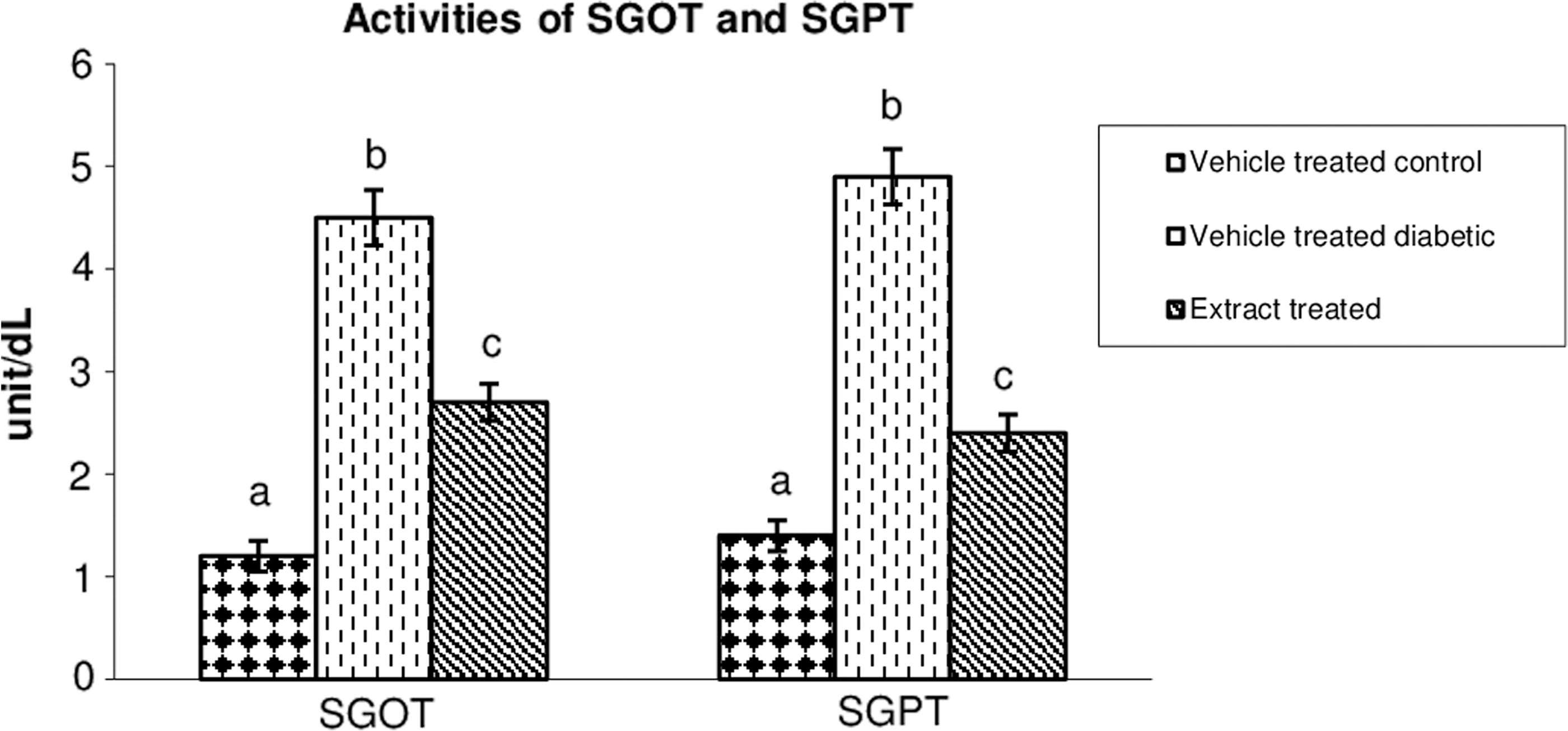
Assessment of SGOT and SGPT activities after the treatment of with hydro-methanolic extract of Musa balbisiana flower at 10 mg dose to experimental diabetic rat. ANOVA followed by multiple comparison Student's two-tailed t test was performed. Values were expressed as mean ± SEM, n = 6. Bars with different superscripts (a, b, c) differ from each other significantly, p < 0.05. SGOT, serum glutamate oxaloacetate transaminase; SGPT, serum glutamate pyruvate transaminase.
Gene Expression Study by Quantitative Real-Time Polymerase Chain Reaction
Hepatic Bax gene showed a significant upregulation in the expression (p < 0.05) in STZ treated diabetic group when compared with vehicle treated control. Oral administration of hydro-methanolic extract for 28 days to the diabetic group exhibited a significant recovery in the concerned gene expression towards the vehicle treated control.
Similarly, Significant downregulation of the hepatic Bcl-2, GLUT-4, and Hex-I gene expression was noted in diabetic group in respect to vehicle treated control group. Significant recovery (p < 0.05) toward vehicle treated control group was noted after extract administration for 28 days in the diabetic group (Fig. 5).

qRT-PCR study of gene expression of Bax, Bcl-2, Hex-I, and GLUT-4 in hepatic tissue after treatment with hydro-methanolic extract of Musa balbisiana flower at 10 mg dose to STZ-induced diabetic rat. ANOVA followed by multiple comparison Student's two-tailed t test. Values were expressed as mean ± SEM, n = 6. Values with different superscripts (a, b, c) differ from each other significantly, p < 0.05. qRT-PCR, quantitative real-time polymerase chain reaction; STZ, streptozotocin.
Discussion
Burden of diabetes is spreading globally at alarming rate. An appropriate diet is considered as a milestone for diabetes management. 32 The M. balbisiana flower is a popular vegetable in Eastern South Asia, Northern SouthEast Asia, Malaysia, Indonesia and Srilanka. This experiment was designed to explore the possible corrective effect of hydro-methanolic (3:2) extract of M. balbisiana flower on STZ-induced diabetic rat. Moreover, this study also focused on the phytomolecule–gene interaction that is also empowering the knowledge bank of pharmacogenomics in connection with diabetes management by phytoingredients, which is also the novelty of this work in respect to previous work in this line. 10
The administration of M. balbisiana flower extract has showed decreased FBG level that may be due to the stimulation of pancreatic islet cell for insulin release, which plays a crucial role in controlling FBG. 1 In this study, the supplementation of hydro-methanolic extract of M. balbisiana flower at the dose of 10 mg/100 g body weight/day effectively elevated the serum insulin level in STZ-induced diabetic rats. The possible mechanism of extract may be due to generation of β cells from existing stem cells in Islet, 33 which has been proved by significant diminution in HbA1C after the extract treatment in diabetic group since elevated HbA1c is due to low level of insulin or insulin resistance. 34
Carbohydrate metabolic enzymes play a pivotal role in the maintenance of glucose homeostasis. The activities of these enzymes were decreased in diabetes due to insufficient insulin production by beta cell. 35 The remedial effect of the extract was observed from the elevation in the activities of hexokinase and glucose-6-phosphate dehydrogenase in hepatic and skeletal tissue. This result supported the possibility that high expression of Hex-I gene increased the glucose utilization in cell. Simultaneously, upregulation of gene expression of one of the glucose transporters, GLUT-4 revealed the antihyperglycemic activity of the M. balbisiana flower as treatment with the extract recovered GLUT-4 expression. As a result, glucose can be transported inside the cell and the glucose level is maintained in the blood.
Decreased activities of antioxidant enzymes like SOD, catalase and elevation in oxidative stress end products like CD and MDA levels were noticed in diabetic condition probably due to the direct influence of diabetes on the oxidative stress. 36,37 Oral administration of the extract resettled the antioxidant profile in diabetic rat may be due to the presence of antioxidant in the extract or indirectly, the phytomolecules present in extract may stimulate such antioxidant enzymes. Moreover, one of the most important characteristic of STZ-induced diabetes is hyperlipidemia. Elevation in lipid profile of STZ-induced diabetic rat was resettled toward the vehicle treated control after the extract treatment. This mechanism indicates that the extract of M. balbisiana flower has hypolipidemic activity that may be due to decreased choleterogenesis. 38
The results also indicated that diabetes-induced oxidative stress accelerate the proapoptotic gene expression and decreased the antiapoptotic gene expression, focused in this study. Administration of the extract at the dose of 10 mg/100 g body weight/day exhibited a significant recovery in the apoptotic markers toward the vehicle treated control and thus it recovers the apoptosis. This is probably due to anti apoptotic nature of said plant part.
The activities of SGOT and SGPT are the most important general toxicity markers and are also found in different diseases including diabetic state. 39 SGOT and SGPT activities were recovered in diabetic group after extract treatment. This focused on the safety of using the extract as it reduces the toxicity present during diabetic state. From the outcome, it may be hypothesized that the extract may recover the diabetic status either by resettling the activities of carbohydrate metabolic enzyme and glucose transporter by acting at genomic level, which maintains the glucose homeostasis or by recovering the diabetes-induced β cell degeneration. Another view may be antiapoptotic capacity of the phytomolecule present in the extract that recovers the hepatocytes, mainly involved in carbohydrate metabolic homeostasis.
Conclusion
From the outcome of the study, it may be concluded that hydro-methanolic::3:2 extract of M. balbisiana flower at 10 mg/100 g dose exhibited a potentiality to manage hyperglycemia and oxidative stress in STZ-induced diabetic rats.
Footnotes
Acknowledgment
The research work was funded by Moulana Azad National Fellowship (UGC).
Disclosure Statement
No competing financial interests exist.