
Abstract
Retinopathy is one of the most common complications of diabetes. Approximately 80% of patients with diabetes history for over 10 years suffer from some degree of diabetic retinopathy (DR). Currently available treatments include use of antivascular endothelial growth factor-165 (VEGF165) agents or steroids. However, they are very expensive, involve an invasive procedure that is painful, and show ocular and systemic complications. Currently, the focus for treatment of such disorders has shifted from new drug discovery to repositioning of available drugs because of the cost and time consumption involved in the former. Working on this strategy, itraconazole (ITR) was selected for treatment of DR due to its potent unutilized antiangiogenic activity for the management of DR. An attempt was made to develop a topical, noninvasive nanostructured lipid carrier (NLC) owing to the potential to carry entrapped drug across the membranes. ITR-NLCs were prepared using high-pressure homogenization by applying Box–Behnken design for optimization. Surface of NLCs was modified by chitosan (CS) coating. ITR-NLCs were examined for antiangiogenic potential and their VEGF165 targeting efficiency. Drug-loaded NLC showed desired particle size, zeta potential, and polydispersity index. In VEGF-induced DR rats, ITR and CS-ITR-NLCs were found to exhibit an antineovascularization effect by targeting VEGF165. The developed CS-ITR-NLC proved to be an effective topical therapy for management of DR, offering the advantages of cost-effectiveness, higher patient compliance, and better tolerance.
Introduction
Diabetic retinopathy (DR) is the leading cause of vision impairment and blindness. 1 However, delivery of drug to the retina is a challenging task. It has been reported that intravitreal administration leads to direct delivery of drugs to the posterior segment of the eye. 1 However, the retinal diseases cannot be controlled by a single intravitreal injection. To achieve therapeutic activity, multiple injections of drug to the retina are administered, in turn leading to several adverse effects such as endopthalmitis and cataract development along with retinal as well as vitreous hemorrhage. 2,3 To overcome these limitations, nanocarriers have been developed to improve drug penetration, leading to optimum ocular bioavailability. 4
Itraconazole (ITR) is an antifungal agent used for the treatment of fungal infections. Recently, ITR has also been demonstrated to exhibit a potent antiangiogenesis activity. 5 It inhibits the endothelial cell proliferation, capillary tube formation, migration, and angiogenesis. Due to this property, ITR is being explored for its potential as a cancer therapeutic. 5,6 Many antivascular endothelial growth factor-165 (VEGF165) agents such as pegaptanib (Macugen®), ranibizumab (Lucentis®), bevacizumab (Avastin®), and aflibercept (Eylea®) are used for the treatment of ocular antiangiogenesis, but their use is associated with significant risks and may affect the patient's quality of life. 7,8 They are very expensive, involve an invasive procedure, and show ocular and systemic complications. Prolonged treatment for chronic diseases such as DR often requires repeated injections, which can lead to severe ocular complications such as endophthalmitis, cataract, vitreous hemorrhage, and retinal detachment. 9,10 Systemic exposure of anti-VEGF agents has established that the systemic VEGF inhibition is associated with the adverse effects, including myocardial infarction, stroke, and hemorrhage. For this reason, ITR and other drug candidates such as steroids are being explored as they are potentially safer and more effective antiangiogenic leads. Being a Biopharmaceutical Classification System class IV drug, ITR is highly lipophilic and exhibits poor penetration through ocular barriers because of its large molecular weight, that is, 705 Da. Moreover, it is highly bound to the protein albumin. Diffusion across the membranes therefore is limited. Lipid-based nanocarriers have been reported to overcome the barriers. In these carriers, the nanostructured lipid carrier (NLC) is the second generation of solid lipid nanoparticles. NLC has an imperfect crystal structure so that the problems of drug loading and instability of system are overcome to a much larger extent compared with that in the solid lipid nanoparticles. 11
Formulation development, especially for the posterior segment of the eye, is a challenging task because static (conjunctival epithelium, corneal epithelium, sclera, Bruch's membrane, choroid, and retinal pigmented epithelium) and dynamic barriers (lacrimation, choroidal and conjunctival blood flow, and lymphatic drainage, as well as efflux to efficiently reject foreign substances and pathogens) limit penetration of therapeutic molecules into the ocular tissues. 12 This causes low bioavailability of drugs that are administered topically through eye drops.
In the present study, ITR-loaded NLC formulation, which can be topically administered to the eye, is reported. ITR-NLCs were prepared by a hot high-pressure homogenization technique, and surface modification was carried out by coating with the cationic polymer chitosan (CS). Cationic NLCs were evaluated for their particle size, entrapment efficiency (EE), and antiangiogenic potential.
Materials and Methods
ITR was procured from BMR Pharma and Chemicals Pvt. Ltd. (India). Tripalmitin was obtained as a gift sample from Gattefosse (Colorcon, India). Tween 80, Transcutol HP and CS were purchased from Sigma Aldrich (India). Cell culture media, Dulbecco's modified Eagle's medium (DMEM), and phosphate-buffered saline solution were purchased from Hi Media (India). Ketamine and chlorpromazine were procured from Cipla Pharmaceuticals (India). All other reagents were of analytical reagent grade and were used without any further purification.
Methods
Screening of the carriers
Solubility and partition coefficient studies of ITR in various solid lipids
Selection of solid lipid was done on the basis of a modified affinity study.
13
Double distilled water (1 mL) was transferred to 5 mL of screw capped glass vial and the excess amount of drug was added. To this mixture, 100 mg of lipid of interest was added and stirred on shaker water bath at 70°C for 1 h at 100 rpm. It was cooled to room temperature and centrifuged at 10,000 g for 10 min to separate the solid lipid from water. The congealed solid lipid and water were suitably diluted with mobile phase, centrifuged at 5,000 g for 5 min, and the percentage of drug entrapped was estimated in both the phases using ultrafast liquid chromatography (UFLC) (n = 3). The partition coefficient (PC) was calculated by the formula given in Equation 1:
where Di is the initial amount of drug taken, and Dw the amount of drug in aqueous phase.
Chromatographic measurements were made on a UFLC (Shimadzu LC2010A HT, Japan) model, which consisted of LC20AD solvent delivery modules, an SPD-M 20A PDA detector, and a Rheodyne injector (model 7125, Rheodyne, Shimadzu, Japan) valve fitted with a 20 μL loop. The system was controlled through a system controller (SCL-10A) and a personal computer using Shimadzu chromatographic software (LC Solution, Release 1.11SP1) installed on it. UFLC analysis was carried out by using a BDS C18 (250 × 4.6 mm, 5 μm) column with a mobile phase of acetonitrile–triethylamine (0.1%) pH 3 in 90:10 v/v ratio at a flow rate of 1.0 mL/min. The detection wavelength was selected as 264 nm and retention time was found to be 3.7 min.
Solubility of ITR in liquid lipids and surfactants
ITR solubility was also determined in various liquid lipids and surfactants. One milliliter of oil and surfactant of interest were taken into 2 mL of centrifuge tube separately. An excess of drug was added to each vial containing oil as well as surfactant and mixed using a vortex mixer (Yorco Instruments, Delhi, India). Afterward, it was kept on an isothermal shaker (IKA®KS4001, Germany) for 72 h at 37°C and 150 rpm to reach equilibrium. The mixture was centrifuged after 72 h at 5,000 g for 30 min and supernatant was filtered using a 0.22 μm membrane filter (Hi Media, India) and analyzed for drug content using UFLC (n = 3). 14
Preparation of NLC
ITR-loaded NLCs were prepared by using the hot high-pressure homogenization method (Stansted, Pressure Cell Homogenizer, United Kingdom), with tripalmitin as lipid. Briefly, ITR was dissolved in a mixture containing solid lipid and liquid lipid at 60°C–70°C. Tween 80 and Transcutol HP were dissolved in water maintained at the same temperature as lipid phase. Aqueous phase was added to the lipid phase and subjected to probe sonication for 15 min. The obtained pre-emulsion was subjected to the high-pressure homogenizer applying seven cycles 70 mPa at 70°C.
Surface modification of NLC
Surface modification was carried out by using 20% acetylated CS. CS (0.5% w/v) was dissolved in 0.1% v/v acetic acid. NLCs were added drop by drop to this solution under continuous stirring for 30 min at 250 rpm.
Experimental design
The Box–Behnken statistical design with 3 factors, 3 levels, and 17 runs was used for the optimization study using Design-Expert software (Design-Exper®10; Stat-Ease, Inc., Minneapolis, MN). Total lipid ratio (mg) (A), percent surfactant concentration (B), and number of cycles of high-pressure homogenizer (C) were selected as independent variables and they were set at high, medium, and low levels on the basis of the results of initial trials. Table 1 summarizes the coded values of different variables. In accordance with the design, 17 NLC formulations were prepared and characterized for particle size (Y1) and EE (Y2), which were chosen as response parameters. This design explicates the main effects, interaction effects, and quadratic effects of the independent variables on the formulation characteristics.
Variables and Their Constraints in the Box–Behnken Design
The polynomial equation generated from the experimental design is given in Equation 2.
where Yi is the dependent variable, b0 is the intercept, and A, B, and C are the coded levels of independent variables. The polynomial equation was statistically validated using analysis of variance (ANOVA), by regression coefficient values. Statistical analysis was considered significant when the p-values were ≤0.05.
Furthermore, three-dimensional (3D) response surfaces were plotted for determining the effects of factors on the measured responses. 3D response surface plots are useful in explaining the relationship between independent variables and dependent variables.
Mean particle size, polydispersity index, and zeta potential
The mean particle size, polydispersity index (PDI), and zeta potential were measured by photon correlation spectroscopy (Zetasizer; Malvern Instruments, Malvern, United Kingdom). The study was performed at 25°C ± 1°C temperature and the detection angle of 90 degrees. Each value was measured in triplicate.
Entrapment efficiency
EE of ITR-NLC was calculated indirectly by determining the amount of unentrapped drug in supernatant obtained after the centrifugation process.
15
NLC dispersion (10 mL) was centrifuged (Remi, Mumbai, India) at 10,000 g for 30 min, washed twice with water, and the supernatant was collected. The supernatant was diluted suitably with mobile phase. The amount of unentrapped drug was determined by UFLC, and the percentage of encapsulation efficiency of NLC was calculated by using the following formula (n = 3):
Transmission electron microscopy
Morphological characteristics of NLC and CS-ITR-NLC were studied using transmission electron microscopy (TEM; Zeiss Libra 200C). The sample was prepared by placing a drop of diluted formulation on to a 200-mesh copper grid coated with carbon film. The thin film was left for drying up to 1 h and observed for morphological characteristics.
Compatibility of NLC and ITR by differential scanning calorimetry
Thermograms were recorded by means of differential scanning calorimetry (DSC) for the identification of interaction between excipients used in NLC and ITR. For calorimetric measurements, standard aluminum pans with accurately weighed 1–2 mg samples were tightly sealed. Samples were heated at a scanning rate of 10°C/min over a temperature range between 40°C and 400°C. An empty pan was used as reference. An inert atmosphere was maintained by purging with nitrogen.
In vitro drug release
In vitro drug release studies were performed using the dialysis bag technique. Dialysis membrane having a molecular weight cutoff of 12,000–14,000 (Dialysis membrane-150; HiMedia, Mumbai, India) and a pore size of 2.4 nm was used. The dialysis membrane was soaked in a buffer overnight for activation. The experiments were carried out under sink conditions. Each formulation (2 mL), that is, ITR-NLC, CS-ITR-NLC (0.2 mg/mL), and ITR solution (0.2 mg/mL), was loaded into a dialysis bag and sealed. The bags were placed in a beaker containing 100 mL of simulated tear fluid (STF) pH 7.4 that was used as release medium. Temperature was maintained at 37°C and it was stirred at 100 rpm. The release medium (1 mL) was withdrawn at predetermined time intervals up to 12 h and the same volume of fresh medium was replaced to maintain the sink conditions. The aliquots were analyzed by UFLC. Percentage cumulative drug release was calculated. The graph was plotted between percentage cumulative drug release and time. Drug release data were subjected to different models to study the possible mechanism of drug release of NLC dispersion. The experiment was carried out in triplicate.
Ex vivo corneal permeation study using goat cornea
Goat cornea was used for study permeation across the corneal membrane. Whole eyeballs of goat were procured from a certified slaughter house in Ooty, Tamil Nadu, India, and transported to a laboratory in cold conditions in normal saline maintained at 4°C. The corneas were carefully removed along with 5–6 mm of surrounding scleral tissue and washed with cold saline. The washed corneas were kept in freshly prepared cold solution of STF of pH 7.4. The study was carried out by using Franz‐diffusion cell in such a way that the corneum side was continuously remained in intimate contact with formulations (1 mL) in the donor compartment. The corneal area available for diffusion was 0.64 cm2. The receptor compartment was filled with STF (pH 7.4) and maintained at 34°C ± 0.5°C to maintain corneal temperature. 16 The receptor medium was stirred on a magnetic stirrer. Samples (2 mL) were withdrawn at different time intervals and replenished with an equal volume of STF. The experiment was continued for 4 h and permeability coefficient was calculated. Each experiment was carried out in triplicate, and the mean and standard deviation were calculated. 17,18
Ex vivo chorioallantoic membrane assay
The ex vivo chorioallantoic membrane (CAM) assay was performed according to standard protocol. The Giriraja breed hen's fertilized eggs were procured from the Indian Veterinary Institute, Poultry Department, Bangalore. All the eggs were wiped with 70% alcohol before incubation. Then, the fertilized eggs were incubated at 37°C in a humidified incubator (Rotex, India). On the 11th day of incubation, all the eggs were grouped separately, with minimum six eggs in a group, as mentioned below.
Group 1: Control (1 μg of VEGF165 [produced in Molecular Biomedicine Laboratory, Department of Biotechnology, Sahyadri Science College, Shimoga, Karnataka, India] alone)
Group 2: ITR (5 μg) +1 μg of VEGF165
Group 3: Blank NLC +1 μg of VEGF165
Group 4: CS-coated ITR-NLC +1 μg of VEGF165
A rectangular window was made in the egg shell and Whatman filter paper (3 mm diameter) impregnated with or without the compound was placed on the CAM and the window was sealed using sterile vegetable wrap (Pacific India, India). Windows were opened after the 13th day of incubation, inspected for changes in the microvessel density in the area under the microscope, and photographed using the Sony steady shot DSC-W610 camera. 19,20
Animal studies
All animal experiments were carried out after approval from the Institutional Animal Ethics Committee (IAEC Approval No: NCP/IAEC/CL/40/10/2017), Sahyadri Science College, Shimoga.
Rat aortic ring assay
Rat aortic ring assay was performed by following the protocol reported earlier with minor modification to validate the angiopreventive activity of ITR and NLC. 21
Separation of aorta: The thoracic aorta was surgically harvested from Swiss albino rats weighing 150–155 g under sterile conditions and immediately washed with sterile physiological phosphate-buffered saline (PBS) thrice and serum-free DMEM. Fibro adipose tissue and fat bodies around the vessel were accurately removed by fine microdissecting forceps and scissors.
Culture of aortic rings: Aorta was chopped into 1-mm-wide tiny rings. The rings were washed with PBS three times and DMEM containing 100 U/mL of penicillin, 100 μg/mL of streptomycin, and 0.25 μg/mL of amphotericin B. The wells of 96-well plates were filled with 100 μL of extracellular matrix (ECM) gel at 4°C and gelled by keeping at 37°C for 30 min. The rings were embedded in each well (1 ring/well) by using ECM gel.
Treatment with ITR and formulation: The wells with aortic rings were categorized into four groups as being treated with VEGF165 and formulation (Table 2). The rings were cultured in DMEM with 20% FBS for 6 days at 37°C in a humidified CO2 incubator.
Angiomodulation: After the indicated time period, endothelial sprouting from aortic rings was photographed with EVOS® FL cell imaging (Thermo Scientific). The distance from the ring to the edge of the cell front (in mL), reflecting cell proliferation, migration, and tube formation, was assessed and the effect of ITR and NLC on endothelial sprouting was documented.
Design for Rat Aortic Ring Assay
VEGF165, vascular endothelial growth factor-165.
Antineovascularization study on rat
Corneal neovascularization was induced by alkali injury using a standard protocol method with little modification. 22 The Swiss albino rats were examined and found to have no ophthalmic diseases before alkali burn. Animals were grouped into four treatment groups, each group consisting of six animals (n = 6).
Group 1: control animals (1 μg of VEGF165)
Group 2: ITR 5 μg + 1 μg of VEGF165
Group 3: blank NLC +1 μg of VEGF165
Group 4: CS-coated ITR-NLC 5 μg + 1 μg of VEGF165
Corneal alkali burn was generated in the right eye of animals from all groups after general anesthesia by a combined intraperitoneal injection of ketamine (1 mL/Kg) and chlorpromazine (1 mL/kg; Cipla Pharmaceutics, India). A piece of Whatman filter paper (3 mm diameter) soaked in 4 mL 1 M NaOH solution, was applied to the center of the cornea for 40 s. The alkali-treated cornea was then irrigated with 60 mL of normal saline. Animals were treated with 1 μg of VEGF165 on the right eye. The area of corneal neovascularization was quantified by photographic documentation after 48 h of alkali burn and VEGF165 induction. Animals from Group 4 and 2 were given treatment with compound ITR and CS-coated ITR-NLC along with VEGF165 (1 μg). Neovascular inhibitory effect of compound ITR and NLC was assessed by photographic documentation 24 h after treatment.
Results and Discussion
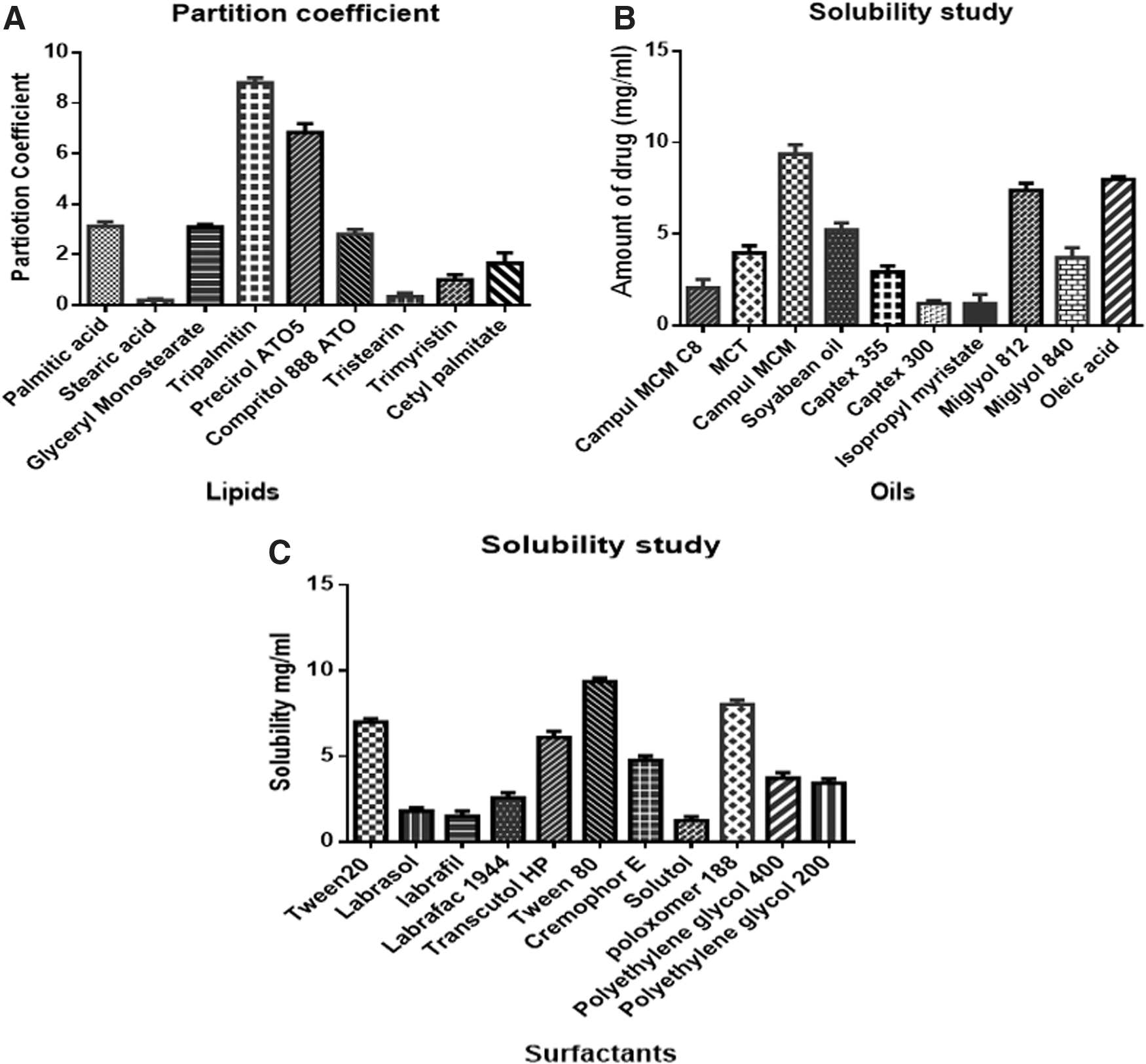
Solubility Studies of ITR in Various Solid Lipids, Liquid Lipids, and Surfactants and Partition Studies of ITR in Solid Lipids
Solid lipids were screened based on their affinity for the drug. Among various solids lipids, tripalmitin showed higher partitioning for ITR. Hence, tripalmitin was selected for the preparation of NLC. Among the liquid lipids, ITR showed higher solubility in Capmul MCM (9.36 ± 0.51 mg/mL) compared with other liquid lipids. Hence, as a liquid lipid, Capmul MCM was chosen for the preparation of NLC. Surfactants play a major role in the preparation of NLCs as they help in stabilization of formulation. Among surfactants, ITR showed higher solubility in polysorbate 80 (9.37 ± 0.2 mg/mL), and in case of cosurfactants, it showed higher solubility in Transcutol HP. The results are shown in Figure 1.
Formulation and Optimization
Preliminary batches of ITR-NLC were prepared to identify the effect of various factors on their size and EE. The parameters studied include the amount of solid and liquid lipids, surfactants, time, and homogenization cycles. Based on the preliminary formulation studies, three major variables affecting the particle size and EE were identified: amount of solid and liquid lipids, surfactants, and homogenization cycles.
Experimental design
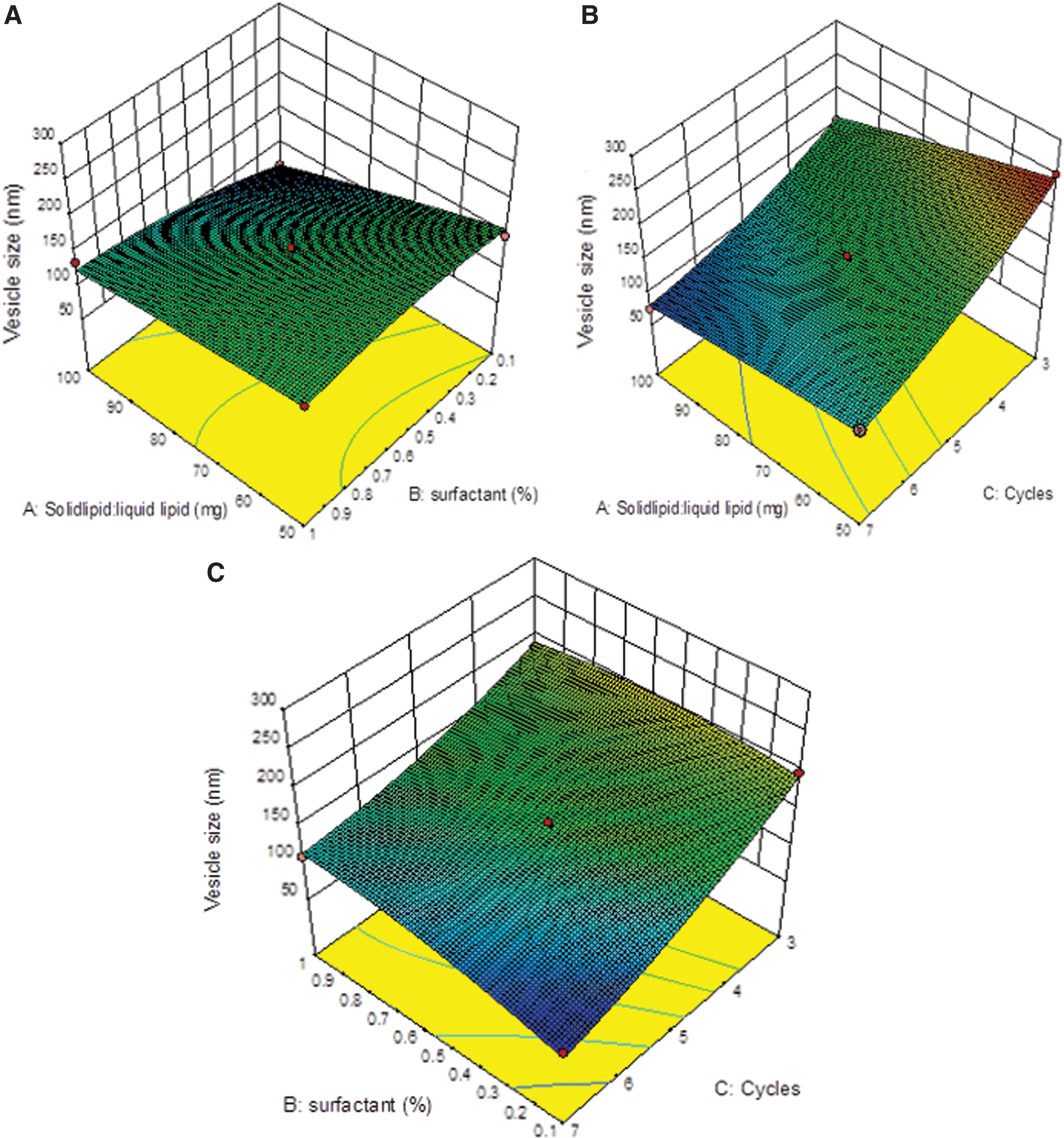
Seventeen experimental runs were conducted using the Box–Behnken design while two responses were measured. All the responses obtained from the 17 formulations were fitted to various models in a statistical design. It was observed that the best fitted model for particle size (Y1) and EE (Y2) was quadratic. The values of R 2, adjusted R 2, and predicted R 2 are given in Table 3. The ANOVA for the experimental data results is shown in Table 4. For dependent variables (Y1 and Y2), it was revealed that the models were significant for all responses. Response surface analysis plots were prepared depicting the effect of significant independent factors on the observed responses of particle size and EE (Figs. 2 and 3).

Summary Results of Regression Analysis for Responses
R 2, regression coefficient.
Analysis of Variance Parameters for the Measured Responses
DF, degrees of freedom; F, ratio of two mean square values; MS, mean of square; P, probability; SS, sum of square.
Effect on particle size
The independent variables A (solid lipid: liquid lipid concentration) and B (polysorbate 80 concentration) had a positive effect on vesicle size (Y1) and EE (Y2), and C (number of homogenization cycles) had a negative effect on responses. This indicates that vesicle size increases with an increase in solid lipid and liquid lipid ratio as well as with surfactant concentration. In case of C, a decrease in homogenization cycles was found to result in an increase of vesicle size. The polynomial equation was generated for the response Y1 and Y2. The polynomial equation obtained is shown in Equation 4.
where A and B represent the coded values of the solid lipid: liquid lipid concentration and surfactant concentration. The positive value in the polynomial equation represents that the response increases with the factor and vice versa. The value of the square of correlation coefficient (R 2) was found to be 0.9788, indicating an excellent fit.
Entrapment efficiency
The EE varied from 85% to 99.5% for various factor-level combinations. The independent factor affecting EE is shown in Equation 5.
The positive value in the polynomial equation represents that the response increases with the factor and vice versa. The higher value before A indicates a higher influence of lipid concentration on EE. Influence of B and C observed from the response surface plot, and EE was increased in the midrange of the factor. The increase in number of homogenization cycles was found to decrease in EE. In case of response Y2 in 3D plot (Fig. 3), higher EE was observed by increasing the lipid concentration. As the lipid portion increased relative to the drug, more amount of drug was seen to be entrapped into the lipid matrix. Addition of liquid lipid increases the solubility of drug, and hence, EE increased.
Optimization and Validation
The desirability function was obtained using Design-Expert software to obtain the optimized formulation. The formulation was optimized on the set criteria of minimum particle size and maximum EE. Therefore, a new batch of NLCs with the predicted levels of formulation factors was prepared to confirm the validity of the optimization process. The optimized formulation was achieved with the formulation composition having solid lipid: liquid lipid total concentration 89.74 mg, surfactant 0.35%, and 6.5 homogenization cycles. Mean particle size of 70.55 nm and EE of 98% ± 1.02% were achieved, which represented a good agreement with the predicted values that were 77.6 nm for vesicle size and 96.59% for EE. The PDI of developed formulation was 0.186 and zeta potential was—17.2 mV. After coating with CS, CS-ITR-NLC showed particle size of 86.75 nm, PDI 0.4, and zeta potential of +25.6 mV, respectively. The PDI should be low as it is indicative of uniform particle size. Value less than 0.5 indicates very good uniformity in particle size. 23,24
TEM Analysis
The morphological observation of CS-coated NLC and ITR-NLC was performed by TEM (Fig. 4). ITR-NLCs were found to have a smooth spherical structure, while CS-coated NLC particles showed slightly larger particles, which indicated the presence of coating of CS around the NLC.

Transmission electron microscopy morphology of
DSC Analysis
DSC analysis was performed to observe the physicochemical changes of drug-loaded NLC. The DSC thermogram of ITR endothermic peak was observed at 171°C, but in case of CS-ITR-NLC, disappearance of ITR peak was observed, which indicated solubilization as well as encapsulation of drug in solid lipid matrix (Fig. 5).

Differential scanning calorimetry thermograms of ITR; CS-ITR-NLC; physical mixture.
In Vitro Drug Release
The pattern of drug release from various NLCs is shown in Figure 6a. ITR-SOL showed 99% drug release within 2 h, but in case of ITR-NLC and CS-ITR-NLC, sustained release of ITR was observed. In case of CS-ITR-NLC, the release of ITR took longer time indicating adhesion of CS by formation of hydrophilic matrix layer around NLC. The release kinetics was calculated for drug release and it was observed that release of ITR from ITR-NLC was found to have a higher R 2 value (0.9832) for the Peppas model. It showed that the release of ITR from the ITR-NLC was due to drug diffusion. Whereas the drug release from CS-ITR-NLC was found to have higher R 2 (0.9896) for the Higuchi model, which shows that the release of ITR from the CS-ITR-NLC was concentration dependent.
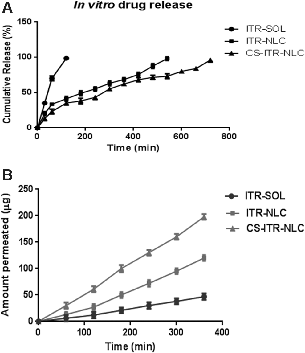
Permeation Studies
The results of the permeation study are shown in Figure 6b. Permeability of ITR from CS-ITR-NLC in the form of suspension was observed to be significantly higher (p-value <0.0001), that is, 198 μg. The steady-state flux for ITR-SOL, ITR-NLC, and CS-ITR-NLC was found to be 0.66 ± 0.54, 1.69 ± 0.8, and 2.7 ± 0.44 μg/min cm2, respectively, with R 2 value of 0.9944, 0.9880, and 0.9988, respectively.
Ex vivo CAM Assay Using VEGF165-Induced Model
Chick CAM model is widely used in the study of angiogenesis and demonstrates a functional assay to screen agents for antiangiogenic activity. 25,26 The VEGF family was involved in vascular leakage and angiogenesis in DR. 27 HET-CAM assay in Figure 7 shows that ITR and drug-loaded formulation lead to reduction of abnormal vessel growth in a VEGF165-induced model, which indicates drug and formulation targeting VEGF165 and thereby reducing angiogenesis. Compared with the control, NLC-treated eggs showed higher reduction in blood vessel growth. CS-ITR-NLC showed a still better antiangiogenic effect compared with the ITR. Hence, ITR and CS-ITR-NLC were found to inhibit the proliferation of abnormal blood vessel formation by downregulating VEGF165 (n = 3).
Antiangiogenesis study of prepared formulation and ITR (ex vivo chorioallantoic membrane assay).
Rat Aortic Ring Assay
The effect of ITR and ITR-NLC on microvessel sprouting from vascular tissues was studied using an ex vivo rat aortic ring assay. VEGF has a potent role in angiogenesis, which plays direct effects on vascular endothelial cells, including endothelial cell proliferation and survival, tubulogenesis, and vascular permeability. 28 Rat aortic ring study in Figure 8 shows the absence of sprouting of vessel growth, while tube formation could be observed in ITR- and CS-ITR-NLC-treated aorta (n = 3). ITR inhibits the sprouting of blood vessels; on the contrary, blank NLC allows sprouting of blood vessels and tube formation. It indicates that ITR and drug-loaded NLC are able to block the VEGF-dependent microvessel sprouting from vascular tissues.
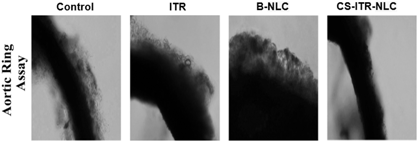
Aorta images after treatment with drug and formulations.
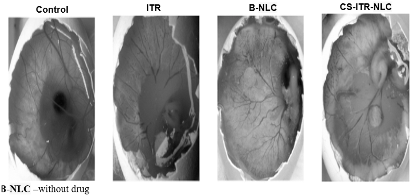
Antineovascularization Study on Rat
To study the antineovascularization effect of ITR on rat, the VEGF165-induced model is reported as the best model to study the inhibitory effect of antiangiogenic agents. 29 –31 CS-coated ITR-NLC showed a high reduction in neovascularization in the VEGF165-induced model, as shown in Figure 9. It may be attributed to higher penetration and mucoadhesive property of CS-ITR-NLC behind the cornea along with less precorneal drainage due to the viscosity of formulation owing to the pressure of CS. The control (untreated) and blank-NLC-treated rat cornea showed highest neovascularization. CS-ITR-NLCs were thus found to possess targeting efficiency, capacity to increase contact time with tissues, and capability to enhance drug absorption, thereby increasing the ocular bioavailability of drug.
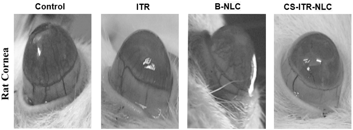
Rat corneal images for antineovascularization.
The permeability studies indicated very good permeation and retention of CS-ITR-NLCs behind the cornea. This indicated very good targeting potential of formulation at the site of action (i.e., behind the cornea). This was further proven through the in vivo results of antineovascularization study on rats, in which it was clearly observed that the developed formulation showed very good antiangiogenic potential behind the cornea. Furthermore, it could be used as an effective alternative to the intravitreal injections of various drugs.
Overall data from the present study suggest repositioning ITR may be a better management of DR by targeting VEGF165. However, further studies need to be carried out, such as formulation development, molecular-level studies, dose to be optimized, transport of drug across the barriers using confocal microscopy, in vitro blood–retinal endothelial cell line studies, and more nanotoxicological studies to determine the safety of formulated drug-loaded NLCs to confirm the potential antiangiogenic activity of CS-ITR-NLC.
Conclusion
From the results obtained, it was concluded that after ITR-NLCs were coated with CS, their effectiveness as carriers for ocular drug delivery increased. The ITR-NLC was coated with the CS surrounding the surface uniformly, which resulted in the positively charged NLC dispersions. These, in turn, provided a longer retention time by interacting with the negatively charged mucous membrane of the eye. Eventually, an improved penetration rate was achieved with the presence of CS. The most notable advantage was that CS-ITR-NLCs were found to possess superior mucoadhesive property compared with ITR-NLC, which, in turn, was beneficial for bioavailability from the ocular drug delivery system. Taking these findings into account, CS-coated NLCs of ITR represent a promising treatment for DR.
Footnotes
Acknowledgments
The authors acknowledge the help and kind support of Dr. Prabhakar and Mr. Vikas, Department of Biotechnology, Sahyadri College of Science, Shimoga, India. The authors would like to thank Department of Science and Technology–Fund for Improvement of Science and Technology Infrastructure in Universities and Higher Educational Institutions (DST-FIST), New Delhi, for their infrastructure support to our department.
Disclosure Statement
No competing financial interests exist.