
Abstract
The drug-discovery process is expensive and lengthy, and has been causing a rapid increase in the global health care cost. Despite extensive efforts, many human diseases still lack a cure. To improve the outcomes, there is a growing need to implement novel approaches into the early stages of the drug-discovery pipeline. A specific such effort has focused on the development of novel disease models such as cellular models (genetically modified cell lines, spheroids, and organoids) and whole-animal models (small animal models and genetically modified large animal models). The whole-animal screens are advantageous as they can provide system-level information, off-target effects, complete absorption, distribution, metabolism, excretion, and toxicity architectures, and early in vivo toxicity, which help to prioritize compounds before using them for human trials. Such multivariate analysis helps to improve the translational potential of drug compounds. Drug testing in large animals is expensive and time consuming. A solution is small animal models that have simplified biological system with intact physiology and sufficient homology with human genes. In recent times, many such models have constantly been developed and tested to identify new disease mechanisms. Caenorhabditis elegans is one such small animal model that has been considered for large-scale drug testing. In this review, we will discuss the current state-of-the-art technologies, including two platforms developed in my group that have enabled high-throughput and high-content screening using C. elegans disease models.
Introduction
Identification of small-molecule modulators for lead optimization with high translational properties is one of the most essential tasks in modern drug discovery due to escalating downstream costs during development and high clinical failure rates. 1 Meticulous screens and stringent selection criteria demand the development of new model systems and high-content screening (HCS) strategies to recapitulate disease complexity better through drug absorption, distribution, metabolism, excretion, and toxicity. For this purpose, there is a great interest in the development of new drug screening methods based on in vivo small animal models such as Caenorhabditis elegans, Drosophila melanogaster, and zebrafish.
As one of the best-studied small animal models, C. elegans has been used to model various disease situations, delineate molecular pathways, and understand disease mechanisms. 2,3 C. elegans, with highly conserved genomics (with ∼38% homology with human disease genes), has great potential for high-throughput drug and genetic screens to advance the development of effective chemical compounds. 4 C. elegans has many advantages, including a short lifespan, well-characterized genetics, a cellular architecture with ∼1,000 cells, a simple neuronal circuit with 302 neurons, and an optically transparent body throughout its development. New discoveries are made with precise genome editing in C. elegans using CRISPR technologies for human-related diseases such as neurodegenerative, 5 –10 rare disease, 11 and protein homeostasis. 12,13 With the development of cell-specific protein expressions to mimic human disease pathology, the animals develop more subtle degenerative phenotypes, which are difficult to phenotype by conventional methodologies.
The existing screening systems commonly utilized to screen C. elegans models are classified into two major types: (1) stationary methods such as microplate readers and (2) flow methods such as flow cytometers. However, these methodologies operate either at low resolutions or at low throughputs. For example, high-throughput image-based screening is possible only at low resolutions where gross cellular phenotypes are identified using objectives with low magnifications and poor optical resolutions. Higher magnification (high-resolution) imaging in plate readers cannot achieve high throughputs as it requires time-consuming multiple stage motions for finding individual animals in the large area of the wells with randomly oriented animals. 14,15 In addition, randomly oriented overlapping animals in multiwell plates make the image analysis very challenging to identify the phenotypes located across the animals' cross sections. In contrast, flow-based analysis platforms such as COPAS Biosort from Union Biometrica can image animals at high speeds but using very low-resolution and one-dimensional fluorescence intensities along the animal body length. 16
In recent times, microfluidic technologies have provided the state-of-the-art screening platforms using precise fluid controls in micrometer-scale channels. 17 –19 Various microfluidic designs have been developed to immobilize C. elegans for high-resolution imaging, 20 –23 although at low throughputs. Our laboratory has pioneered several such microfluidic technologies 20,21,24 –27 as well as ultrafast imaging methods, 28 pushing the technological barriers that limited throughput. We are now moving forward to bring these innovations to the marketplace. Specifically, we have developed advanced microfluidic immobilization technologies that can provide highly sensitive analysis capabilities to detect subtle phenotypes and screen in vivo models in C. elegans at high throughputs. 20,21,24 –28 Two of our recent platforms, described in this article, have enabled detection of subtle phenotypes, distributed in three dimensions (3D) and expressed at low levels in different developmental stages. High-speed imaging methods of small animal models are attracting both academic and industrial communities for high-content and high-throughput screening for chemical and genetic modulators of animal physiology.
The Technical Challenge
The implementation of microfluidic technologies for high-throughput and high-resolution (high-content) imaging of C. elegans models requires overcoming major challenges. Specifically, such systems need to (1) be compatible with automated platforms such as liquid handling and robotics systems, (2) eliminate cumbersome interfaces such as multiple inputs and complex tubing manifolds, (3) provide a sufficient number of samples (animals) per test in a limited space, and (4) immobilize the animals close to the imaging optics and orient them in their optically favorable side for high-resolution imaging. Complex channel geometries with multiple fluid inputs have been demonstrated for high-resolution applications such as imaging 22,25,29 –31 and laser axotomy 20,21,24,32 with submicrometer resolutions. However, these device designs are only suitable for single tests (multiple animals from a single population) with serial sample processing. These technologies cannot scale up to accommodate multiple samples simultaneously on a single chip with a footprint that is compatible with existing automation industry standards. To address this gap, we have recently developed a microfluidic technology for high-content and high-throughput screening of C. elegans disease models with subtle fluorescence phenotypes. Most recently, we also developed an ultrafast fluorescence imaging method that was implemented as the first 3D flow cytometry that can image C. elegans flowing at 1 m/s without the need for immobilization. With these two inventions, we can finally perform high-throughput and high-content in vivo screening using C. elegans.
Innovative Solutions for High-Throughput and High-Content Imaging of in Vivo Models
To develop a microfluidic technology that can overcome all the challenges mentioned earlier, we took a new approach to develop a hybrid device structure that can close the gap between the macro- and microworld of microfluidic structures. First, the exterior of the device has a standard multiwell format for interfacing with the existing commercial automated platforms of the macroworld. Second, the interior of the device consists of microfabricated channels that are directly connected with the multiple wells to handle a large number of animals for each test/well. The standard multiwell format (having 96 independent sample wells) of the microfluidic device enables parallel sample preparation using multichannel sample loader equipment. Third, the platform has a single-input and single-output fluid interfaces for simple connectivity and flow-control operation, eliminating cumbersome interfaces. Finally, the microfluidic immobilization channels are designed with tapered geometry and multiple heights to achieve an aspect ratio (width to height ratio) ∼1.0 to maintain the animals in their natural lateral orientation 33 when pushed inside the narrow channel using a pressure cycle. The animals' lateral orientation is best suited for optical imaging of the cellular processes such as the neuronal processes located along the ventral cord of the animals.
This novel approach provided the first high-content and high-throughput screening platform for 96 different C. elegans populations, overcoming the current limitations of low resolution, random positioning, ease of use, and optically unfavorable orientation of the animals using plate-based methods. The schematic of our general approach is illustrated in Figure 1a. 25 Microfluidic devices are made of polydimethylsiloxane (PDMS) and are fabricated using soft lithography. 33,34 The PDMS microchannels present 40 parallel traps bonded to a thin glass for high-resolution imaging using large magnification and high-resolution objectives with short working distances. The on-chip PDMS wells connected with the PDMS microchannels underneath the wells house the animals and provide structural support to the chip during immobilization pressure cycles. Multiple channels across 96 wells trap ∼4,000 animals in total within <3 min using a two-piece gasket system that applies pressure through a single common input (Fig. 1b). The applied pressure can then keep the animals trapped in an optically favorable orientation and at predetermined channel locations. The bottom gasket is mounted on a microscope stage to image approximately 4,000 channels using an automated image-acquisition software, which can be easily performed thanks to the predetermined locations of the animals. The software automatically controls the stage movement, focuses on the sample, and takes images. We can then analyze the images using a custom-written image analysis software to identify fluorescence phenotypes from each animal. Degeneration and aggregation phenotypes are scored and multiple parameters are saved into multidimensional arrays for statistical analysis.
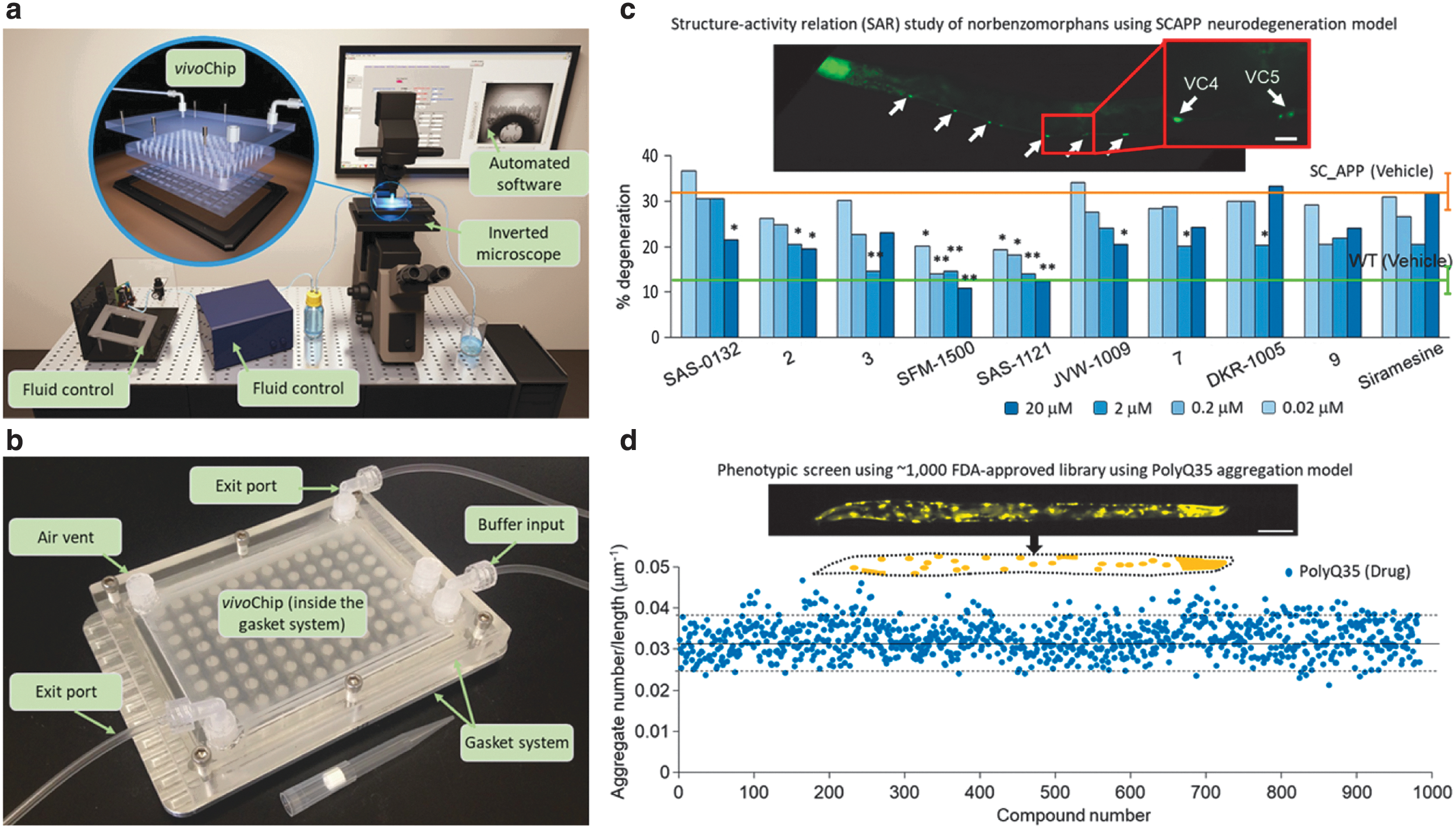
The microfluidic-based imaging platform (vivoChip) for high-throughput and high-content imaging of Caenorhabditis elegans disease models.
We already used the 96-well microfluidics platform to perform an automated HCS of thousands of C. elegans in two different large-scale screens. In our first pilot HCS, for example, we tested ∼1,000 FDA-approved compounds and identified four hits that reduced aggregation significantly in a polyglutamine aggregation model (Fig. 1d) relevant for Huntington's disease in humans. 25 In another HCS, we studied structure–activity relations of various compounds in a C. elegans disease model with a single copy of the human amyloid precursor protein (SC_APP) gene integrated into the genome (Fig. 1c). Specifically, we tested uniquely designed norbenzomorphans structures in the SC_APP animals to identify the efficacy of different structures using in vivo neurodegeneration phenotypes. 26
The microfluidic immobilization platform (vivoChip) can successfully perform high-resolution phenotyping. However, vivoChip can only test a limited number (30–40) of animals per population, and the chip can be used only a few times due to the accumulation of debris or foreign particles that can easily clog the channels. The limited usage and high fabrication cost of this platform can eventually be a bottleneck for primary screens with large-size compound libraries. Therefore, the vivoChip platform is best suited for secondary screens with a limited number of compounds that require high-resolution phenotyping for subtle changes or multivariate analysis with many parameters.
Most recently, we have developed a second screening platform with an ultrafast fluorescence imaging modality (Fig. 2a) to reduce the cost and eliminate the restriction on the number of animals per population. 28 This is a flow cytometry platform, which does not require animal immobilization and can image hundreds of animals within <1 s and with 3.5 μm resolution in all 3D (Fig. 2b). The new fluorescence imaging technique, called line excitation array detection microscopy, can image C. elegans moving at speeds >1 m/s without motion blur, 1,000 × faster than the currently available 3D cytometers. 35,36 We performed a small-scale drug screen and confirmed our recent findings that the compound dronedarone prevented aggregation with a dose response in the polyglutamine aggregation model. The whole-animal flow cytometer now provides the potential to screen a 10,000 compound drug library in under a day when combined with fast population delivery microfluidic systems. 27
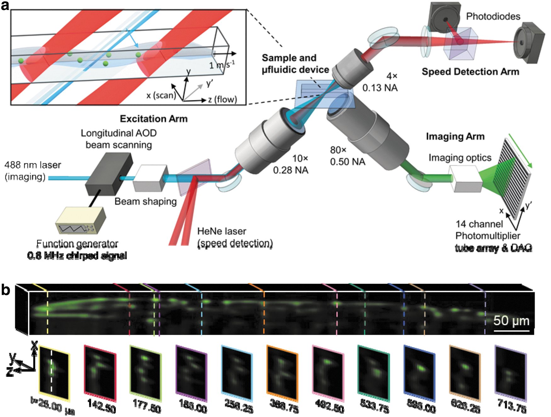
Schematic of LEAD microscopy that can provide unprecedented fluorescence imaging speeds of 0.8 million frames per second and can image whole animals in 3D in <1 ms per animal while flowing inside a microfluidic device.
Taken together, we developed two new approaches for high-content and high-throughput screening of various C. elegans human disease models. Such platforms are easily extendable to other model systems such as other small animal models (Drosophila and zebrafish) and 3D cellular models (organoids and spheroids). Advancements in screening technologies can reduce drug-discovery cost and identify potent compounds with a higher translational possibility to become a drug to reduce patients suffering.
Future Directions for in Vivo Screening Platforms for Drug Discovery
The future of in vivo screening platforms is bright and growing with an accelerated pace thanks to new advanced technologies, development of new disease models, integration of technologies with automated platforms, and computer-assisted automated analysis. These technologies will soon be enabling rapid phenotypic screening of larger drug libraries to identify off-target effects in disease biology and mechanisms that were missed during targeted screens. Disease models with patient-derived tissue samples, disease implants in whole animal, and targeted genome editing using clinical information are booming in the communities. With improved knowledge from patient-specific diseases and the heterogeneity in the disease pathologies are helping to design more relevant models to screen for better drug candidates on isogenic models. The integration of automated platforms for culture, imaging, and image analysis have advanced multivariate analysis with more subtle phenotypes obtained using high-resolution images. Similar to the health care industry, screening technologies are heavily benefited from artificial intelligence research, improving the image analysis quality, automate hit identification, and identify new traits from population data.
In conclusion, the screening technology field combined with the development of other accessories will contribute to improving the health care industry. Collectively, small-animal screens with other model systems such as patient-derived cellular models can aid in identifying better compounds and lead candidates in the drug-discovery process. Along with the improvement in the models, the new technologies will improve the human health, reduce the suffering of patients, and discover the cure of new diseases.
Footnotes
Disclosure Statement
The author is a co-founder of Newormics LLC and inventor on pending patents on high throughput imaging and microfluidics platforms.