




Abstract
Inducible pluripotent stem cells (iPSCs) are being used to model brain disorders across the continuum of neurodevelopment, neurodegenerative, and neuropsychiatric disease allowing for the mechanistic unraveling of the neurological disease state. Subsequently, there is a diverse array of cell model systems that can be used for target validation, pharmacodynamic endpoint development, and high-throughput/content assay development and screening. However, to successfully model neurological disorders with iPSCs, the disease-relevant neuron must be first identified, and it is critical to have the appropriate neuronal progenitor cell derivation and neuron differentiation protocols available to produce desired neuronal phenotypes. Moreover, special considerations are necessary if adaptation to high-throughput/content assay systems is anticipated. Discussed here are the three-dimensional embryoid body–neural rosette and two-dimensional monolayer methodologies to derive iPS neural progenitor cells and neurons with a specific focus on cortical neurons. Outlined are some of the commonalities, advantages, and disadvantages associated with both methodologies.
Introduction
Inducible pluripotent stem cells (iPSCs) can be derived from normal or diseased patient somatic cells, which are engineered to revert to an embryonic state. First described in 2007, adult human fibroblasts were reprogrammed back to a pluripotent state using defined factors. 1 This study provided the ground work for the potential for the transduction of other somatic cells to an embryonic-like state. Loh et al. described the reprogramming of peripheral blood mononuclear cells (PBMCs) as a viable source for iPSCs. 2 PBMCs provide the advantage of ease in which the cells can be harvested from the patient and are minimally invasive compared with tissue biopsy punch for establishing fibroblast lines. Other potential sources for establishing patient-derived iPSC lines include but are not limited to urine-derived cells, 3 –5 pancreatic islet beta cells, 6 and keratinocytes. 7,8 These cells arm researchers with a unique tool to study cells that contain the patient's pathology, and several studies have used these iPSC-derived populations to design drug and biomarker screens. 3,9 –11
Before the discovery of iPSCs, researchers relied on in vivo or in vitro animal models, which may or may not be “translatable” depending on disorder being studied, and primary human tissue culture and established cell lines. iPSCs can be artificially induced to produce a wide array of cells depending on the study at hand, which can include but is definitely not limited to cardiomyocytes, 12 retinal epithelial cells, 13 and neurons. 14 –16 Even though many cell lineages can be derived from iPSCs, the real power and impact of iPSC-based research is most evident within the study of neurological disorders, where the ability to study the living human brain is virtually impossible as most tissue is obtained from autopsy, which provides better insights into end-of-life neurological disease rather than neurological disease initiation and progression, and methodologies for deriving neural progenitor cells and cortical neurons will be further discussed. While iPSC technology is still relatively new, it has produced several neurological disease models that have enabled researchers to study Alzheimer's disease, 17,18 amyotrophic lateral sclerosis, 19,20 schizophrenia, 15 and autism spectrum disorder 21,22 under more physiologically relevant conditions. Subsequently, there is a diverse array of cell model systems that can be used for target validation, pharmacodynamic endpoint development, and high-throughput/content assay development and screening. In this article, the differentiation of neural progenitor cells and cortical neurons by three-dimensional embryoid body (EB)–neural rosette (NR) and two-dimensional monolayer methodologies will be further compared, outlining the commonalities, advantages, and disadvantages associated with both methodologies with thoughts and consideration on robustness, ease of use, and reproducibility for drug screening applications.
Modeling Neurological Diseases with i PSCs
To model neurological disorders with iPSCs, however, the disease-relevant neuron (i.e., dopaminergic, motor, cortical, glutamatergic, GABAergic, or sensory neurons) must be first identified, and it is critical to have the appropriate neuronal progenitor cell (NPC) derivation and neuron differentiation protocols to produce desired neuronal phenotypes. For example, the stimuli or patterning cues needed for neural induction of NPCs capable of producing cortical neurons focus on the exposure to single or dual SMAD inhibitors (SMADi), 14,15,23 as well as retinoic acid, which further potentiates neural differentiation processes. 24 Conversely, the patterning cues for motor neurons involve neural induction to neuroepithelia with SMADi (LDN193189, SB413542) and a GSK3β inhibitor (CHIR99021) for a week before the addition of retinoic acid and purmorphamine to induce a motor neuron phenotype. 25 Liu et al. 26 provide a comprehensive review of commonly used agents in NPC induction (e.g., SMADi LDN193189 and SB431542, transforming growth factor-β family inhibitors, GSK-3 β inhibitors, MAPK signaling pathway inhibitors, and Sonic hedgehog pathway agonists) and also provide insights on epigenetic modifiers that help accelerate iPSC reprogramming (i.e., valproic acid, ascorbic acid, and retinoic acid).
Unfortunately, NPCs are often underappreciated as a crucial intermediate iPSC population to differentiated neurons. Critically, NPCs provide a cell population for scaling up and cell banking allowing for continuous research. This advantage is highlighted when comparing methodologies that directly convert iPSCs to neurons without allowing for a stopping point in the early neuroectoderm processing (i.e., NPC stage). The conversion of iPSC to sensory neurons (i.e., nociceptors) 23 provides an excellent example. Multiple patterning cues (i.e., LDN193189 [SMADi], SB431542 [TGFβi], SU5402 [VEGFR2i], DAPT [γ-secretase inhibitor], and CHIR99021 [GSK3βi]) were used in combination to directly convert several iPSC lines into sensory neurons as confirmed by ISL1 and BRN3A expression. 23 While there is evidence of a transient neural stem cell (NSC) stage, as verified by the co-expression of Nestin and Pax6 (NSC markers), there was no “hardened” NPC or NSC intermediate for further evaluation or to cell bank for long-term research. Thus, using this methodology, neuron derivation from iPSC will need to be continuous, which is more labor and time intensive compared with NPC-derived neuronal differentiation. NPC derivation protocols that produce verifiable NPC populations offer an advantage as they allow NPC cell banking, which confers the ability for continuous in vitro experimentation with the same NPC population and to limit “lot-to-lot variability,” both crucial elements for high-content/throughput screening.
Generation of NPCs —EB versus Monolayer Methods
Historically, NPCs have been derived through three-dimensional aggregates (EBs) 15,27 ; however, more recently, NPCs have been generated through shorter monolayer derivations. 14,28 Since dysfunctional forebrain neurons cause a variety of neurological diseases, including Alzheimer's disease, Huntington's disease, and schizophrenia, the strategies for generating NPCs for forebrain cortical neurons will be highlighted, as will their potential of these NPCs for scalability, cell banking, and prolonged use for drug screens.
EB and NR selection for NPC Generation
When cultured on low attachment dishes, iPSCs will spontaneously form three-dimensional aggregates in the absence of growth factors in a minimal media, such as Essential 6 (E6) (Fig. 1). Neural induction is initiated by the addition of neural induction basal media (e.g., DMEM/F12+Glutamax, N2, and B27 without retinoic acid supplementation) and the SMADi LDN193189 and SB43154215 (Fig. 1). EBs are pooled together into one well of an extracellular matrix-coated plate (e.g., Matrigel™ or poly-ornithine/laminin) permitting cell attachment and are then monitored for the development of NRs, which pattern the development of early neural tube formation and can express the early neuroectodermal markers, Pax6 and Sox1. 29,30 After the NRs have matured, they can be mechanically isolated or dissociated using the NR-specific dissociation reagent (e.g., Neural Rosette Selection Reagent [NRSR]; StemCell Technologies).
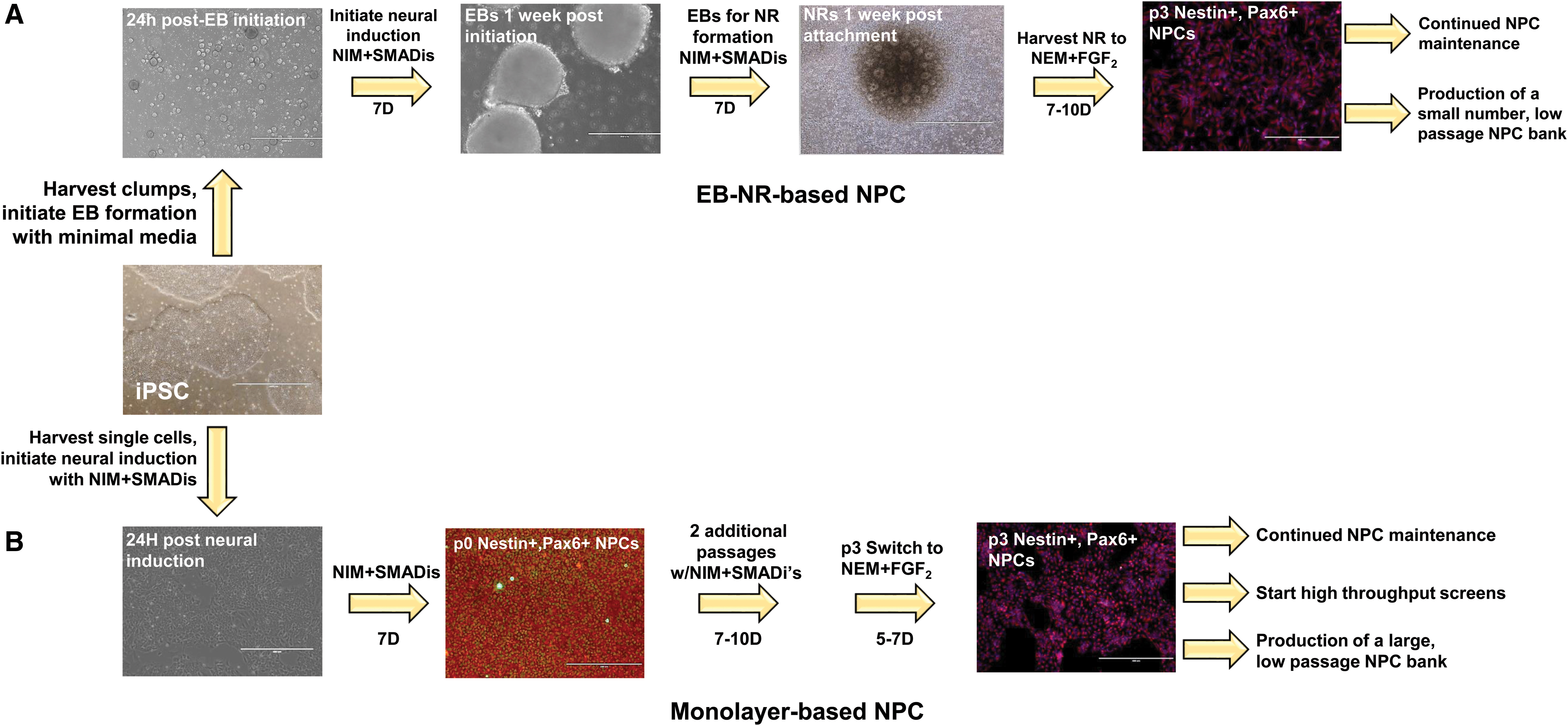
Timeline of EB-NR-based and monolayer-based NPC for high-throughput studies and cell banking. Starting with high-quality iPSCs, NPCs can be derived from either EB-NRs
While mechanically isolating NRs is a cheaper method in terms of necessary reagents, NR identification is time consuming and requires specialized expertise of cell morphologies under bright-field microscopy to accurately identify and isolate NRs without disturbing non-NR cells (i.e., neural crest cells, nonneuronal cells). Even though NRSR will add cost, it is a quick and reliable method to isolate NRs. Nonetheless, when accurately performed, and directly compared, both methods enrich for Pax6+ and Nestin+ NPCs indicating that the harvested NRs are expressing NPC markers. However, there is some evidence that using NRSR-based methods have a higher potential of carrying over Oct4+ cells (i.e., iPSC marker) versus manual NR selection, but this is dependent on researcher expertise. 16 Regardless of the particular harvesting method selected, a challenge will be to isolate NRs while simultaneously avoiding inadvertent carryover of nonneuronal cells.
Once NRs are selected, they are plated for expansion in NPC maintenance media, comprising neural induction basal media (without the SMADis), with the addition of FGF2 and laminin. At this point, the cells are considered to be at passage zero (p0) NPC. Subsequent NPC maintenance requires medium feeds every 2 days with NPC growth media and passage with Accutase when cells reach >80% confluency. A limited number (i.e., 1–2 vials) of early passage NPC can be preserved; however, this is highly dependent on the initial number of EBs harvested. Further NPC expansion is an option; however, these NPC populations will be at a later passage, and unfortunately, higher passage NPCs have been reported to yield glial cells (i.e., astrocytes), shifting differentiated NPC cultures from neuronal to glial populations. 27 This “shift” has been reported as early as p10, although there are some newer techniques that can help provide a higher population of neurons from higher passage NPCs. 3
It is important to note that there are variations in EB-NR-based methodologies for deriving NPCs, such as spontaneous neural differentiation 31 and NPC derivations without defined factors. 16,32 In general, however, the EB-NR method is time consuming, requiring ∼10–14 days to achieve p0 NPCs and then potentially another 10–14 days of expansion and subsequent small-scale NPC banking before initiation of neuronal differentiation. Moreover, the EB-NR strategy does not always result in high-quality NPCs, as identified by Nestin and Sox2 expression. 27 Unfortunately, validation of NPC lines derived via EB-NR methodologies cannot be performed until after the NPCs have been passaged and expanded at least two times (i.e., ∼2 weeks), to ensure a population of cells for continued studies. Thus, while these protocols can yield high levels of neurons expressing mature neuronal markers (i.e., Map2, βIII tubulin), the limitations of the EB-derived NPC protocols (i.e., low-yield EBs, low NR induction, high carryover of nonneuronal cells, or low-quality NPCs) create bottlenecks for the generation of consistent NPCs for downstream procedures (Table 1). Moreover, with low expression of NPC markers, fewer neurons may be available for experimentation.
Comparison of Two-Dimensional and Three-Dimensional Neuronal Progenitor Cell Methodologies
NPC, neural progenitor cell; EB-NR, embryoid body–neural rosette; NRSR Neural Rosette Selection Reagent; 2D, two dimensional; 3D, three dimensional.
Two-Dimensional Monolayer Cultures or 1 Week to NPC
Chambers et al. 14 realized the need for improved NPC generation that used defined factors (dual SMADi) and bypassed the necessity of generating complex EBs while maintaining a homogenous NPC population, which was able to differentiate into multiple neuronal subtypes. 14 As a result, a monolayer differentiation protocol was developed. Using a combination of dual SMADis (i.e., Noggin and SB431542) exposure, this protocol yielded >80% of Pax6+ NPCs compared with <10% Pax6+ NPCs when the SMADis were used individually. 14 Other researchers have also successfully replaced Noggin with LDN193189, 33,34 yielding similar high expression of Pax6+ cells. Moreover, large-scale NPC induction to differentiation is also feasible under SMADi-independent conditions using minimal neural progenitor selection media with the addition of recombinant human FGFβ during expansion. 28 Subsequently derived differentiated neurons expressed functional ligand-gated channels and underscored the advantages of using two-dimensional culture systems to generate consistent batches for high-throughput/content screening applications. 28
Overall, the monolayer protocol enables the production of relevant neuronal subtypes in a much shorter period of time (19 days) compared with EB-derived protocols (30–50 days) 14 (Fig. 1). This is an advantage, as it enables researchers to begin studies earlier, without the need for time-consuming maintenance steps. The monolayer protocol also provides researchers with a methodology to create a large NPC bank at low passage number for long-term experimentation, a critical attribute for high-throughput/content applications. Using high-quality iPSCs, any format (i.e., multi-well plate or tissue culture flasks) can be used for neural induction. However, it may be a better strategy to start with a smaller scale induction to confirm the expression of NPC markers (i.e., Nestin, Sox2, and/or Pax6) after 1 week and then expand the remaining NPCs according to the cell density to surface area ratios. Moreover, by using lower passage NPCs to generate neurons, glial (i.e., astrocytes) populations will be minimized during neuronal differentiation, ensuring a high neuronal percentage in vitro.
Comparison of EB to Monolayer Derivation Methods
Although monolayer-based NPC protocols are efficient as well as less labor and time intensive with respect to generating NPCs, there is a bias toward EB-derived NPCs possibly reflecting a “first to bench-side” advantage. However, newer studies have directly compared the two methods to see if one is superior. Muratore et al. 16 undertook a thorough comparison not only between EB versus monolayer protocols but also examined coating matrices, manual versus Aggrewell™ (creates uniform size EBs), and sorting versus manual picking of NRs. Using EBs, 93% of derived expressed Map2 (mature neuron marker) at day 40 compared with 45% of the neurons were generated through the monolayer protocol. 16 Unfortunately, in their studies, there was minimal optimization in the post-processing of NPCs (i.e., continued passaging, ensuring amenable cell densities for neuronal differentiation, generation of a cell bank) and continuous neuronal differentiation from iPSCs was being performed.
However, a more comprehensive, and recent, study of EB and monolayer protocols was performed by Chandrasekaran et al. 35 In this study, five hiPSC lines with different genetic backgrounds were used. 35 After inclusion of dual SMADi exposure, high-quality NPCs and neurons were derived from both protocols. Moreover, there was comparable ranges of expression of the TBR1 (cortical neuron marker), with 9%–22% and 7%–22% for monolayer and EB-derived NPCs, respectively. 35 Although the EB-derived cortical NPCs displayed elongated neurite outgrowth and differentiated mature neurons earlier, patch clamp analysis revealed no significant difference in the electrophysiological outputs and reactivity of neurons derived from either protocol. 35
Nonetheless, both the embryoid aggregate and monolayer methodologies can produce viable NPC intermediate populations; however, the variation of NPC derivation protocols and the yield of differentiated neuronal populations/percentages suggest that protocols need to be refined and standardized. Moreover, while there are clear advantages of deriving NPCs through an EB body (i.e., more mature neuronal phenotype early, longer neurites), 35 there are significant disadvantages including step intensive protocols that may yield less than optimal NPC populations (i.e., low number of EB formations, carryover of nonneuronal phenotypes) and that may not be amenable to cell banking. These disadvantages can be avoided with the two-dimensional monolayer system that (1) yields higher number of cells for neural induction, (2) enables monitoring for nonneuronal cell morphologies during differentiation, and (3) can be scaled up in less time to create a large-scale NPC banks for downstream applications.
Commericialized KITS: A Way to Cut Down on lot-to-lot Variabilities between Neural Inductions
Arguably, however, is that the most exciting advance in the field of iPSC and NPC technology is the ability to use patient-derived cell populations in drug screens. Thus, it is vital to optimization NPC derivation protocols that are transferable across laboratories, research groups, and assay platforms. Several reviews are available that discuss the importance of rigor and reproducibility of iPS neurons for targeted drug screens. 36 –39 Essentially, NPC derivation protocols should meet exacting requirements for reproducibility, consistent yields of NPC and iPS neurons, scalability for high-throughput and content screens, and cost/time effectiveness. 36 The two-dimensional monolayer of NPC induction appears to be more aligned with the needs of high-throughput/content applications.
There are a variety of commercially available kits to derive NPCs with main advantages focusing on quality control and lot-to-lot reproducibility within, as well as, across laboratories. Using a commercial source for neural induction is one way to ensure that the reagents and protocols used are uniform and should minimize quality control issues, a critical component of high-throughput/content activities. StemCell Technologies offers a variety of reagents and kits for neural induction and neuronal differentiation. An advantage of this media and systems is that they are compatible with EB or monolayer neural induction. StemCell Technologies also offer other downstream media that are compatible with the NPCs differentiation into a variety of neuron subtypes and glial cells enabling the generation of multiple brain relevant cell types.
Other commercially available kits are available through ThermoFisher and include Gibco Pluripotent Stem Cell Neural Induction medium. This kit also can produce NSCs in 7 days without the need of an EB and produces NSCs that are Sox1/Sox2+. However, unlike StemCell's SMADi kit, this kit will only produce about 15%–50% Pax6+ NSCs (
The Future of i PS Cortical Neuron Development
Existing cortical NPC derivation and neuron differentiation methodologies are powerful tools for neurodegenerative research, as they provide models for disease progression as opposed to the end stages of the disease. With recent advances in high-throughput screening, using NPCs to generate neurons for targeted drug screens are being developed. Other exciting advances in the field include directly converting fibroblasts (or even peripheral blood T cells 40 ) into induced NPC (iNPC) 41 –43 and induced neurons (iNs). 44 These techniques offer an advantage of bypassing the experience needed to derive and maintain iPSC cultures as well as provide a means for potentially providing a therapeutic approach in neurodegenerative disease. While this is an exciting avenue for the field, the techniques will still need to be fine-tuned to allow for scalability (efficiencies for iNPCs are 0.02%–0.04% 41 and ∼2%–8% for iNs 44 ) for targeted drug screening applications but should still be considered for future studies. Finally, a well-designed experiment needs to not only consider the final output but also should include strategies that ensure robustness of cells generated, ease of use, reproducibility, the ability to create a cell bank for further analysis and future studies, and if the derivation is producing the proper neurons needed for the studies.
Footnotes
Acknowledgments

Disclosure Statement
No competing financial interests exist.