




Abstract
The MTS [3-(4,5-dimethylthiazol-2-yl)-5-(3-carboxymethoxyphenyl)-2-(4-sulfophenyl)-2H-tetrazolium] assay is one of the most commonly used assays to assess cell proliferation and cytotoxicity, but is subject to interference by testing compounds. Hydralazine, an antihypertensive drug, is commonly investigated in multiple fields such as heart failure, cancer, and blood pressure research. This study reported interference of the MTS assay by hydralazine and a simple modification overcoming this interference. Vascular smooth muscle cells were cultured in the presence or absence of hydralazine (0, 10, 50,100, and 500 μM) for 2 or 24 h. Cell numbers were analyzed using MTS, trypan blue exclusion, or microscopic assays. A modified version of the standard MTS assay was established, in which an additional step was added replacing the test medium, containing hydralazine, with fresh culture medium immediately before the addition of the MTS reagent. Culture with hydralazine at concentrations of 50, 100, and 500 μM for 2 h increased absorbance (p < 0.05) in the standard MTS assay, whereas microscopy suggested no change in cell numbers. Culture with 500 μm hydralazine for 24 h increased absorbance (p < 0.05) in the standard MTS assay, however, trypan blue exclusion and microscopy suggested a decrease in cell numbers. In a cell-free system, hydralazine (≥10 μM) increased absorbance in a concentration-dependent manner. The modified MTS assay produced results consistent with trypan blue exclusion and microscopy. In conclusion, a simple modification of the standard MTS assay overcame the interference of hydralazine and may be useful to avoid interference from other tested compounds.
Introduction
In viable cells, NAD(P)H-dependent oxidoreductase enzymes expressed in mitochondria are capable of converting tetrazolium into colored formazan. 1 The amount of the formazan is proportional to the metabolic activity of mitochondrial enzymes produced by live cells. The amount of produced formazan can be quantified through absorbance measured by a spectrophotometer and used to estimate the cell numbers. Different types of tetrazolium can be used for this purpose. 1 –6 The MTS assay uses 3-(4,5-dimethylthiazol-2-yl)-5-(3-carboxymethoxyphenyl)-2-(4-sulfophenyl)-2H-tetrazolium. 1 MTS can be converted to formazan products that are directly soluble in cell culture medium, 1 which is in contrast with the MTT [3-(4, 5dimethylthiazol-2-yl)-2, 5-diphenyl-tetrazolium bromide] assay in which the formazan products are insoluble, and a proper method to solubilize the formazan product is needed. 1,7 The MTS assay is therefore often described as a “one-step” MTT assay. The standard MTS assay protocol is very simple, since the MTS reagent is directly added to the cultured cells and then the absorbance measured after a defined period of incubation.
Accurately measuring cell proliferation and cytotoxicity is important in many research fields and the MTS assay is one of the most commonly used assays for this purpose. Hydralazine is an antihypertensive drug and commonly investigated in multiple fields such as heart failure, 8 cancer, 9,10 and blood pressure research. 11 The effect of hydralazine on the MTS assay has not been previously reported. The aim of this study was to examine the effect of hydralazine on cell proliferation and cytotoxicity and the MTS assay. A modified version of the standard MTS assay was established, and tested, in which an additional step was added replacing the test medium, containing hydralazine, with fresh culture medium immediately before the addition of the MTS reagent.
Materials and Methods
Cell Culture
The primary vascular smooth muscle cells (VSMCs) were isolated from mouse aorta 12 and cultured as previously described. 13 In brief, cells were cultured in Dulbecco's modified Eagle's medium (Life Technologies Australia Pty Ltd, Melbourne, VIC, Australia) supplemented with 10% fetal bovine serum (Sigma-Aldrich, Sydney, NSW, Australia), 100 U/mL penicillin, and 100 μg/mL streptomycin (Sigma-Aldrich) at humidified atmosphere at 37°C in an incubator containing 5% CO2. The cells were split with 0.05% trypsin/0.02% ethylenediaminetetraacetic acid when they reached 80% confluency and subcultured for further passages. The use of animal tissues was approved according to the principles consistent with the 2013, Eighth Edition “Australian Code for the Care and Use of Animals for Scientific Purposes” (National Health and Medical Research Council of Australia).
MTS Assay
The standard MTS assay
Cells were seeded at a concentration of 1 × 10⁵ cells/mL, 200 μL/well, into 96-well flat-bottomed tissue culture plates in eight replicates. The MTS assay was carried out using the MTS Cell Proliferation Colorimetric Assay Kit (BioVision, Inc., Milpitas, CA) following the manufacturer's instruction. In brief, after the cells were cultured with hydralazine (0, 10, 50, 100, and 500 μM) or phosphate-buffered saline (PBS) for 2 or 24 h, 20 μL of MTS reagent was added into each well, and the cells were further incubated for 2–4 h at 37°C in standard culture conditions. Then the absorbance was detected at 490 nm with a microplate reader. 14
The modified MTS assay
This was the same as the abovedescribed method except that the culture medium in the well was aspirated and replaced with 200 μL fresh, prewarmed standard cell medium immediately before the addition of 20 μL of MTS reagent. The aspiration was conducted gently with a glass pipette that was connected to an electric vacuum pump. The glass pipette touched only a small spot on the bottom of a well at the end of the aspiration procedure. The liquid in the well was completely aspirated, and cell loss beyond the small spot where the glass pipette touched was minimal.
Trypan Blue Exclusion Assay
Two milliliters of VSMCs (1 × 10⁵/mL) was placed in a flat-bottom 6-well plate for 24 h. Four microliters of PBS or various concentrations of hydralazine were added to give a final concentration of 0, 10, 50, 100, and 500 μM hydralazine. Twenty-four hours later, the cells were trypsinized. Ten microliters of cell suspension was mixed with 10 μL trypan blue, and cell numbers were counted using an automated Cell Countess cell counter (Thermo Fisher Scientific, Melbourne, VIC, Australia). 14
Imaging Using Light Microscopy
Images of the cell culture plates were captured using a contrast-phase microscope (Thermo Fisher Scientific).
Statistical Analyses
SPSS (version 25) was used for all statistical analyses. Data are presented as mean ± standard deviation. Comparison of means was performed by one-way analysis of variance with Bonferroni post hoc test. Differences were considered to be statistically significant at p < 0.05.
Results
Overestimate of Live Cell Numbers by the Standard MTS Assay
In the cells incubated with hydralazine for 2 h, the results obtained from the standard MTS assay showed that hydralazine dose dependently increased the absorbance (Fig. 1A). This suggested that hydralazine dose dependently increased cell numbers. The results suggested that incubation with 500 μM hydralazine for 2 h increased the cell number to 8.8 times that of the control (0 μM hydralazine) (Fig. 1A). The observations using a light microscope (Fig. 1B) suggested that the cell numbers in each group were similar, suggesting that the MTS method did not accurately estimate the cell numbers.

Discrepancy of live cell numbers of VSMCs after incubation with hydralazine for 2 h.
Similar findings were obtained from VSMCs incubated with hydralazine for 24 h (Fig. 2). The standard MTS assay suggested that incubation with 500 μM hydralazine for 24 h increased the cell number to two times of that of the control (0 μM hydralazine) (Fig. 2A), whereas incubation with 100 μM hydralazine for 24 h did not affect the cell number (Fig. 2A). These findings contradicted the results from the trypan blue exclusion assay (Fig. 2B), which suggested that both 100 and 500 μM hydralazine decreased the live cell numbers. The results from the trypan blue exclusion assay were confirmed by observations using a light microscope (Fig. 2C), which showed that both concentrations of hydralazine (100 and 500 μM) caused cell death.

Discrepancy of live cell numbers of VSMCs after incubation with hydralazine for 24 h.
The Direct Reaction of MTS with Hydralazine in a Cell-Free System
The possibility of a direct reaction between MTS and hydralazine was then investigated. In the absence of cells, the absorbance of MTS significantly increased in the presence of hydralazine in a dose-dependent manner (Fig. 3), suggesting the direct reaction of the colorless MTS with hydralazine to produce a colored compound. The reaction was detectable within 2 h and incubation of MTS with hydralazine for 3 h or 4 h did not increase the absorbance further (Fig. 3).
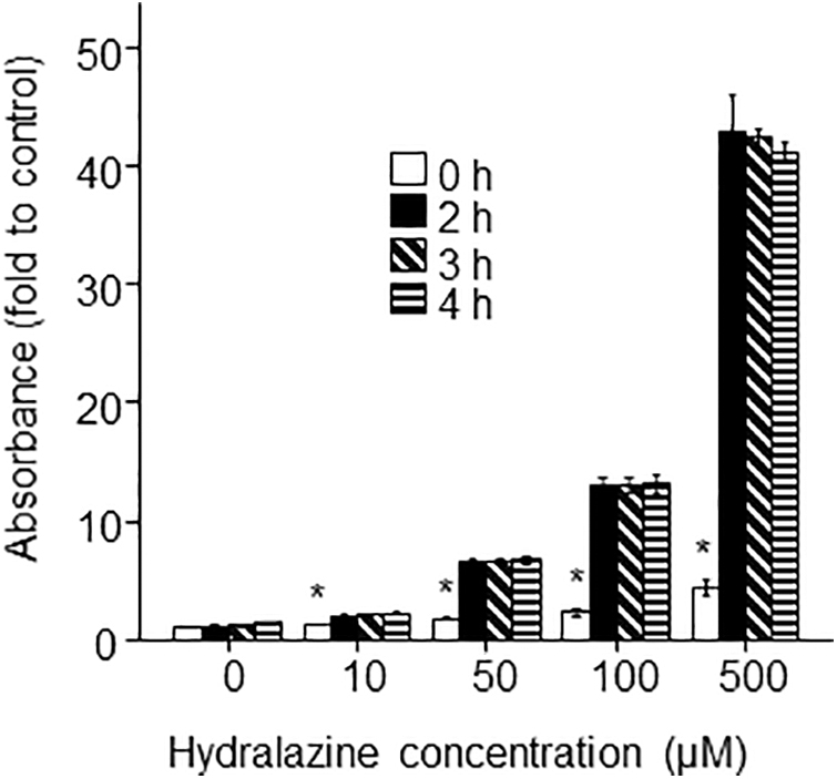
The reactivity of hydralazine with MTS. Four microliters of PBS or various concentrations of hydralazine were added to 200 μL of cell culture medium without cells in wells of a flat-bottom 96-well plate to give final concentrations of 0, 10, 50, 100, and 500 μM hydralazine. Twenty microlitersuL of MTS was then added and absorbance was measured using a plate reader 2 h after addition of MTS. The absorbance value in the well containing the medium only was regarded as background and subtracted from the absorbance of other wells. The absorbance of the control group (0 μM hydralazine at 0 h) was regarded as onefold (baseline). Error bars represent standard deviation. The means among groups were analyzed using one-way ANOVA followed by Bonferroni post hoc tests. *p < 0.001 compared with any other groups with the same concentration of hydralazine.
Modified MTS Method Can Accurately Reflect the Live Cell Number of VSMCs Incubated with Hydralazine
The manufacturer's instruction recommends directly adding MTS to the wells containing cells. Given that hydralazine can directly react with MTS, the assay protocol was modified by replacing the used culture medium in the wells with prewarmed fresh culture medium immediately before the addition of MTS. The modified MTS assay suggested that incubation of VSMCs with hydralazine for 2 h did not affect the absorbance (Fig. 4), consistent with observations under the light microscope (Fig. 1B). Also, the results from the modified MTS assay suggested that incubation of VSMCs with 100 and 500 μM hydralazine for 24 h significantly decreased the cell number (Fig. 5), which was consistent with the results from the trypan blue exclusion assay (Fig. 2B) and the microscopy observations (Fig. 2C).

The live cell numbers of VSMCs after incubation with hydralazine for 2 h measured by the modified MTS assay. Two hundred microliters of VSMCs (1 × 10⁵/mL) was placed in each well of a flat-bottom 96-well plate for 24 h. Four microliters of PBS or various concentrations of hydralazine were added to give final concentrations of 0, 10, 50, 100, and 500 μM hydralazine in the wells. Two hours later, the medium was aspirated and replaced with a prewarm (37°C) 200 μL culture medium. Twenty microliters of MTS was then added and absorbance was measured using a plate reader 2 h after the addition of MTS. The absorbance value in the well containing the medium plus MTS in the absence of cells was regarded as background and subtracted from the absorbance of other wells. The absorbance in the control group (0 μM hydralazine) was regarded as 100%. N = 8. Error bars represent standard deviation. The means among groups were analyzed using one-way ANOVA followed by Bonferroni post hoc tests.
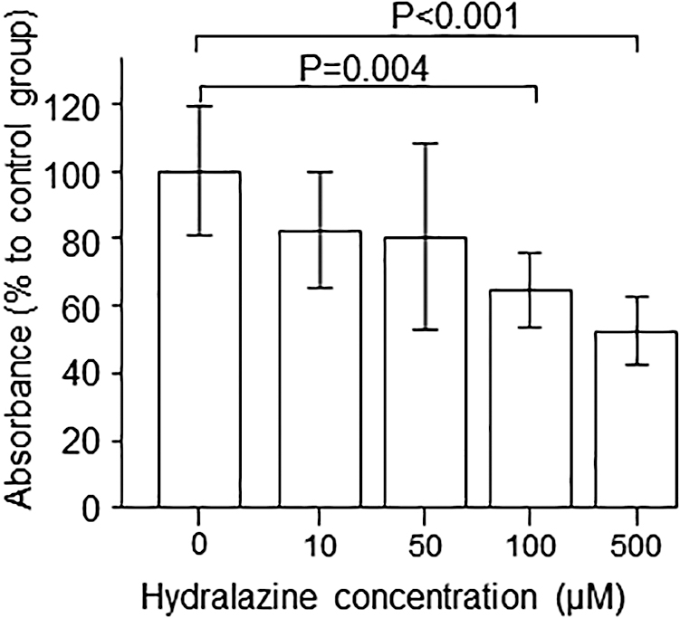
The live cell numbers of VSMCs after incubation with hydralazine for 24 h measured by the modified MTS assay. Two hundred microliters of VSMCs (1 × 10⁵/mL) was placed in each well of a flat-bottom 96-well plate for 24 h. Four microliters of PBS or various concentrations of hydralazine were added to give final concentrations of 0, 10, 50, 100, and 500 μM hydralazine in the wells. Twenty-four hours later, the medium was aspirated and replaced with a prewarm (37°C) 200 μL culture medium. Twenty microliters of MTS was then added and absorbance was measured using a plate reader 2 h after the addition of MTS. The absorbance value in the well containing the medium plus MTS in the absence of cells was regarded as background and subtracted from the absorbance of other wells. The absorbance in the control group (0 μM hydralazine) was regarded as 100%. N = 8. Error bars represent standard deviation. The means among groups were analyzed using one-way ANOVA followed by Bonferroni post hoc tests.
Discussion
This study reported a simple modification of MTS assay that overcame the interference of the testing reagent hydralazine. A discrepancy in the results of the standard MTS assay with those from the trypan blue and microscopic assays was identified. Further investigation discovered that hydralazine directly reacted with the MTS reagent in the absence of cells. A simple modification of the MTS assay protocol, by replacing the used culture medium immediately before the addition of the MTS, was established. The results from the modified MTS assay were in agreement with the results from both the trypan blue exclusion and microscopic assays.
This study used an MTS Cell Proliferation Assay Kit from BioVision, Inc., which is a colorimetric method for quantification of viable cells. The method is based on the reduction of MTS tetrazolium compound by viable cells to generate a colored formazan product that is soluble in cell culture media. This conversion is thought to be carried out by NAD(P)H-dependent dehydrogenase enzymes in metabolically active cells, 1 in particular mitochondrial succinate dehydrogenase. 14,15 The formazan dye produced by viable cells can be quantified by measuring the absorbance at 490–500 nm.
MTS can be directly converted into a colored product by hydralazine in a cell-free system in a dose-dependent manner. This reaction completed within 2 h, as an increase in the incubation time further to 3 or 4 h did not increase the absorbance further. Hydralazine is a reducing reagent that can reduce many oxidizing compounds such as lipid-derived electrophiles 16 and superoxide and peroxynitrite. 17 These findings demonstrate that hydralazine interferes with the MTS assay. It is worthwhile to investigate whether other reducing reagents such as vitamin E and vitamin C can interfere with the MTS assay similarly in the future.
In the presence of the cells, the standard MTS assay suggested that incubation with 50 μM hydralazine for 2 h or 500 μM hydralazine for 24 h increased absorbance (i.e., cell number), which could not be confirmed from the microscopy and trypan blue assays. When cells were incubated with 100 or 500 μM hydralazine for 24 h, the trypan blue exclusion assay and microscopic observation suggested that there was reduction in cell numbers compared with the nonhydralazine control, suggesting the cytotoxicity of hydralazine. The false results of the standard MTS assay may be due to the inability of the cultured cells to completely absorb and metabolize the large amounts of hydralazine within 24 h, in contrast to the incubation with 50 μM hydralazine for 24 h. The modified MTS assay replicated the results of the microscopy and trypan blue assay, suggesting it overcame the interference by hydralazine.
The MTS assay can be potentially affected by other factors. The assay would be expected to be unreliable when testing compounds that affect mitochondrial function, as mitochondrial succinate dehydrogenase is a key enzyme in the MTS conversion. 14,15 Indeed, incubation of the breast cancer cell line MCF-7 with isopropyl-β-thiogalactoside has been reported to decrease cell numbers by 60% as measured with the trypan blue exclusion assay, however, the MTS assay found an increase by 15% in absorbance. 18 In that study, isopropyl-β-thiogalactoside incubation increased mitochondrial counts by threefold, 18 which is likely responsible for the inaccuracy of the MTS assay.
Reliable measurement of cell viability, proliferation, growth inhibition, and death is important in screening for drug treatment efficacy in vitro. 18 To avoid inaccurate result, supplementation of the MTS assay with other nonmetabolic assays such as the trypan blue exclusion assay, flow cytometry, and microscopic observations, is recommended. 19
In summary, this study suggests an important limitation of the standard MTS assay protocol. A modification of the protocol, where the used culture medium in wells containing cultured cells is replaced with a fresh medium immediately before the addition of MTS reagent, rectifies this problem and results in accurate cell number estimation.
Footnotes
Disclosure Statement
No competing financial interests exist.
Funding Information
This work was funded by grants from the National Health and Medical Research Council of Australia (1062671). J.G. holds a Practitioner Fellowship from the National Health and Medical Research Council of Australia (NHMRC; 1117061) and a Senior Clinical Research Fellowship from the Queensland Government, Australia.