




Abstract
Age-related macular degeneration (AMD), a multifactorial age-related retinal hypoxic disorder resulting in irreversible loss of vision, is the foremost cause of blindness in the United States. Current treatment strategies involve multiple intraocular injections of antivascular endothelial growth factor (VEGF) agents into the vitreous of eye. In addition to the challenges of drug localization and targeted delivery, the need of frequent injections into the eye raises patient compliance issues, and thus call for development of sustained drug delivery systems. In this study, a sustained drug delivery system was prepared by loading an antihypoxia-induced factor (HIF) agent, honokiol (HON), into methoxy poly (ethylene glycol) polycaprolactone (MPEG-PCL) polymer. These HON-MPEG-PCL micelles were characterized by evaluating size, ζ potential, in vitro drug release profile, and morphology by transmission electron microscopy. The cytotoxic nature of developed micelles was assessed on human retinal pigment epithelial cell line (ARPE-19) cells by cytotoxicity assay. The cellular uptake and HIF and VEGF expression levels were determined in in vitro settings. Micelles formed had a particle size of 30.8 ± 0.8 nm with the poly dispersity index of 0.19 ± 0.0004 and ζ potential was found to be −5.46 ± 0.49 mv. Entrapment efficiency was calculated to be 64 ± 0.135%. In vitro drug release showed sustained release of drug from the formulation. Result from in vitro cytotoxicity study confirmed noncytotoxic nature of HON-MPEG-PCL micelles compared to HON drug solution. Furthermore, enzyme-linked immunosorbent assay studies performed showed the periodic downregulation of HIF and VEGF, which are major growth factors involved in underlying mechanism of AMD. The results showed successful development of HON-MPEG-PCL micelles, which may be useful for the effective treatment of AMD.
Introduction
Age-related macular degeneration (AMD) is a multifactorial age-related ophthalmic disorder that is characterized by substantial, progressive, and irreversible loss of central vision due to macular degeneration. 1 AMD is the leading cause of blindness in United States with an estimated 10 million people being affected by this disease. 2
Out of three stages of AMD, early AMD is diagnosed by the presence of medium-sized drusen and intermediate AMD has large drusen, pigment changes in the retina, or both, while late AMD (dry or wet) has vision loss from damage to the macula. Based on pathophysiology, AMD can be broadly classified into dry AMD and wet AMD. Dry AMD (geographic atrophy) has a gradual breakdown of the light-sensitive cells in the macula that convey visual information to the brain and of the supporting tissue beneath the macula. These changes cause vision loss.
In wet AMD (neovascular AMD), abnormal blood vessels grow underneath the retina. Because these new blood vessels are abnormal, they tend to break, bleed, and leak fluid, damaging the macula and causing it to lift up and pull away from its base. This can result in a rapid and severe loss of central vision. Dry AMD is the nonvascular form of AMD and is characterized by degeneration of retinal pigment epithelial (RPE) cells and photoreceptors, while wet AMD is the vascular form of AMD and is characterized by choroidal neovascularization (CNV). 3 Wet AMD is an acute disorder and rapidly escalates leading to blindness, which affects ∼10%–15% of individuals with AMD, but accounts for ∼90% of all cases of severe vision loss from the disease. 4
Hypoxia-induced factor or HIF is responsible for oxygen hemostasis and is involved in maintaining and ensuring cell survival under hypoxic conditions. 5 Under normal circumstances, there exists a balance between proangiogenic factors, for instance, vascular endothelial growth factor (VEGF) and antiangiogenic factors such as pigment epithelium-derived factor. Studies have shown that hypoxia plays a role in the upregulation of proangiogenic factors. 6 Ischemic tissue is one of the important pathological factors that results in upregulation of HIF, which initiates a cycle of events leading to upregulation of many proangiogenic factors causing CNV, a clinical hallmark of wet AMD and serious complication of AMD that can lead to blindness in AMD. 7 As a follow-up event to damage to retinal epithelial layer, secretion of cytokines and growth factors such as VEGFs, HIF, ion channel dysfunction, and abnormal lipid metabolism lead to oxidative damage of cells. 8 To compensate for the decrease in blood supply at the retinal region, neovascularization occurs, which may lead to increase in risk of fluid deposition, inflammation, vascular occlusion, CNV, and haemorrhage. 9 Thus, an approach based on targeting HIF, a master regulator of many angiogenic factors leading to CNV, can prove to be potentially beneficial in treatment of wet AMD. 10 Honokiol (HON) is a lignin isolated from the bark of Magnolia and this biphenolic phytochemical has been shown to have potent anti-HIF properties. 11 Studies have shown that HON inhibits HIF resulting in a significant decrease in CNV. 12
Based on pathophysiology, anti-VEGF therapies have been approved by Food and Drug Administration (FDA) to target CNV such as bevacizumab/Avastin, aflibercept/VEGF trap eye, pegaptanib/Macugen, and ranibizumab/Lucentis. 13 Use of these anti-VEGF medications has helped achieving good result for the treatment of AMD. However, a study reports the need of repetitive ranibizumab and bevacizumab administration either on monthly basis or in some cases may require the setup of personal dose regimen based on severity or progression of disease. 14 The physiology of the eye is complex and achieving therapeutic dose at posterior side of eye can be a complicated affair offering fair challenges. While the eye's anterior segment is easily assessable for topical delivery, multiple clearance mechanism, physical barriers, transient residence time, and corneal epithelium impermeability make it harder for drugs to cross these barriers 15 and reach the posterior segment of eye, which is a desirable target location in the case of AMD. Furthermore, systemic delivery of drug requires high doses, which puts patients at risk of several toxic side effects. 16 Intravitreal route bypasses all these physiological barriers to achieve the desired therapeutic concentration in the eye. Studies have reported that increased or repetitive intravitreal injections may not be a very practical approach and can compromise patient's compliance, in addition to putting patients at risk of infections, endophthalmitis, vitreous floaters, intraocular inflammation, retinal detachment, and cataract. 17,18 Thus, the development of sustained release drug formulation to treat AMD is an ideal drug delivery system.
The vast applications of this technology in medical field have potential to revolutionized health care facilities provided to patients. Encapsulated drug in nanoparticles has shown to decrease toxic effects along with achieving high efficiency and targeted delivery. 19 Furthermore, the use of micelles for ocular drug delivery offers advantages such as increased solubility, surface area, and drug dissolution. Methoxy poly (ethylene glycol) polycaprolactone or MPEG-PCL polymer is an FDA-approved biocompatible, amphiphilic polymer. It combines hydrophilic polyethylene glycol (PEG) and hydrophobic PCL, which imparts them the advantage of encapsulating wide range of drugs. These well-researched polymers are widely used in the biomedical field for drug delivery purposes and have properties like biocompatibility and controllable biodegradable nature. 20 These polymers, because of their slow degradation rate, are favored for sustained release drug delivery system. Research has shown these polymers are capable to localize in inflamed areas, which helps to achieve targeted actions. These polymers have been widely used in the field of nanotechnology, pharmaceutics, and medicinal chemistry. 21 Outer covering is constituted by the PEG part of polymer, which provides “stealth” effect, thus making them less immunogenic. 22
The objective of this work is to formulate sustained release formulation of HON-MPEG-PCL and to evaluate the formulation using physiochemical methods and cell-based assays. Thus, due to the sustained release profile, the residence time of the HON will be longer after injection at the target site, which in turn will decrease the dosing frequency. Therefore, loading micelles with HON has the potential to show promising therapeutic effects by achieving sustained release localized effect with reduced toxicity and side effects.
Materials and Methods
Materials
HON [CN: H13091G] was purchased from TCI chemicals (USA). Methoxy poly (ethylene glycol)-block-poly (ɛ-caprolactone) mPEG-b-PCL in different ratio combinations (2K–2K, 5K–2K, and 5K–10K) [CN: 900649-500MG, 900671-500MG, and 900672-500MG, respectively] was purchased from Sigma Aldrich (St. Louis, MO). High performance liquid chromatography (HPLC) grade Acetone [CN: A949-4] was purchased from Sigma Aldrich. HPLC grade Methanol [CN: A452-4] was ordered from Fisher Scientific. Dimethyl sulfoxide (DMSO) [CN: D1391] and polyvinyl alcohol (Mw 100,000, 87% hydrolyzed) [CN: S93328] were also ordered from Fisher chemicals and Tween-80 was purchased from Sigma Aldrich. Slide-A-Lyzer® Dialysis Cassette (MWCO, 10,000 Da) [CN: S93328] was purchased from Thermo Scientific (IL). Human retinal pigment epithelial cell line (ARPE-19) derived from human retinal pigment epithelial cell line was ordered from ATCC© CRL2302™ (American Type Culture Collection). Trypsin (0.05%) [CN: 25300054] was purchased from Thermo Fischer Scientific (Lansing, MI). Fetal bovine serum (FBS) [CN: 10437028] along with penicillin-streptomycin (10,000 U/mL) [CN: 15140122] were ordered from Gibco Thermo Fisher Scientific. Incomplete media (i.e.) Dulbecco's Modified Eagle Medium (DMEM) F12 medium [CN: ATCC® 30-2006™] was also bought from ATCC (VA). Furthermore, phosphate-buffered saline (PBS) [CN: MT21040CV] was purchased from Corning cellgro (Manassas, VA). MTT reagent [3-(4,5-dimethylthiazole-2-yr)-2,5-diphenyltetrazolium] bromide salt reagent [CN: 5224] was ordered from Tocris Bioscience (MN). Nucleus stain DAPI (4’,6-diamindion-2-phenylindole) [CN: 5748] was ordered from Tocris Bioscience. Cell Mask™ deep red plasma stain was bought from Molecular probes, Invitrogen™ Thermo Fisher Scientific. For enzyme-linked immunosorbent assay (ELISA) studies, an Invitrogen Bioscience™ human HIF [CN: EHIF1A] kit and VEGF-A Platinum ELISA Kit [CN: 50-182-08] were ordered from Fisher Scientific.
Cell Culture
Cell culture studies were performed using ARPE-19 (ATCC CRL2302) cells. These cells derived from a human retinal pigment epithelial cell line were grown in complete media, formulated by adding 10% FBS and 1% 10,000 U/mL pen—strep antibiotics in DMEM F12 medium. The cell cultures were allowed an incubation period of 24 h at 37°C temperature supplied with 5% carbon dioxide (CO2).
Synthesis of HON-MPEG-PCL Micelles
The HON-MPEG-PCL micelles were synthesized using solvent evaporation technique 23,24 (Fig. 1). Briefly, micelles were prepared by using 1:10 drug to polymer weight ratio. For preparing the organic phase, 2 mg of HON drug was solubilized into 2 mL of organic solvent mixture of DMSO: acetone (1:9 v/v). In the next step, 20 mg of MPEG-PCL polymer was added to the prepared organic solution of drug. Four milliliters of filtered distilled water was used as an aqueous phase. The 2 mL organic phase was added into 4 mL aqueous phase dropwise using a 23G syringe under magnetic stirring. The emulsion formed was left stirring on magnetic stirrer (Thermo Fisher) for 24 h at 700 rpm to allow time for formation of micelles and evaporation of organic solvents. Next day, the formulation was centrifuged at 5,000 rpm for 15 min to remove any free drug precipitates followed by centrifuging supernatant at 18,000 rpm for 20 min at room temperature to obtain the micelle pellet. The micelle pellet obtained after centrifugation was resuspended in saline for further studies. Blank micelles were prepared following the same protocol, except for the addition of drug (HON). The formulation was filter sterilized to be used for in vitro and future in vivo studies.
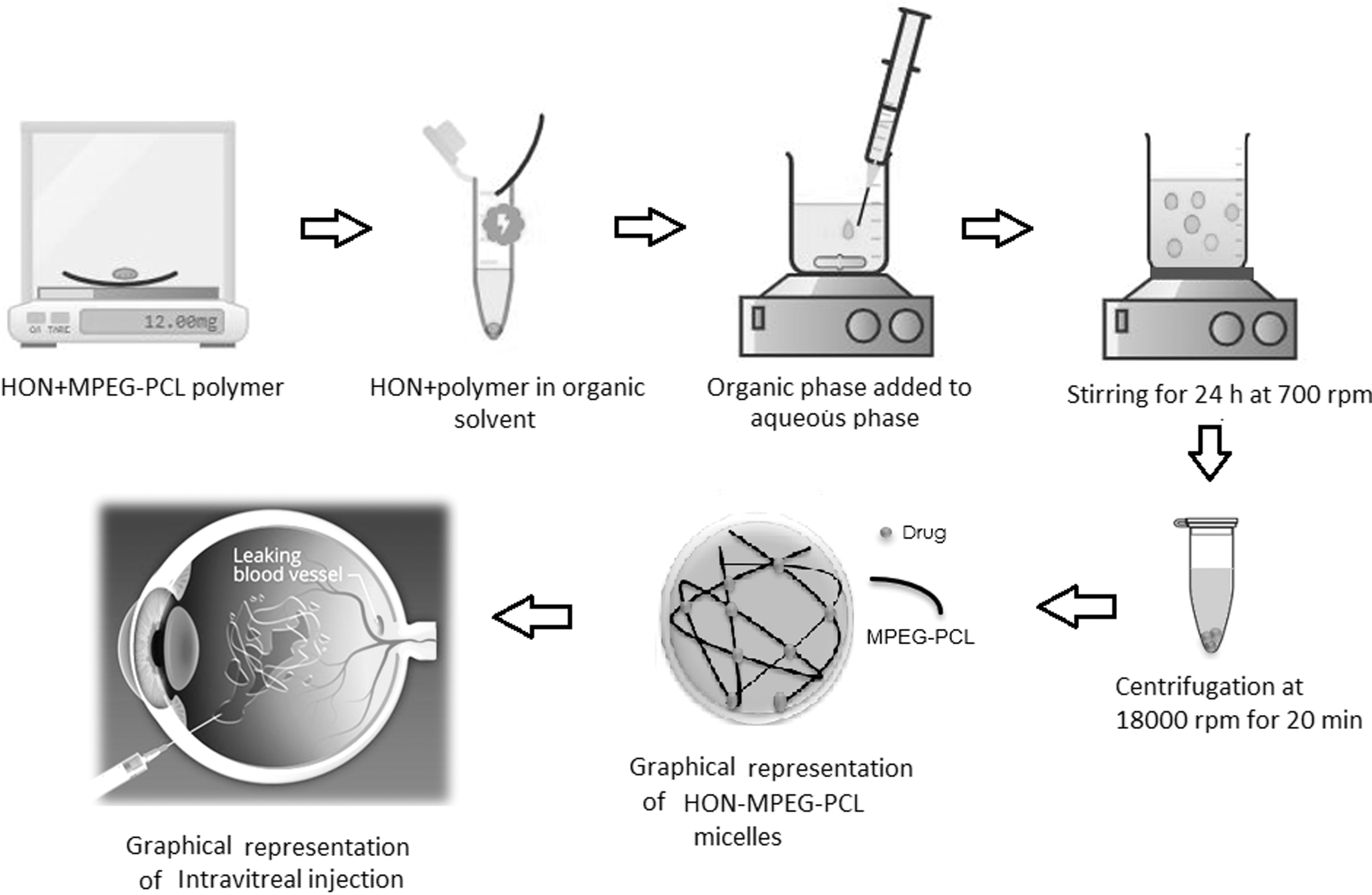
Graphical presentation of preparation of HON-MPEG-PCL micelles and intravitreal injection into the eye for treatment of AMD. AMD, age-related macular degeneration; HON, honokiol; MPEG-PCL, methoxy poly (ethylene glycol) polycaprolactone.
Differential Scanning Calorimetry
Differential scanning calorimetry (DSC) Q-20 (Q series Q-20-2288-DSC Software; TA Instruments, New Castle, DE) was used as a thermal analytical method to confirm the loading of HON in HON-MPEG-PCL micelles. In addition, the DSC technique was used to evaluate the physical state of drug after entrapping in micelles because that affects the release pattern. HON drug, MPEG-PCL (5K–2K) polymer, and HON-MPEG-PCL micelles were analyzed. An appropriate quantity of all three samples (5 mg) was individually placed in an aluminum pan, which was later hermetically sealed. These samples were heated gradually at the rate of 10°C/min from 30°C to 300°C in a nitrogen atmosphere. The flow rate of nitrogen was 50 mL/min. Empty aluminum pan was used as control. 25
Particle Size and ζ Potential
Dynamic light scattering (DLS) principle was used for the determination of nanoparticle size. A total of 633 nm He–Ne laser was used as a light source and scattering angle was 90° 26 (Nano ZS90; Zeta Sizer Software Ver. 7.10; Malvern Instruments Ltd., UK). Each sample was diluted 1:10 using filtered distilled water. Samples were run in triplicate at 25°C temperature and results were reported as an average of size in nm along with poly dispersity index (PDI).
Zeta potential measurements provide information about the particle surface charges. Smoluchowski equation was used to obtain ζ potential of micelles using particle's electrophoretic mobility. Samples were appropriately diluted (1:10) by adding 100 μL of micelle formulation into 900 μL of distilled water. Zeta cuvettes were used to analyze samples in triplicate at 25°C (Zeta Sizer software Ver. 7.10; Malvern Instruments Ltd.).
Transmission Electron Microscopy
The morphology of HON-MPEG-PCL was characterized using transmission electron microscopy (TEM) (JEOL JEM 1400 electron microscope with Gaton camera, Peabody, MA). For taking images, samples were prepared using Cu-film square grids. Formulation was diluted using filtered distilled water (1:10); 5 μg of sample was taken on grid. Samples were allowed to settle for ∼20 min. Furthermore, samples were negatively stained using 2% w/v phosphotungstic acid. Four hundred K × magnification was used to take images at an accelerating voltage of 120 kV. 27
Entrapment Efficiency
Entrapment efficiency (EE) was analyzed to check the drug loading capacity of HON-MPEG-PCL micelles. The formulation was centrifuged at 5,000 rpm for 15 min to separate free drug solution or polymer. Supernatant obtained was collected and centrifuged further at 18,000 rpm to obtain pellet, which was further washed thrice with filtered distilled water to get rid of any free drug or polymer. Samples were prepared by re-constituting micelles with methanol using 1:10 dilution. One hundred microliters of formulation was diluted into 900 μL of methanol. Samples were analyzed in triplicate using ultraviolet (UV) spectrophotometer at 292 nm wavelength (Model: S-2150UV; Cole Parmer Instrument Company). Calibration curve equation (R 2 0.9993) was used to quantify the results.
% EE was calculated as following equation
28
:
In Vitro Drug Release Studies
In vitro drug release studies were conducted to determine the sustained release action of HON-MPEG-PCL micelle formulation. This experiment comprised comparison between drug release profile of free drug and prepared micelle formulation. Release media were formulated by adding 1 L of phosphate buffer along with 0.1% v/v of Tween-80 (pH 7.4). 29 Approximately 0.5 mL of both the drug solution and HON-MPEF-PCL micelles was injected into dialysis cassette using syringe having a 23G needle. These cassettes were suspended into the release media. One hundred milliliters of release media was taken in a beaker and sample filled cassette was immersed in this release media under magnetic stirring at 150 rpm and 37°C. Samples were collected at different time periods. For each 1 mL sample drawn at each time interval, it was replaced with fresh 1 mL phosphate buffer release media. Samples were drawn at 0.25, 0.75, 1, and 2 h for the 1st day. Later samples were drawn every 24 h for 10 days (240 h). Samples obtained were diluted with methanol and analyzed under UV spectrophotometer at 292 nm. Finally, results were quantified by plotting a graph.
Cytotoxicity Study
ARPE-19 cells were used to perform MTT analysis for calculating cytotoxicity of prepared HON-MPEG-PCL formulation in comparison with pure drug solution. On the first day, cells were seeded in a 96-well plate at a density of 5,000 cells/well. These cells were supplemented with 200 μL of complete media composed of DMEM F12 and 10% FBS and were later allowed to grow in incubator supplied with 5% CO2 at 37°C for 24 h. Next day, the cells were treated with formulation and drug samples at a different concentration ranging from 0.001, 0.01, 0.1, 1.0, 10, to 20 μM in triplicate. After treatment, cells were allowed to incubate for 4 h. After the 4-h treatment, cells were removed and replaced with fresh complete media. Again, cells were incubated for 24 h. After 24 h, complete media were removed and MTT reagent 100 μL (1 mg/mL) was added to the 96-well plate, which was left to incubate for 4 h. After 4 h, each well was replaced with 100 μL of DMSO after removing MTT agent. DMSO was used to dissolve formazan crystals. The crystal displays cell viability by showing purple color. The cell viability was quantified by analyzing the 96-well plate through UV spectrophotometer at a wavelength of 595 nm (Spectra MAX 190; Molecular Devices, CA). Cells treated with just DMEM F12 were used as negative control and 0.1% Triton X100 was considered the positive control. Results were quantified by plotting a graph between the concentrations versus viability, while considering the cell viability of negative control as 100% viable.
Cellular Uptake
Cellular uptake capacity of the formulation HON-MPEG-PCL was measured to determine the localized action of formulation in the cell. This was achieved using the confocal microscopy. ARPE-19 cells were seeded at 2 × 10⁵ density per well in a three = well plate and incubated for 24 h to achieve confluency. Free drug solution, dye-loaded coumarin HON micelles, and blank micelles were used as treatments. Furthermore, cells were washed thrice next day with PBS to remove any suspended dead cells. Each well was treated with a different treatment for about 15 min. DAPI was used to stain nucleus, while Cell Mask deep red plasma stain was used for cell membrane staining. The results were analyzed under confocal microscope FV1200 (Olympus, Tokyo, Japan) at 400K magnification for a period of 80 min.
Anti-HIF ELISA
This experiment was performed to study the anti-HIF activity of HON. Cells were seeded at a density of 5 × 10³ cells/mL in a 96-well plate. The plate was incubated for a period of 24 h in an incubator supplied with 5% CO2 at 37°C to achieve confluency. Next day, cells were treated with HON drug solution and HON-MPEG-PCL micelles at a concentration of 10 μm. The treatment was given for a period of 24, 48, 72, and 96 h. Invitrogen ELISA HIF kit was used to calculate the expression of HIF protein. Experiment was performed following the given protocol within kit and plate was studied at 450 and 550 nm in UV spectrophotometer (Spectra MAX 190; Molecular Devices). Graph was plotted by taking Control as expressing 100% HIF expression and results were analyzed.
Anti-VEGF ELISA
The effect of HON-MPEG-PCL micelles and HON drug solution on expression of VEGF was studied by seeding APRE-19 cells in a 96-well plate at a density of 5,000 cells/well. The 96-well plate was incubated for 24 h, supplied with 5% CO2 at 37°C. After 24 h, cells were treated with different treatment groups for the period of 24, 48, 72, and 96 h. Invitrogen human VEGF ELISA ready to use kit was used to analyze the expression of VEGF in samples collected at different time points. The plate was analyzed in a UV spectrophotometer at 450 nm wavelength (Spectra MAX 190; Molecular Devices). Complete media and incomplete media were taken as negative and positive control. Graph was plotted by taking control as 100% expression of VEGF.
Statistical Analysis
All samples were run in triplicate and data are represented as mean ± standard deviation. Analysis of variance and GraphPad Prism (version 6) were used to analyze data statistically. p-Value <0.05 and <0.01, <0.001 were considered significant.
Results
Differential Scanning Calorimetry
DSC was performed to confirm loading of HON in micelles. This thermal analysis was performed on HON drug, polymer MPEG-PCL (5K–2K), and formulation (HON-MPEG-PCL), which helped to identify the physical state of a drug in polymer that can in turn dictate the drug releasing pattern of the formulation. In case of HON drug, the peak was appeared at 84.85°C, while for the polymer, the characteristic peak appeared at 55.16°C; however, the absence of these characteristic peaks in DSC spectra of HON-MPEG-PCL formulation, which showed peak at 112.67°C, confirmed the entrapment of HON drug in HON-MPEG-PCL micelles (Fig. 2).

DSC spectra of Honokiol drug, MPEG-PCL polymer and HON-MPEG-PCL micelles. DSC, differential scanning calorimetry.
Particle Size and ζ Potential
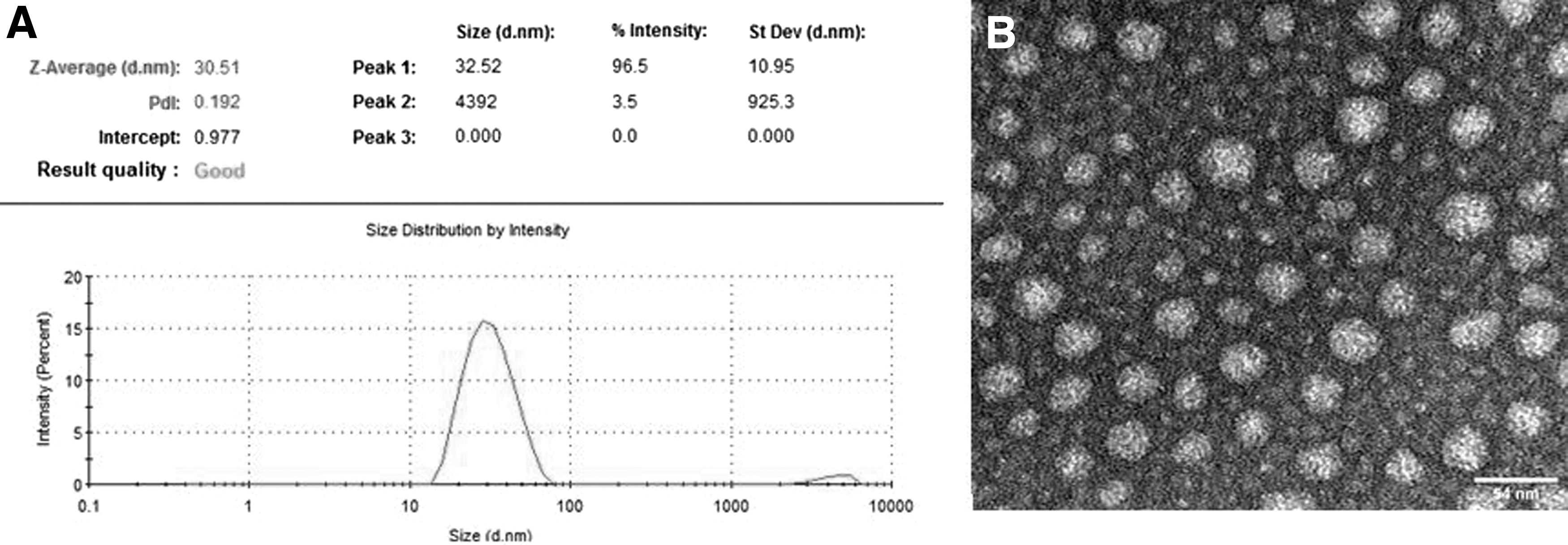
Preformulation trials were conducted using different variations of MPEG-PCL polymer composition, including MPEG-PCL (5K–2K, 5K–5K, and 10K–5K ratio). These studies concluded MPEG-PCL (5K–2K) polymer in a 1:10 drug to polymer ratio to be optimum for micelle formulation (Table 1). From various MPEG-PCL with varied molecular weight ratios of MPEG and PCL as 2K–2K, 5K–2K, and 5K–10K, the 5K–2K ratio was optimized because of its smaller particle size, which helps micelles pass through RPE cells. Moreover, the smaller PDI value also confirmed uniformity of micelles in a formulation of MPEG-PCL (5K–2K) compared to other two polymers. Particle size of micelles along with PDI was determined using DLS. Size was found to be of order 30.8 ± 0.8 nm and PDI was 0.19 ± 0.0004 (Fig. 3). The low PDI value obtained from results was indicative of homogeneity and monotonous distribution of micelles in a formulation. The smaller particle size of micelles and lower value of PDI made formulation optimum for ocular delivery. The mean ζ potential was determined using Malvern Zeta sizer Nano ZS90 and was calculated to be −5.46 ± 0.49 mV for the HON-MPEG-PCL micelles.
Size, Poly Dispersity Index, and % Entrapment Efficiency of Various Methoxy Poly (Ethylene Glycol) Polycaprolactone Micelles for Preformulation Study Trials
EE, entrapment efficiency; MPEG-PCL, methoxy poly (ethylene glycol) polycaprolactone; PDI, poly dispersity index.
Transmission Electron Microscopy
TEM was performed to check the morphology of micelles. The TEM images obtained after analyzing Cu grid containing samples showed micelle size to be around 30 nm. The obtained result falls in line with previous result obtained from the DLS studies, which confirms the size of micelles. TEM image (Fig. 3) further confirmed the uniformity of preparation along with spherical shape.
Entrapment Efficiency
EE of HON-PEG-PCL micelles was obtained by resuspending the micelles in methanol in 1:9 ratio. EE was calculated using the equation previously developed by dissolving varying concentrations of HON in methanol at 292 nm (λmax) and plotting a calibration curve r value. The reading was obtained by analyzing samples in UV spectrophotometer at 292 nm (λmax). EE was calculated to be 64 ± 0.135%.
In Vitro Drug Release
Sustained release action of HON-PEG-PCL micelles was analyzed through in vitro release studies. Dialysis cassettes containing with 0.5 mL of both drug solution and formulation were immersed in 100 mL of release media comprising PBS along with (0.1% v/v) Tween-80 to create sink condition. For the 1st day, samples were withdrawn at 0.25-, 0.5-, 1-, and 2-h time point. From the following day, samples were withdrawn after every 24 h for a total period of 240 h. Each time, the removed volume (1 mL) was replaced with fresh release media. To quantify drug release pattern, samples were diluted in 1:2 ratio of methanol. Samples were mixed with methanol and the amount of released HON in the media was quantified in triplicate using UV spectrophotometer at 292 nm wavelength (Model: S-2150UV; Cole Parmer Instrument Company). Equation of calibration curve was (R 2 0.9993). “Cumulative quantity of drug” describes the total amount of drug released when it is all added together by considering the amount released at that time point and previous time points. Finally, graph was plotted between cumulative drug release (%) as a function of time. As shown in graph, after 24 h, ∼24% of the HON was released from the formulation, while 50% HON was released from the drug solution in release media. One hundred percent of HON was released from drug solution in 48 h, while only 79 ± 0.004% of HON was released from formulation quantified in release media at the end of a 240-h period (Fig. 4). This confirmed the sustained release action of HON-MPEG-PCL micelles.
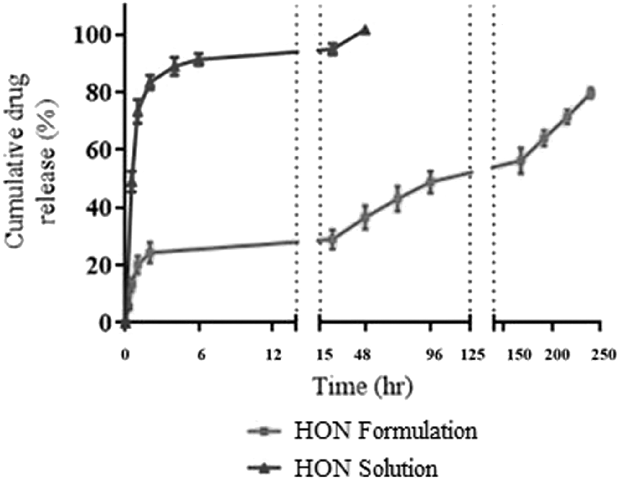
Comparison of cumulative drug release percentage of HON-MPEG-PCL micelles and HON solution for period of 240 h (10 days) at 37°C in release media (i.e.), phosphate buffer saline (pH 7.4) + 0.1% (v/v) Tween-80 (mean ± SD, n = 3).
Cytotoxicity Study
The cytotoxicity of the HON-MPEG-PCL formulation was evaluated using MTT assay performed on human epithelial retinal pigment (ARPE-19) cell line. Cells were treated in triplicate with serum-free media having varying concentrations of HON drug solution, blank micelles, and HON-MPEG-PCL micelles (0.001, 0.01, 0.1, 1, 10, and 20 μM) for the period of 24 h. Results obtained were analyzed by taking negative control as 100% cell viable. Blank micelles were 88% cell viable at highest concentration of 20 μM. For all concentrations of micelle formulation, cell viability was calculated to be above 80%, showing formulation to be biocompatible. Lowest concentration (0.001 μm) of HON-MPEG-MPCL micelles showed cell viability of 97 ± 0.028%, while highest concentration (20 μm) of formulation was 84 ± 0.024% cell viable. HON drug solution showed a concentration-dependent decrease in cell viability with 20 μm concentration, showed only 18 ± 0.001% cell viability. The low cell viability with higher concentrations of drug solution showed that drug is toxic to cells, while blank micelles and HON-MPEG-PCL micelles were cell compatible with cell viability >80% at higher concentrations as shown in Figure 5.
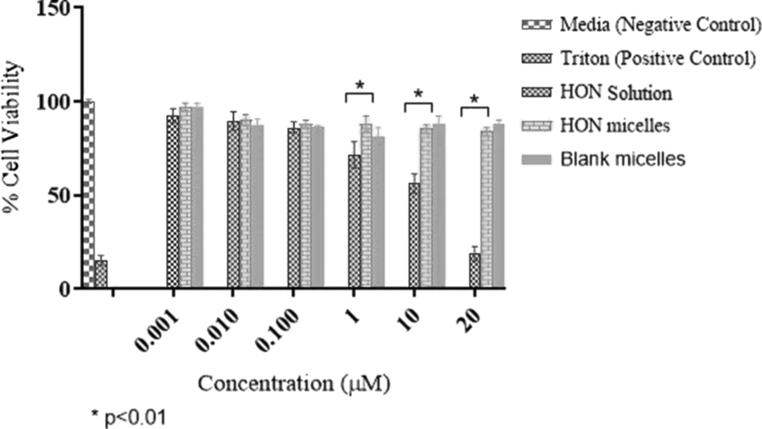
Cell viability plots of ARPE-19 cells after treatment with HON solution, HON-MPEG-PCL micelles, and blank micelles (mean ± SD, n = 3, *p < 0.01). ARPE-19, human retinal pigment epithelial cell line.
Cellular Uptake
The cellular uptake of prepared formulation was checked to understand the micelle interaction with cells. This study helps to evaluate the effect of size and shape on micelle uptake by cells and surface characteristics of micelles. This experiment is performed by preparing fluorescent emitting formulation. The fluorescent loaded micelles were prepared using the same protocol as HON-MPEG-PCL micelles except micelles were loaded with fluorescent dye instead of drug. ARPE-19 cells were treated with these fluorescent-containing micelles for the period of 20 min. As shown in figure, the cell uptake increases in a time-dependent manner. DAPI was used to stain nucleus blue color; cell membrane was stained with cell mask dye exhibiting red florescence. Green florescence represents cytoplasm of cell membrane, which takes up micelles labeled as green in Figure 6.
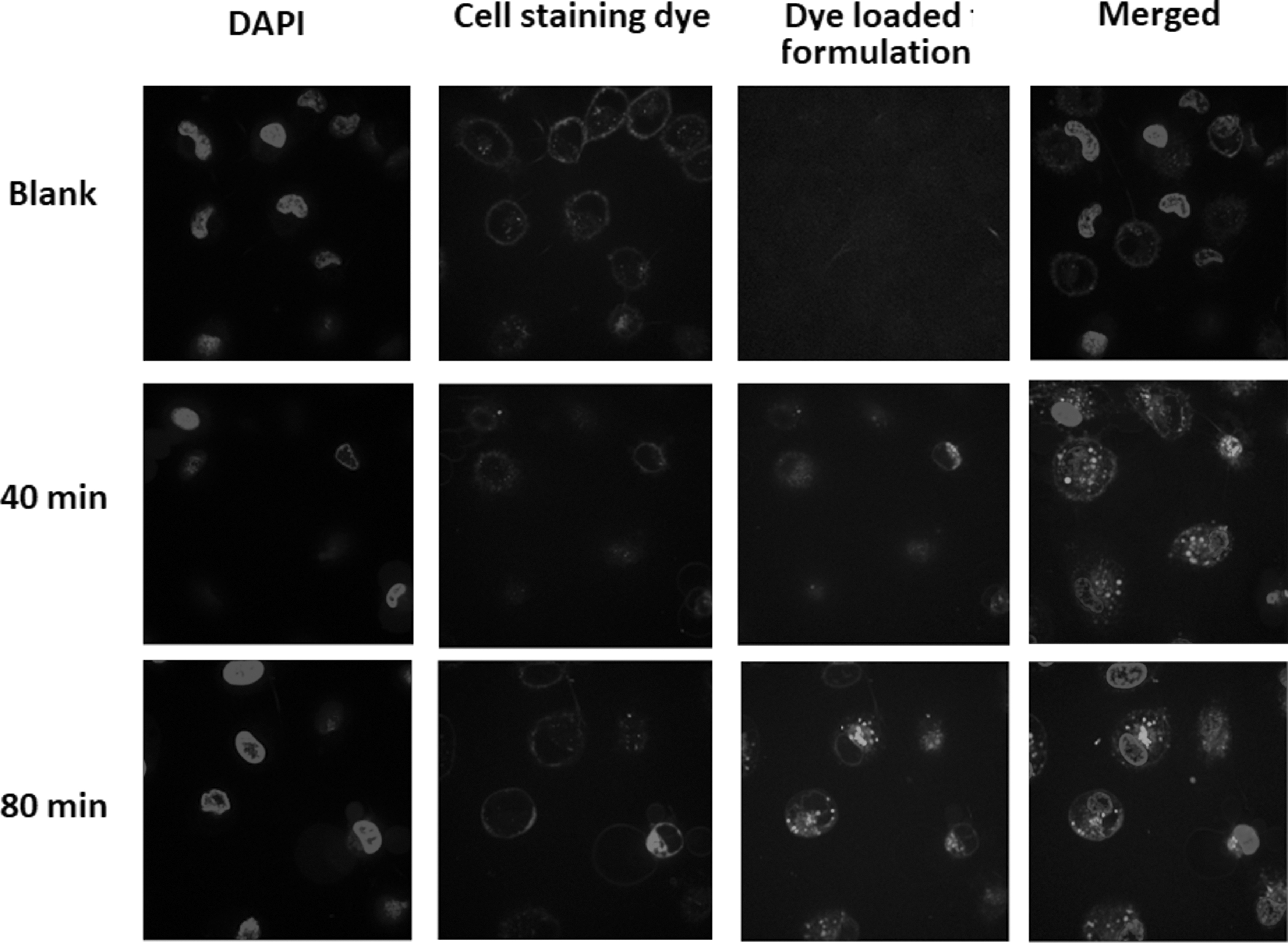
Cellular uptake of dye-loaded MPEG-PCL micelles (37°C) in ARPE-19 cells at 40 and 80 min observed using confocal microscopy. DAPI, 4’,6-diamindion-2-phenylindole.
Anti-HIF ELISA
The effect of HON drug solution and HON-MPEG-PCL micelles on HIF expression was studied using ELISA technique. Human HIF ELISA ready to use ELISA Kit was used to for this purpose. Samples were taken at 24, 48, 72, and 96 h and percentage decrease in HIF expression was calculated by taking control as 100%. The data values of % reduction in HIF expression levels for HON solution were 70.59 ± 2.9, 65.85 ± 2.0, 55.74 ± 3.6, and 41.19 ± 1.9 at 24, 48, 72, and 96 h, respectively. The data values of the % reduction in HIF expression levels for HON micelles were 63.91 ± 2.1, 67.71 ± 2.3, 72.45 ± 1.8, and 83.35 ± 2.7 at 24, 48, 72, and 96 h, respectively. At the 96-h time point, formulation performed better with 83.35 ± 2.7% reduction (p < 0.01) in HIF level. This can be explained by initial burst of free drug, but its effect decreased overtime due to metabolism. On the contrary, formulation showed a sustained release effect, and its effect increased over a period of 96 h (p < 0.01) as shown in Figure 7.
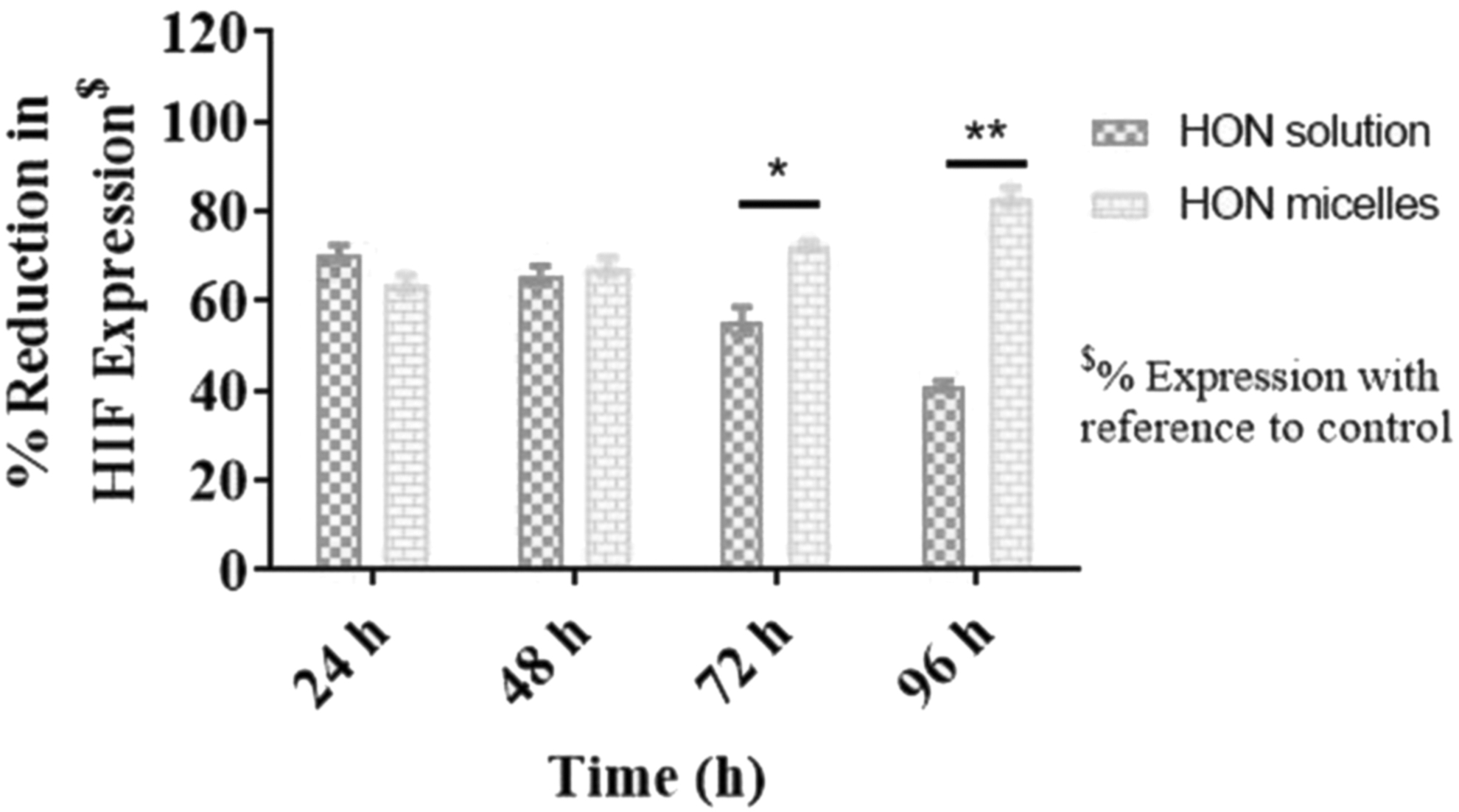
Percentage of decrease in HIF expression in ARPE-19 cells treated with HON solution and HON-MPEG-PCL micelles for different time points determined using ELISA assay method (mean ± SD, n = 4) *p < 0.05, **p < 0.01. ELISA, enzyme-linked immunosorbent assay; HIF, hypoxia-induced factor.
Anti-VEGF ELISA
The anti-VEGF ELISA was further performed to check the effectiveness of HON-MPEG-PCL micelles to downregulate expression of VEGF. By taking into consideration the physiological role of HIF to increase expression of proangiogenic factors, including VEGF. Its downregulation on treatment with HON-MPEG-PCL micelles in ARPE-19 cells shown in Figure 7 was expected to decrease level of VEGF expression as well. To analyze this, the effect of HON-MPEG-PCL micelles to decrease expression of VEGF was compared with HON drug solution in human retinal pigment epithelial cells (ARPE-19) after treating cells for 24, 48, 72, and 96h. Percentage of reduction in VEGF expression was calculated compared to control as 100%. HON-MPEG-PCL micelles (10 μM) were able to reduce 46 ± 0.5% expression of VEGF compared to HON drug formulation, which decreased VEGF expression by only 13 ± 0.4% (p < 0.01) after 96 h of treatment (Fig. 8). This showed that the formulation was successfully able to downregulate expression of VEGF responsible for CNV.

Percentage of decrease in VEGF expression in ARPE-19 cells treated with HON solution and HON-MPEG-PCL micelles for different time points determined using ELISA assay method (mean ± SD, n = 4) *p < 0.05, **p < 0.01. VEGF, vascular endothelial growth factor.
Discussion
Currently, the most employed treatment for AMD includes the use of anti-VEGF agents to counter CNV, which is the underlying cause of AMD. These treatments are expected to work by selectively inhibiting the binding of VEGF to its receptor. However, research has shown that only VEGF downregulation is insufficient to treat AMD. 10 Therefore, in this study, both the proteins are targeted. Studies regarding systemic delivery of these agents have shown limited clinical efficiency as side effects such as proteinuria and thromboembolism have been reported. Therefore, the local delivery of these agents is preferred to overcome these side effects. 28 However, there are some clinical challenges associated with this route of delivery as well such as the need for repetitive administration that is associated with retinal detachment, hemorrhage, edema, and so on. This study, therefore, was aimed to develop a sustained drug delivery system. This was achieved by loading of the drug in a polymer that has shown sustained release action. MPEG-PCL polymer is an FDA-approved biocompatible and biodegradable system. 20 HON used in a traditional herbal medicine has demonstrated anti-HIF activity and anti-VEGF activity. The HON-MPEG-PCL micelles were prepared by solvent evaporation method (Fig. 1) and in vitro studies were performed to check efficacy of these micelles by characterizing them and performing cell line studies on human pigment retinal epithelial (ARPE-19) cells. Size of micelles was calculated to be 30.8 ± 0.8 nm and PDI value was found to be <1 (0.19 ± 0.0004), which showed the uniformity and monotonous distribution of micelles (Fig. 3). TEM images obtained further showed the micelles to be spherical in shape. Compatibility of micelles formed was tested by running DSC. Appearance of characteristic peaks of drug and polymer in the formulation shows the incompatibility of both components. However, on successful entrapment of drug in polymer, these peaks disappear. The thermal analysis performed on the formulation confirmed encapsulation of HON into the polymer successfully with no individual peaks of drug and polymer (Fig. 2). To correlate in vivo drug release pattern of formulation, in vitro drug release experiment was performed. 30 Release media for this study were prepared by adding 0.1% v/v Tween-80 solution to create sink conditions. 29 The experiment was conducted at pH 7.4 and comparison of formulation and drug solution's release pattern was studied. One hundred percent of the drug was released within 48 h, showing no extended release pattern, while only 79 ± 0.004% of the formulation was calculated in release media after 144 h. This showed that formulation had the capability to act in sustained release manner, while initial burst of drug release in first 24 h at site of action would result in most percentage of the drug metabolized without providing any clinically significant effect (Fig. 4). The MTT analysis performed to check the safety of formulation MPEG-PCL micelles showed the formulation (HON-MPEG-PCL micelles) to be less cytotoxic compared to HON drug solution with cell viability calculated to be >80% at higher doses of 10 μM (p < 0.05) and 20 μM (p < 0.01). The low cytotoxic effect of formulation can be attributed to the usage of FDA-approved PEG-PCL polymer that forms the outer matrix of formulation. Sustained release effect of this formulation further exposes cells to the small increment of total dose at a time that explains the better safety profile of formulation compared to drug solution (Fig. 5). The cellular uptake profile of the formulation was further studied by analyzing the samples in confocal microscopy.
The uptake of formulation by cells can be explained by endocytosis process. The time-dependent increase of green florescence observed in images Figure 6 further provides explanation for sustained release action of formulation (Fig. 6). In AMD, hypoxic conditions, increased accumulation of free radicals, and decreased ability of RPE cells of the eye to get rid of free radicals lead to accumulation of debris. This results in activation of HIF, which further favors the increase expression of proangiogenic factors, including VEGF. Its upregulation results in CNV causing blindness. Thus, the HON formulation was aimed to decrease the expression of HIF, which was further expected to decrease VEGF expression. 31 This effect was studied by performing HIF and VEGF ELISA analysis. Human retinal pigment epithelial cells (ARPE-19) were treated for time of 24, 48, 72, and 96 h with different treatment groups at a concentration of 10 μM. In the case of HIF ELISA, initially, drug solution was able to suppress HIF expression better than formulation; however; at later time points, formulation performed better than drug solution with 83.35 ± 2.7 reduction in HIF expression p < 0.01 (Fig. 7). Wearing off the drug effect can be explained by the rapid metabolism of drug in comparison with formulation. Anti-VEGF ELISA further performed showed downregulation of VEGF expression as well (Fig. 8). Formulation was able to significantly reduce the expression of VEGF compared to drug solution p < 0.01. This showed the dual effect of formulation in downregulating two important genes playing major role in pathophysiology of the disease. The in vitro data obtained were promising for the prepared micelles as it could reduce the expression of HIF and VEGF with sustained release profile; however; in vitro data cannot be translated to in vivo. Hence, further in vivo study needs to be done to confirm in vivo efficacy and safety of developed formulation upon intravitreal instillation in animal model.
Conclusion
HON-MPEG-PCL micelles were developed and characterized successfully. In vitro studies were performed on human retinal pigment epithelial cells (ARPE-19) to check the effectiveness of prepared formulation. The formulation was able to show sustained release action that can help to overcome patient compliance issues associated with the need of multiple intravitreal injections to treat AMD. Finally, the formulation showed potential to serve dual purpose of downregulating expression of HIF and VEGF, two important proteins involved in underlying pathology of AMD.
Footnotes
Acknowledgments
The authors would like to acknowledge the Lisa Muma Weitz Laboratory for Advanced Microscopy and Cell Imaging, USF Health, University of South Florida (Tampa, FL), for providing facility for microscopy and imaging, and Department of Chemistry, College of Arts and Science, University of South Florida, for providing facility for DSC study.
Disclosure Statement
There is no conflict of interests.
Funding Information
No funding was received.