




Abstract
This study focuses on the development of topical formulation of methoxsalen using Babchi oil as formulation component that can be applied at body surfaces providing sustained delivery and enhanced penetration of methoxsalen leading to significant epidermal localization and better anti-psoriatic activity. The combination of psoralens, that is, methoxsalen (synthetic) and Babchi oil (natural) has been developed into nanoemulgel formulations. A total of four nanoemulsion formulations was developed using Babchi oil as oil phase and Tween 80 as surfactant by high-pressure homogenization method. The prepared nanoemulsions were characterized for entrapment efficiency, mean droplet size, and zeta potential. Based on characterization results, the optimized nanoemulsion formulation(s) were incorporated into the carbopol gel base to make a nanoemulgel. The prepared nanoemulgel formulations were analyzed for pH, drug content determination, spreadability, viscosity, ex vivo skin permeation, and in vivo studies. The nanoemulsions showed droplet size between 51.3 and 146.7 nm, entrapment efficiency of 92.76%–98.10%, and zeta potential of −28.1 to −54.89 mev. The nanoemulsions showed varied in vitro drug release. In ex vivo skin permeation, nanoemulgel (NG2) showed increased penetration and localized accumulation of methoxsalen across the skin compared with plain gel. Ex vivo results were substantiated by in vivo results showing significant amelioration of hyperproliferative skin symptoms. The promising results suggested that nanoemulgel system is a suitable carrier for the topical delivery of methoxsalen–Babchi oil.
Introduction
Psoriasis is a chronic, T lymphocyte-mediated autoimmune inflammatory condition identified by abnormal, rough, and red-colored blotches on the skin owing to epidermal hyperproliferation. 1,2 Several existing therapies like phototherapy, topical, systemic treatments are available to treat mild to extreme psoriasis. The major drawback of a wide range of anti-psoriatic drugs used by topical route has side effects like low permeability through transcutaneous membrane resulting in low bioavailability thereby reducing its clinical importance. 3,4 Methoxsalen is a photoactive furocoumarin derivative used in psoralen plus long-wavelength ultraviolet A irradiation (PUVA) therapy. Under UV light (365 nm), psoralen reacts with pyrimidine bases of DNA forming stable cyclobutane rings. This process determines the inhibition of cell functions, especially in rapidly dividing cells. Hence, psoralens are widely used for the treatment of vitiligo, psoriasis, and mycosis fungoides.
Conventional topical preparation of methoxsalen is also available but when applied topically, it produces hyperpigmentation and phototoxic response and reduces the psoriasis score with a low range owing to poor penetration and ionization of drug at physiological pH. Therefore, this approach was replaced with oral-PUVA but the oral administration of methoxsalen causes severe conditions like depression, nervousness, cold sores, and so on. Thus, the treatment therapy has to be adjusted as per the patient's clinical condition to achieve maximum efficacy. Besides, bath-PUVA is preferred when psoriasis affects diffusely vast body surface minimizing the systemic risks. This topical treatment involves bathing a patient with an aqueous solution of methoxsalen (0.3–5 mg/L) for 5–15 min under UV irradiation. 5 –9 Various literature has been reported for lipoidal nanocarriers in topical treatment suggesting reduced dose, improved percutaneous absorption, and improved bioavailability of lipophilic drugs with nanoemulgel delivery through topical route.
Nanoemulgel is a nanoemulsion integrated into the hydrogel matrix having good penetration. Nanoemulgel formulations act as a drug reservoir in which drug releases from the inner phase to the outer phase across the skin. The mechanism depends on network polymer and crosslink density. The system can enhance the solubility of poorly soluble drugs through the finely dispersed oil phase and also increases the thermodynamic activity of drug compounds and favors the drug partitioning through the skin. It also improves patient compliance because it is nonsticky, spreads easily on topical delivery in comparison with cream and very sticky ointment. Moreover, nanoemulgel is prepared for better delivery of lipophilic and poorly soluble drugs. 10 –14
The lipophilic nature of methoxsalen favors the nanoemulgel system and releases it in a sustained manner at the target site. Babchi oil (natural psoralen) is an essential oil, derived from Psoralea corylifolia, used in the treatment of various skin disorders like vitiligo, psoriasis, leprosy, and so on, and has various other pharmacological benefits including being antibacterial, antifungal, anti-inflammatory, and anti-tumor. Major chemical constituent of oil contains psoralens, corylifolin, corylin, and bavachin. 15,16 The objective of this research study was to develop a topical nanoemulgel formulation of methoxsalen for the treatment of psoriasis. In this study, nanoemulsion formulated with natural psoralen (Babchi oil) and nonionic surfactant were explored to examine their suitability as a vehicle for the topical delivery of methoxsalen. In addition, the optimized nanoemulsion formulation was incorporated into nanoemulgel formulation to assess the sustained effect, improved penetration, and activity enhancement of methoxsalen for better anti-psoriatic potential.
Materials and Methods
Materials
Methoxsalen was obtained as a gift sample from Inga Laboratories (Mumbai, India). Carbopol 934, Carbopol 940, Tween 80, ethanol (99%), triethanolamine, and glutaraldehyde were obtained from Central Drug House (Delhi, India). Babchi oil and Imiquad cream (Glenmark Pharmaceutical Ltd., Mumbai, India) were purchased from a local store in Pitampura (Delhi, India). All the chemicals used were of analytical grade.
Methods
Solubility determination of methoxsalen
The solubility of methoxsalen was analyzed by the shake flask method in different vehicles. An aliquot of drug compound was added into 2 mL of oil and surfactant components to be tested, then water was mixed by using a vortex mixer at temperature maintained at 25°C ± 1°C. The vials containing the drug sample were then kept in an isothermal shaker for 48 h to maintain equilibrium. Afterward, the sample was subjected to centrifugation (4,000 rpm) for 15 min and the above layer was filtered by a membrane filter (0.45 μm). The supernatant was collected and diluted with methanol and the amount of drug dissolved in varying supernatants was analyzed using UV spectroscopy at 247 nm. 17
Preparation of nanoemulsion
Oil-in-water (O/W) type nanoemulsion was prepared with methoxsalen and four formulations were prepared, from F1 to F4. The amount of drug was fixed (0.05 mg/mL), whereas oil phase was used in varying ratios in all nanoemulsion formulations. The accurately weighed amount of drug was taken and dissolved in the oil phase (Babchi oil) and surfactant (Tween 80) in 5:1, 7:1, 5:2, 7:2 ratio by homogenization process. The water phase was then immediately added. The prepared emulsion was introduced in ultrasonic homogenization for 15 min (with on/off at 30 s pulse) using Ultrasonic Homogenizer (Sonics-Vibracell) to obtain a transparent dispersion system. The emulsion was placed in a beaker containing ice to neutralize heat generated during the process. The prepared nanoemulsion was further used for nanoemulgel formulation. 18
Evaluation of Nanoemulsion
Physical appearance
The prepared nanoemulsion formulations (F1–F4) were analyzed manually for physical appearance and emulsion instability parameters such as creaming, cracking, and coalescence.
Morphological analysis
Prepared nanoemulsion formulations (F1–F4) were subjected to transmission electron microscopy (TEM) (Philips CM 12 electron microscope) with accelerating voltage of 100 kV. Nanoemulsion preparation was dried on a carbon-coated microscopic grid for staining. After that, the visualized specimen was observed under a microscope at higher magnification.
Entrapment efficiency
For this, 2 mL of nanoemulsion preparation was taken and subjected to ultracentrifugation at 15,000 rpm for 2 h. The sediment was separated and ethanol (5 mL) was added to the same. The amount of methoxsalen was analyzed by UV-Vis spectrophotometer at 247 nm.
19
The entrapment efficiency was calculated relative to the actual drug amount added using the following equation:
where, Cf = amount of drug present in the formulation, Cs = amount of drug present in the sediment, and Ct = total amount of drug.
Mean droplet size and zeta potential
The parameters were used to determine droplet size and potential using a Zeta sizer (Nicomp). One gram of the sample was dissolved in purified water to make homogenous dispersion. The sample was injected into a photocell of zeta sizer and the results were recorded. 20
In vitro drug diffusion studies
The diffusion study of nanoemulsion was determined by the membrane diffusion method. Drug-loaded nanoemulsion formulation (1 mL) was filled in a dialysis bag that was placed in a release medium (phosphate-buffered saline, pH 6.6) at 37°C. One milliliter of sample aliquot was taken out from the bags at 0, 0.5, 1, 2, 3, 4, 6, 8, 10, 12, and 24 h while maintaining sink conditions. All the samples were subjected to drug content analysis by UV spectroscopy at 247 nm. The examination was performed in triplicate and mean values were taken for final calculations. 21
Preparation of Nanoemulgel
For nanoemulgel preparation, the first hydrogel was prepared using Carbopol 934 and Carbopol 940 (1:1 ratio) and was mixed in purified water. The gel was kept overnight and after that pH (6.6) and texture of hydrogel were maintained using a small amount of triethanolamine and glutaraldehyde, respectively. The prepared nanoemulsion was then incorporated into prepared hydrogel by simple agitation. Prepared nanoemulgel formulation was further tested for various in vitro and in vivo parameters and stability parameters at room temperature. 22,23
Evaluation of Nanoemulgel
pH determination
Suitable sample (2 g) of prepared nanoemulgel formulation was taken and dissolved in phosphate-buffered saline (pH
Drug content determination
The drug content of the prepared nanoemulgel was performed by adding ethanol (5 mL) to the nanoemulgel formulation, and then subjecting the mixture to sonication for 15 min. The solution was filtered and the absorbance of the filtrate was recorded at 247 nm in UV spectrophotometer. 24
Spreadability test
Two glass slides of standard dimension were taken and 1 g of nanoemulgel was placed on the upper slide. The pressure was put to obtain a thin layer of applied nanoemulgel between two slides and the slides were kept standing at 45° angle in way that the lower slide was the clamp on the upper slide at a distance of 0.5 cm in direction of weight. The process was repeated and the time taken was recorded and calculated using the formula as follows:
25
where, S = spreadability, M = weight, L = length of the slide, and T = time taken.
Viscosity determination
A small amount (1 g) was taken and placed at the base plate of Brookfield viscometer at room temperature (25°C). The speed of the spindle (size 63) was maintained at 100 rpm for 30 min. 26
Ex vivo skin permeation studies
The test was performed by Franz diffusion cell with an effective receptor cell volume of 15 mL and a diffusion area of 2.545 cm2 to observe the percutaneous permeation for the developed nanoemulgel formulation on the shaved rat skin. The receptor compartment contained phosphate buffer (pH 6.6) that was maintained at 37°C ± 1°C and constantly stirred by magnetic bead at 100 rpm. The rat skin was kept at room temperature and fixed between donor and receptor where the skin's stratum corneum was faced on the donor side and dermal side was faced on receptor compartment side. The drug-loaded nanoemulgel formulation (1 mL ∼0.05 mg of methoxsalen) was applied to the upper side of the membrane in the donor compartment.
Aliquots (1 mL) were withdrawn from the receptor compartment with a syringe needle at predetermined intervals (0, 0.5, 1, 2, 3, 4, 6, 8, 10, 12, and 24 h) and replaced with the same volume of receptor media to maintain sink conditions. The skin permeation of optimized nanoemulgel formulations was compared with the plain gel of the drug. The amount of the drug was analyzed using UV-Vis spectrophotometer at 247 nm. The cumulative amount of drug permeated (CADP) (μg) and drug flux (μg/cm2 h) was calculated for nanoemulgel formulations and plain gel. 27,28
In vivo studies
Male Wistar rats (100–250 g) were obtained from the All India Institute of Medical Science (AIIMS) (New Delhi, India). The animals were placed in the animal house of ITS College of Pharmacy, Ghaziabad, India. All the animals were kept in cages in a neat and clean room at 22°C ± 2°C, 60%–70% relative humidity with abundant supply of meal and water. All the experimental procedures used in the study were reviewed by Institutional Animal Ethics Committee (Project Proposal No.-ITS/01/IAEC/2019) and the care of animals used in the laboratory was taken as per CPCSEA guidelines, Ministry of Forest and Environment, Government of India. The animals were divided into six animals in each group.
Imiquimod-Induced Psoriasis Model
The experimental design for in vivo activity is given in Table 1. Individual body weight of all the animals of each group was recorded daily and maintained from starting date of study till the last loading and before killing all the animals. The bodyweight of dead animals during the study was also considered for the final calculation. Food intake was taken on each day during the study. Psoriasis was induced in all animal groups except the control group by topical application of Imiquimod (5%) at a dose of 20 mg/cm2 using a plastic spatula on shaved skin (predefined area) on the back of rats up to 7 days. Once the signs of psoriasis developed, the treatment with test formulations (Standard, G1, NG1, and NG2) was applied continuously for 14 days at a dose of 1.25 mg/cm2. At the end of the study (on the 21st day), all the animals were killed and Psoriasis Activity Severity Index (PASI) score was calculated based on redness, scaling, and skin thickening scores on a scale of 0–4 (0, None; 1, Slight; 2, Moderate; 3, Marked; 4, Very marked).
Different Animal Groups for In Vivo Studies
In addition, histological analysis of rat skin was carried out for all treatment groups to observe the anti-psoriatic activity of formulations. The affected skin area was removed, cleaned with normal saline solution, and preserved in 10% formalin for histopathological analysis. The liver homogenate was also used to measure GSH, MDA, and protein content. 29,30
Skin Irritation Study
The test was conducted on albino rat skin. A specified area of rat skin (1 cm2 area) was shaved and a suitable amount of nanoemulgel formulation was applied on rat skin twice a day for 3 consecutive days. After application, any skin reaction or sensitivity was observed and recorded. 31
Results
Solubility Determination of Methoxsalen
Methoxsalen (drug) was found to be highly miscible with the oil phase, Babchi oil (136.35 ± 2.11 mg/mL). The drug compound also showed high miscibility with Tween 80 (7.12 ± 1.95 mg/mL). Buffer solutions are the most commonly used media for lipophilic compounds and methoxsalen showed improved solubility (5.08 ± 1.84) in phosphate buffer (pH 6.6) suggesting the drug's amphiphilic nature (possessing both hydrophilic and amphiphilic nature) (Table 2).
Solubility of Methoxsalen in Various Solvents Saturated for 3 Days at 37°C
Data are given as mean ± SD; n = 3.
Preparation of Nanoemulsion
Four different nanoemulsion (O/W) preparations (F1, F2, F3, and F4) were formulated by homogenization process using different ratios of surfactant, cosurfactant, and oil phase as per composition given in Table 3. Methoxsalen was taken in aqueous medium (0.05 mg/L) in all nanoemulgel formulations.
Composition of Methoxsalen-Loaded Nanoemulsion Formulation
Physical Evaluation of Nanoemulsions
All the nanoemulsion formulations were evaluated for physical appearance by determining size, shape, and surface potential. The nanoemulsions were completely transparent, and postformulation no signs of emulsion instability, that is, cracking, coalescence and creaming, were observed. To determine the morphology of the prepared nanoemulsions, TEM analysis was performed (Fig. 1).
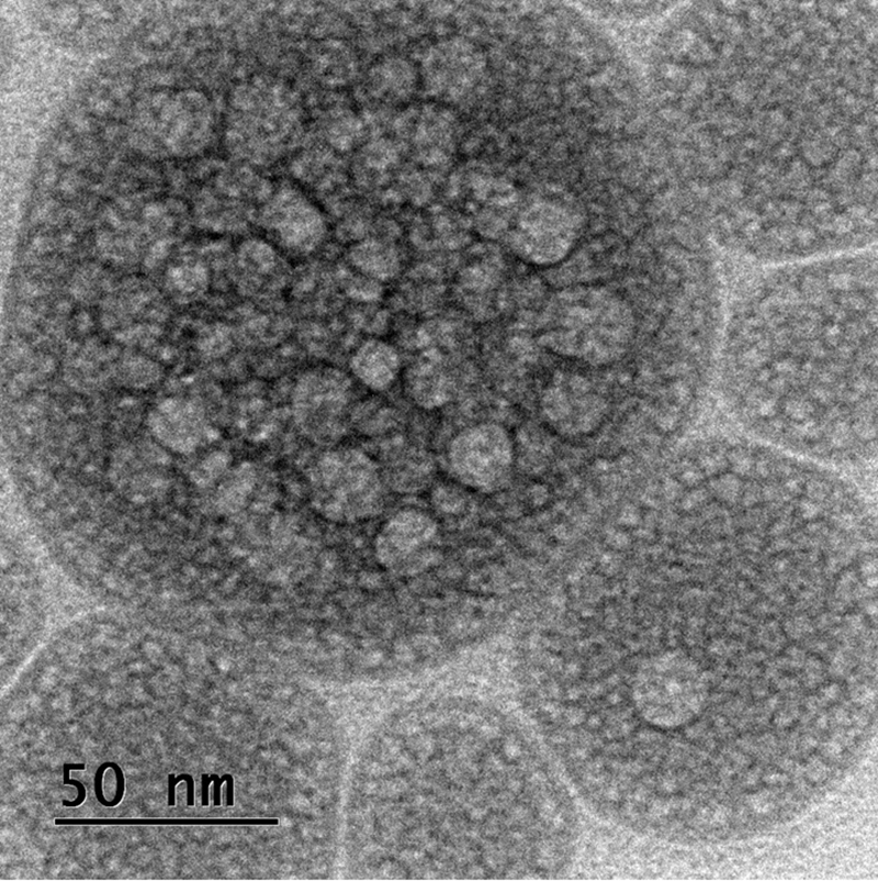
TEM image of nanoemulsion formulation (F4). Image shows spherical shape and uniform distribution. All the droplets had smooth surfaces. TEM, transmission electron microscopy.
The particles demonstrated spherical shape and uniform distribution. The globules had smooth surfaces. The mean droplet size of all nanoemulsion formulations was <150 nm and formulation (F4) was found to have the smallest droplet size. From the results, it was observed that droplet size was decreased at higher oil concentrations with an increasing surfactant concentration significantly by 146.7 ± 3.2, 121.8 ± 2.8, 95.9 ± 5.7, and 51.3 ± 4.1 at a ratio of 5:1, 7:1, 5:2, and 7:2, respectively (Fig. 2) but in these formulations, oil phase concentration did not exert any significant effect on droplet size in Babchi oil formulations.
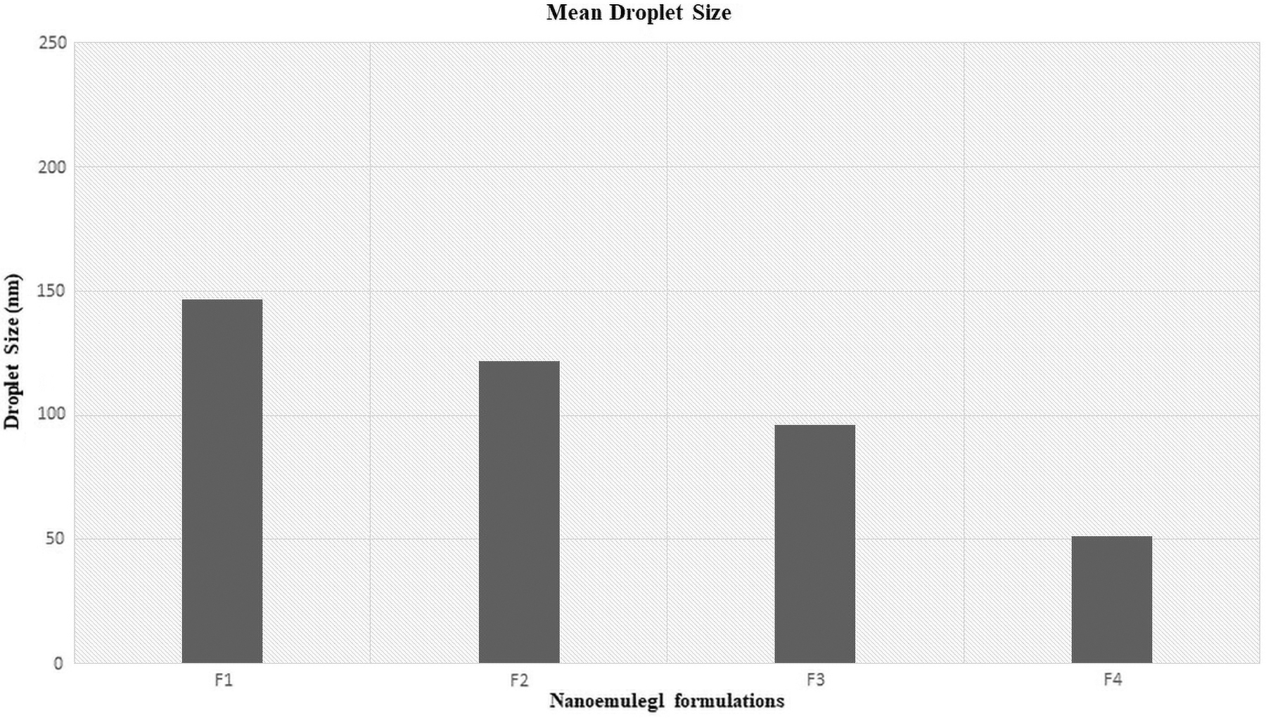
Mean droplet size of developed nanoemulsions. Nanoemulsion formulation F4 demonstrated the lowest droplet size.
The change was seen only at higher surfactant concentration when surfactant-to-oil ratio was changed from 7:1 to 7:2, which exhibited a significant reduction in droplet size (p < 0.05) from 95.9 ± 5.7 to 51.3 ± 4.1 nm. The different concentration of surfactants was found sufficient to prevent coalescence or any instability issue and no phase separation took place in any formulations during the study.
Entrapment efficiency (%) determination revealed that all the nanoemulsion formulations were successful in entrapping >90% content of methoxsalen. In zeta potential analysis, a higher negative value signifies better stability of the system as it prevents the aggregation among droplets owing to electrostatic repulsion potential. 32,33 The zeta potential graph of formulation F4 is given in Figure 3. The data of nanoemulsion characterization are given in Table 4.

Zeta potential of nanoemulsion formulation (F4).
Physical Evaluation of Nanoemulsion Formulations
Data are given as mean ± SD; n = 3.
In vitro drug diffusion studies
The study was performed using the membrane diffusion method for up to 24 h (Fig. 4). The drug release profiles were compared with aqueous dispersion (AD) of methoxsalen. The drug release from the nanoemulsion system was found in the range from 88% to 98% up to 24 h, in a sustained manner (p < 0.05). The F4 nanoemulsion formulation exhibited maximal release of drug apparently because of the smaller mean droplet size of formulation compared with other nanoemulsion formulations. On the contrary, the AD showed drug release within initial hours of drug release study. Nanoemulsion formulations (F1 to F4) showed significant in vitro release compared with AD of the drug over 24 h (p < 0.05).
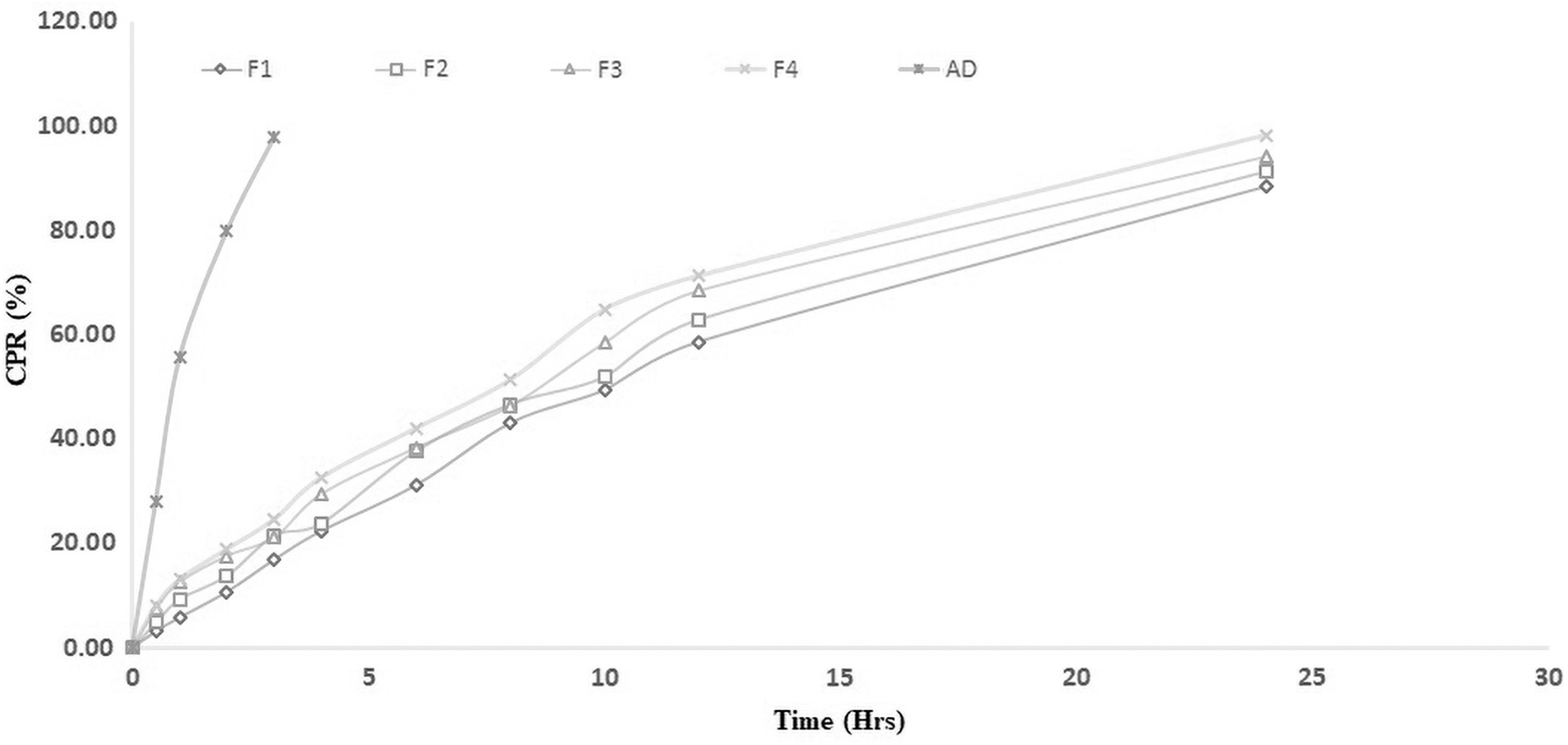
In vitro drug diffusion study comparing drug-loaded nanoemulsion with AD. AD, aqueous dispersions.
Preparation of Nanoemulgel
Based on the results from nanoemulsion evaluation, nanoemulsion formulations F3 and F4 were selected for nanoemulgel preparation. The nanoemulgel was prepared by incorporating selected nanoemulsion formulation into 2% carbopol gel. Then glycerol and triethanolamine were added to the system. The prepared nanoemulgel formulations (NG1 and NG2) were analyzed for ex vivo and in vivo drug permeation against plain gel of the same drug. The plain gel was prepared by adding the drug into the carbopol gel (2%) with simple stirring.
Physical Evaluation of Nanoemulgel
The appearances of plain gel (G1) and nanoemulgel (NG1 and NG2) were milky white. The pH of topical preparation must remain in the range of pH of the skin to avoid any skin irritation. The pH of all the gel formulations, that is, G1, NG1, and NG2 was maintained at 6.57 ± 0.04, 6.59 ± 0.05, and 6.58 ± 0.03, respectively. Rheology measurement was carried out at ambient temperature and NG1 and NG2 were found to be less viscous compared with G1. The viscosity of the nanoemulgel remained the same when the shear rate increased gradually and showed Newtonian flow behavior. 34
Thus, both nanoemulgel formulations demonstrated good consistency. High viscosity inversely affects the spreadability. For semisolid preparations, it is important to have good spreadability as it facilitates even and easy application of gel over the skin surface. 35 The spreadability profiles for nanoemulgel formulations were comparatively good as of plain gel. Drug content analysis revealed uniform dispersion of drugs all over the system in all the prepared formulations and found to be 98.15 ± 1.12, 99.23 ± 1.18, and 98.17 ± 1.14, respectively. The data are given in Table 5.
Characterization of Drug-Loaded Plain Gel Formulation (G1) and Nanoemulgel Formulations (NG1 and NG2)
Note: Drug flux was calculated from drug permeated (cumulative) versus time (hours). Data are given as mean ± SD; n = 3.
G1, drug-loaded simple gel formulation; NG1, and NG2, drug-loaded nanoemulgel formulations; SD, standard deviation.
Ex Vivo Skin Permeation studies
A comparative analysis was carried out for both the nanoemulgel formulations in comparison with that of drug-loaded plain gel (G1) using Franz-diffusion cell on excised skin of Wistar rats up to 24 h. Table 5 and Figure 5a and b represent the result of the skin permeation study. The CADP was found to be higher in NG2 (463 ± 13.09 μg) compared with NG1 (421 ± 11.15 μg), whereas G1 (196 ± 10.03 μg) exhibited the lowest drug permeation value. The percutaneous drug flux (J ss) from the nanoemulgel system NG2 (21.48 ± 0.27 μg/cm2 h) was more than NG1 (18.92 ± 0.31 μg/cm2 h) and two times the flux of drug from G1 (8.37 ± 0.21 μg/cm2 h), respectively. Both the nanoemulgel formulations exhibited zero-order permeation kinetics with a drug diffusion-controlled mechanism. NG2 formulation showed enhanced permeation through stratum corneum, better drug skin accumulation, and sustained drug release.
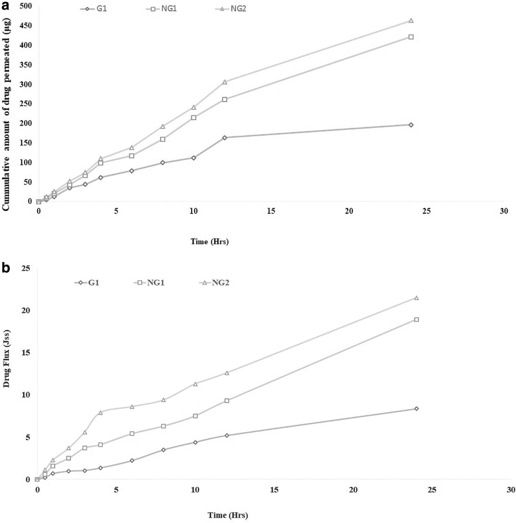
Ex vivo skin permeability study of drug-loaded nanoemulgel formulations (NG1 and NG2) with plain gel (G1).
In Vivo Studies
For In vivo studies, psoriasis was induced using topical application of Imiquimod. It activates immune cells and induces psoriasis-like inflammation. After 7 days of its application on the shaved skin of the rat, the typical symptoms were noticed, which include redness, scaling, and thickening. Treatment was initiated from the 8th day up to 14 days (study of a total of 21 days). Initially, the PASI score was 4 in all treatment groups and reduced to 1 in groups treated with standard and nanoemulgel formulations. PASI score was found to be 3 in the group treated with drug-loaded plain gel. However, the symptoms remained as it is in the negative control group.
All the groups treated with Imiquimod showed the reduction in bodyweight in comparison with control as given in Figure 6. The drop in body weight significantly improved (p < 0.01) by the standard drug methoxsalen (5 mg/kg/day) according to the data mentioned in Table 6. Treatment with standard, G1, NG1, and NG2 after 30 min of Imiquimod successfully recuperated the body weight. Food intake in the presence of the Imiquimod decreased significantly (p < 0.01) compared with the control group as given in Figure 7. Moreover, NG2 and standard drug have a significant difference in the food intake in comparison with the negative control group and other treatment groups as given in Table 6.
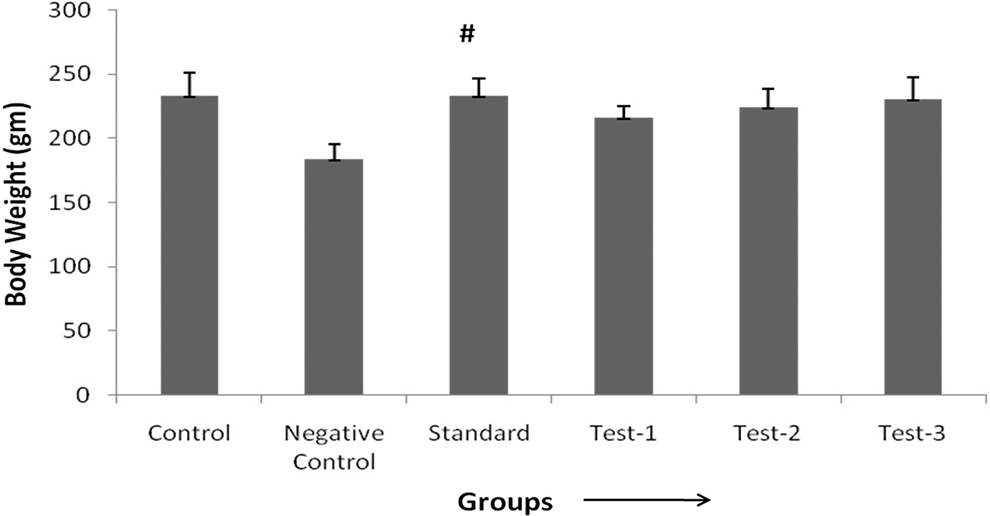
Effect of treatment on rats' bodyweight. #Significant difference when compare with negative control group (p < 0.01).
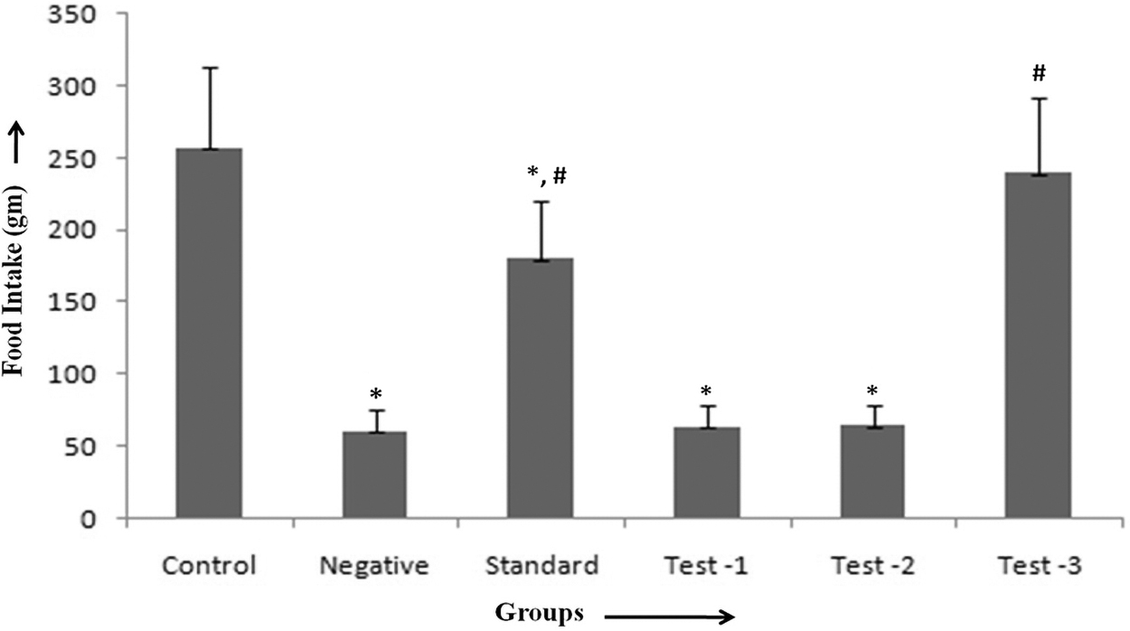
Effect of treatment on food consumption in rats. *Significant difference when compared with control group (p < 0.01). #Significant difference when compare with negative control group (p < 0.01).
Effect of Treatment on Body Weight and Food Intake
All values are expressed as mean ± SD, n = number of rats (6).
Significant difference when compared with control group (p < 0.01).
Significant difference when compare with negative control group (p < 0.01).
Imiquimod (5%) treatment reduced the body weight in comparison with control group rats. Methoxsalen (5 mg/kg/day) treatment as a standard drug showed a significant recovery in the bodyweight compared with the negative control group (p < 0.01). All the treatment groups of Test 1, 2, and 3 also have a recovery effect on body weight but significant results were not observed.
Imiquimod (5%) reduced food consumption in all groups significantly (p < 0.01) in comparison with the control group. Groups that were treated with standard drug and NG2 have a significant difference from the negative control group (p < 0.01).
Estimation of the MDA and GSH Levels in Liver Homogenate
Oxidative stress caused by Imiquimod (5%) enhanced MDA levels in the liver homogenate in comparison with the control group. All the treatment groups, that is, standard drug, G1, NG1, and NG2 have a reductive effect on MDA level and NG2 showed the maximum reduction in MDA level in animals as data given in Figure 8. Application of Imiquimod cream (5%) in all groups reduced the natural antioxidant GSH level in the liver homogenate in contrast to the control group considerably (p < 0.01). The GSH level in the treatment groups (Standard, G1, NG1, and NG2) were compared with the negative control group and there was a significant recovery in the GSH level (p < 0.01) in all treatment groups, and NG2 demonstrated the highest recovery levels among all treatment groups as values given in Figure 9 and Table 7.
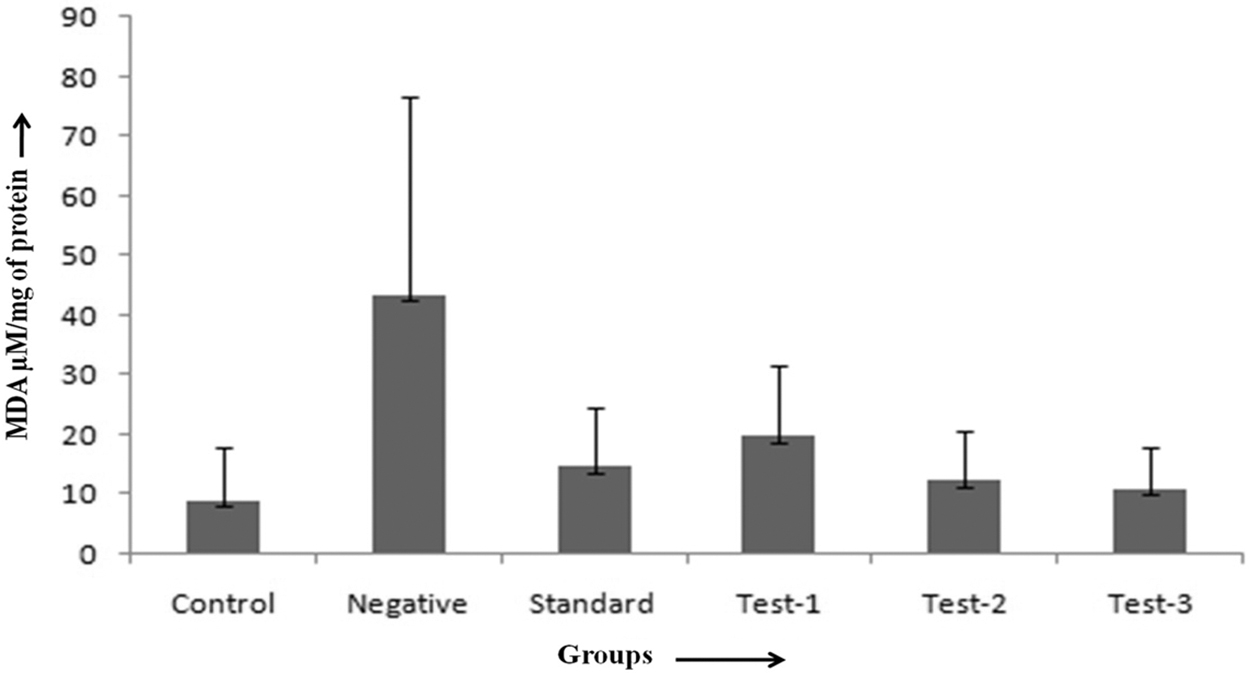
Effect of treatment on MDA content in the liver of rats.

Effect of treatment on GSH level in the liver of rats. *Significant difference when compared with control group (p < 0.01). #Significant difference when compare with negative control group (p < 0.01).
Estimation of the Antioxidant Levels in Liver Homogenate in the Rats
n = number of rats, Data are given as mean ± SD.
Significant difference when compared with control group (p < 0.01).
Significant difference when compared with negative control group (p < 0.01).
Owing to Imiquimod (5%), MDA level increased in the liver homogenate in comparison with control group rats. All the treatment groups of G1, NG1, and NG2 have a recovery effect on the MDA level.
Administration of Imiquimod (5%) in all groups reduced the natural antioxidant, that is, GSH level in the liver homogenate in comparison with the control group significantly (p < 0.01). But when the treatment groups were compared with the negative control group, there was a significant recovery in the GSH level (p < 0.01).
Histopathological Assessment of Skin
The histology of the psoriatic plaques shows acanthosis (epidermal hyperplasia) with inflammatory infiltrates consisting of dermal dendritic cells, macrophages, T cells, and neutrophils. Another prominent characteristic is neovascularization. 36 Application of Imiquimod (5%) produced edematous stroma in all the treatment groups. Imiquimod also triggers keratinocyte proliferation and apoptosis through MyD88 independent pathway, possibly adenosine receptor-mediated cAMP pathway. 37 Negative control shows hyperplasic epithelium but NG1 and NG2 have almost normal histology. The edema and scaling roughly recovered in test 2- and test 3-treated groups. The reason may be the smaller size of globules in both nanoemulgel formulations that contributed toward deep penetration within the tissues of the skin and was found to be more effective (Fig. 10). Label B (negative group) represents the edematous stroma. Test 1-treated group skin (label D) showed the hyperplasic epithelium, but test 2 and test 3 has almost normal histology.
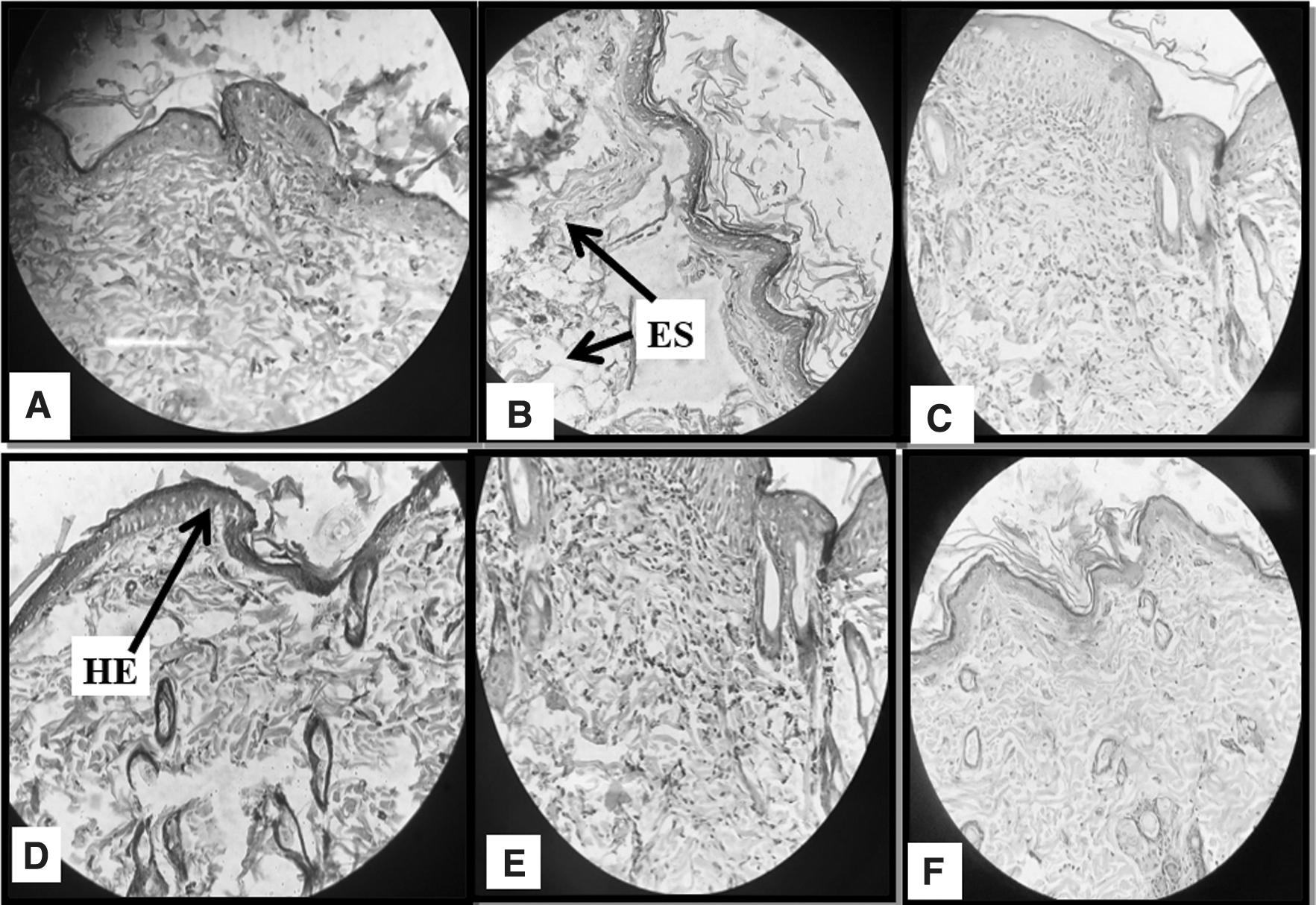
Histopathological analysis of the rat skin, average values expressed (3 sample ± SEM) in each group.
Skin-irritancy study
The methoxsalen-loaded nanoemulgel formulations (NG2) were subjected to skin irritancy study in animals for 3 days. After the 3-day application of the nanoemulgel formulations, no sign of reaction and irritation was observed on the skin of animals, which suggests the suitability of the drug in nanoemulgel formulation for topical application.
Discussion
Psoriasis is a skin condition in which skin cells build up and form scales and itchy, dry patches. It is an immune problem usually triggered by infections, stress, and cold. The classical clinical manifestations are sharply demarcated, erythematous, pruritic plaques covered in silvery scales. The hallmark of psoriasis is sustained inflammation that leads to uncontrolled keratinocyte proliferation and dysfunctional differentiation. The treatments aim to remove scales and stop skin cells from growing so quickly.
Psoralia corylifolia (Kushthanashini) is an Ayurvedic herb, generally used for skin problems especially pigmentation issues. Methoxsalen is derived from furanocoumarins, also known as Psoralens. Upon photoactivation, it conjugates and forms covalent bonds with DNA, which leads to the formation of both monofunctional (addition to a single strand of DNA) and bifunctional adducts (crosslinking of psoralen to both strands of DNA). It has variable metabolism with serum elimination half-life (0.5–2 h) and distribution volume between 1 and 9 L/kg. As its target site is skin, a topical formulation that localizes it in skin, reducing its systemic absorption will improve its action, reducing its systemic toxicity.
Nanoemulgels possess rheological characteristics that are suited especially for topical delivery, and methoxsalen and Babchi oil being lipophilic actives, nanoemulgel is a suitable drug delivery option. This study focuses on formulation and physical–pharmacological evaluation of an emulgel of methoxsalen–Babchi oil mix. Methoxsalen exhibits a positive value for logP, which denotes the drug's higher concentration in the oil phase. Thus, the drug demonstrated a very low solubility profile in water (0.89 ± 1.73 mg/mL). The formulated nanoemulsions showed different droplet size and surface potential based on other components of nanoemulsion. The higher entrapment efficiency (EE) could be associated with low surface tension among droplets that prevents droplets from coalescence, which was confirmed by no phase separation resulting in enhanced drug solubility and its retention in nanoemulsion. 32 In addition, the logP value of methoxsalen is 1.9 and EE was more dependent on the concentration of the oil phase. The higher EE was observed with the increased oil concentration from 0.8% to 3.2% (w/v).
The droplet size was decreased at higher oil concentrations with an increasing significant surfactant concentration and lowest size was obtained by 7:2 ratio at 51.3 nm. The reason may be attributed to the effect of surfactant that adsorbs onto the oil and water interface and minimizes interfacial tension disrupting droplets and reduction in droplet size. The increase in the oil phase concentration at a specific surfactant concentration affected droplet size. The excess amount of oil phase leads to the increase in droplet size. 33
An increase in potential is because of surfactant that prevents droplet aggregation by causing an electrostatic repulsion and provides a net negative surface charge. The in vitro drug release was maximum in formulation with lowest droplet size indicating the direct correlation of droplet size with drug release.
Before evaluation of skin permeation in vivo, the selected nanoemulsions were converted to emulgels. Rheology measurements were carried out at ambient temperature and NG1 and NG2 were found to be less viscous compared with G1. The viscosity of the nanoemulgels remained the same when the shear rate increased gradually and showed Newtonian flow behavior. 34 The spreadability of nanoemulgels was good, owing to the presence of oil phase. The pH was maintained in acidic range to keep the emulgel nonirritant, which was also observed in skin-irritancy study.
To evaluate the effect of developed nanoemulgel, it was evaluated in Imiquimod-induced psoriasis model. Imiquimod activates immune cells and induces psoriasis-like inflammation. After its application (7 days) on the shaved skin of the rat, the typical symptoms were redness, scaling, and thickening. Imiquimod-treated mouse skin closely resembles human plaque-type psoriasis with respect to erythema, skin thickening, scaling, epidermal alterations (acanthosis, parakeratosis), and neoangiogenesis, as well as with respect to the inflammatory infiltrate consisting of T cells, neutrophils, dendritic cell, and plasmacytoid dendritic cell. 37
In animal studies, the nanoemulgel showed restoration of body weight owing to enhanced food intake. It also balanced the MDA and GSH levels, to eradicate the oxidative stress in skin cells. Researches indicate that there is a severity wise increase in MDA levels in psoriatic patients indicating that, the degree of elevation of serum MDA is associated with the progression of psoriasis. 38 The histological evaluation also showed the normalization of skin structures.
Conclusion
Only a limited number of topical agents are available to treat psoriasis because of systemic side effects, thereby increasing the demand for alternative treatments to relieve disease symptoms. The developed topical nanoemulgel formulations containing methoxsalen and Babchi oil in combination significantly improved Imiquimod-induced psoriasis in rats. The nanoemulgel formulation facilitated the drug permeation to reach the deeper tissues of skin at a controlled rate and thus the system was found effective compared with plain conventional gel. The histological analysis also revealed significant results in rats and also does not irritate animal skin. It can be concluded that the nanoemulgel formulations can be a potential topical drug delivery systems to efficiently target mild-to-moderate psoriasis in a sustained manner for reduced frequency of application. It will also reduce the oral and systemic side effects.
Footnotes
Acknowledgments
The authors thank Metro College of Health Science and Research (Pharmacy), Plot No. 41, Knowledge Park III, Greater Noida, Uttar Pradesh, India and I.T.S College of Pharmacy, Murad Nagar, Ghaziabad, Uttar Pradesh, India, for help in conducting the research work and for providing us the platform and infrastructure for preparing this article. The authors also thank I.T.S Dental College, Murad Nagar, Ghaziabad, Uttar Pradesh, India, for carrying out histopathological analysis of animal samples.
Disclosure Statement
No competing financial interests exist.
Funding Information
No funding was received for this article.