
Abstract
Cancer is one of the leading causes of mortality worldwide. As the population increases, there is an enriched thrust for screening a newer delivery system for anticancer agents to treat cancer. Therefore, exosome-mediated cell communication is the hallmark of its participation in cancer metastasis and progression. Furthermore, the inward budding of endosomes is referred to as the multivesicular body. Exosome constitutes phospholipid bilayer-bound vesicles. It transfers bioactive compounds between various cells and the tumor microenvironment. In addition, they were selectively loading oncogenic molecules into exosomes for drug delivery. Exosome act as a potential biomarker in detecting therapeutic targets. Furthermore, exosomes fused with the cell membrane and were used as a diagnostic tool for cancer therapy. Exosomes are used in several ways to inhibit cancer cell growth. It can also carry genetic information and anticancer medications, preventing tumor cells from releasing exosomes. In addition, exosome-based drug delivery is utilized for preclinical and clinical trials, promoting the development of newer anticancer agents that suppress cancer growth. Exosomes further improve the immune system and help to generate new blood vessels. It blocks apoptosis and prevents cancer resistance. Exosomes act as cargo, allowing them to load proteins, chemotherapeutics, RNAs, DNAs, and hydrophobic drugs. Exosomes help to achieve advancement in cancer management through the use of precision medicine. The current review highlighted significant aspects of exosomes for cancer mechanism and prevention. Furthermore, novel strategies aimed to promote the clinical application of exosomes in cancer diagnosis and therapeutic impact. Finally, this review plays a pivotal role for the researchers in the industry and academics working in this field.
INTRODUCTION
Cancer is a primary trigger of mortality worldwide and a severe threat to public health. Worldwide, cancer incidence and death are increasing rapidly. 1 Frequently, cancer cells acquire abilities by succumbing to selection pressures and altering specific and pervasive biological functions, also known as enabling characteristics. They support traits that encourage the development and propagation of cancer. Typically, cancer cells acquire these substances to survive, develop, and prevent being eradicated by the host microenvironment. This review proposes the cancer-derived exosome (CDE) cargo as a unique enabling feature that satisfies the proposed criteria. 2 Exosomes seem more permeable and disposable than synthesized nanoparticles, making them less toxic and immunogenic. 3 On the contrary, extracellular vesicles (EVs) generated from the cell are also absorbent, larger than exosomes, and highly diverse, limiting their use for drug administration.
Exosomes are readily produced for the loading and distribution of pharmaceuticals. Almost every kind of cell may also make exosomes. Exosomes are persistent in physiological secretions due to their minuscule size and may penetrate the blood–brain barrier (BBB) despite being removed by the lungs. Exosomes carry restorative nucleotides and specific medications by adding malignant cell proteins, peptides, or antibodies. Exosomes are among the finest prospects for cancer-targeted treatment due to a combination of these qualities. 4,5
Most eukaryotic cells release exosomes, a subcategory of EVs consisting of a phospholipid bilayer membrane. Their sizes vary from 40 to 160 nm (averaging 100 nm). Previously, they were assumed to become cellular dust rather than a means for the cells to eliminate instead of their own accumulated debris. Exosomes mainly comprise cellular proteins, lipids, glycoconjugates, and nucleic acids. Exosomes are used for various functions, including modifying the extracellular matrix (ECM) and promoting chemical and signal transduction between cells. Exosomes have been implicated in cancer development in multiple ways, including their dual ability to promote and inhibit malignancy. Exosomes are nanovesicles produced by cells with the molecular transfer. In addition, exosomes possess immunogenicity qualities and are used in cancer immunotherapy. In addition, immunosuppressive substances such as programmed cell death ligand-1 (PD-L1) and transforming growth factor (TGF) may be expressed by exosomes.
Exosomes generated by malignancy may reduce CD8+ T cell activation and proliferation while promoting the development of regulatory T (Treg) cells that dampen the immune system. In addition, exosomes have recently been shown to have extraordinary anticancer benefits. Tumor-derived exosome and dendritic cell (DC) exhibited class I molecules and tumor markers. It includes heat shock proteins (HSPs) essential for antigen and T lymphocytes. In addition, in vitro and in vivo studies trigger CD8+ T cell-dependent antitumor responses. 6
Tumor cells generated by exosomes impact immunological evading substantially. Moreover, chemoresistance, ECM remodeling, and the development of the premetastatic environment were also affected. 7 Therefore, exosomes serve as diagnostic indicators and even as carriers for anticancer medications in the clinic. Due to their clathrin-coated membrane, exosomes are very persistent and resistant to degradation enzymes such as RNases, which makes them a powerful diagnostic and therapeutic tool. Exosomes generated significant attention in cancer treatment due to their biocompatibility and better stability. The exosomes have various attributes, including vesicle size, origin, surface characteristics, targeting approach, in vivo investigations, and superior effectiveness of carcinoma cells. Furthermore, exosomes are analyzed as a drug implementation methodology in treating tumors. Exosomes are generated through cells and circulated into physiological fluids. Moreover, exosomes' molecular properties, half-life, retention in circulation, and biological fluids are represented in urine, milk, saliva, pericardial effusion, and blood. 8
SOURCES OF EXOSOMES
Exosomes are produced through different sources, including cell culture supernatants, milk, serum, biological fluid, and blood plasma. 9 Exosomes may carry a variety of payloads and differ in size, surface molecule composition, and organization. Bioactive surface molecules on exosomes are related to their biological genesis. For instance, antigen-presenting molecule (major histocompatibility complex [MHC]-I and MCH-II) complexes and costimulatory molecules are abundantly present in exosomes. To improve consistency and repeatability, exosomes from tumors typically contain immunosuppressive proteins (TNF-related apoptosis inducing ligand [TRAIL], Fas ligand [FasL], TGF-β) and tumor antigens. The efficiency of generating homogeneous exosomes has cutting-edge technology for segregating them and EVs by bulk and surface features. Before purification, essential exosomes are produced from cell culture biological fluids and supernatants. 10
COMPOSITION OF EXOSOMES
The content and fabrication of exosome modification are based on the progenitor cells, their internal conditions, and outside triggers. The exosome content showed that exosomes vary in cells and organisms, having 194 lipids, 764 microRNAs (miRNAs), 1,639 messenger RNAs (mRNAs), and 4,563 proteins. 11,12 Biochemical and proteomic analyses of exosomes have revealed the presence of lipids (phosphatidylserine, cholesterol, ceramide, and sphingomyelin), proteins (integrin, metal tolerance proteins [MTPs], adhesion molecules, enzymes, membrane-bound proteins [MBPs], cytoskeletal proteins, HSP 70/90, Rab 5/7, tetraspanins CD 9/63/81, transferrin receptor, oligomeric proteins, and MHC class I/II), nucleic acids (miRNA, mRNA, long noncoding RNA [lncRNA], circular RNA [circRNA], and mitochondrial DNA [mtDNA]), as well as various substances (Fig. 1). 13 In contrast, lipids comprised the central part. Exosomes contain Notch ligand-like four proteins used for cell transduction pathways. In addition, it also contains normal proteins, interleukins, and Wnt-β-catenin signaling proteins involved in intercellular signaling. 13 –15
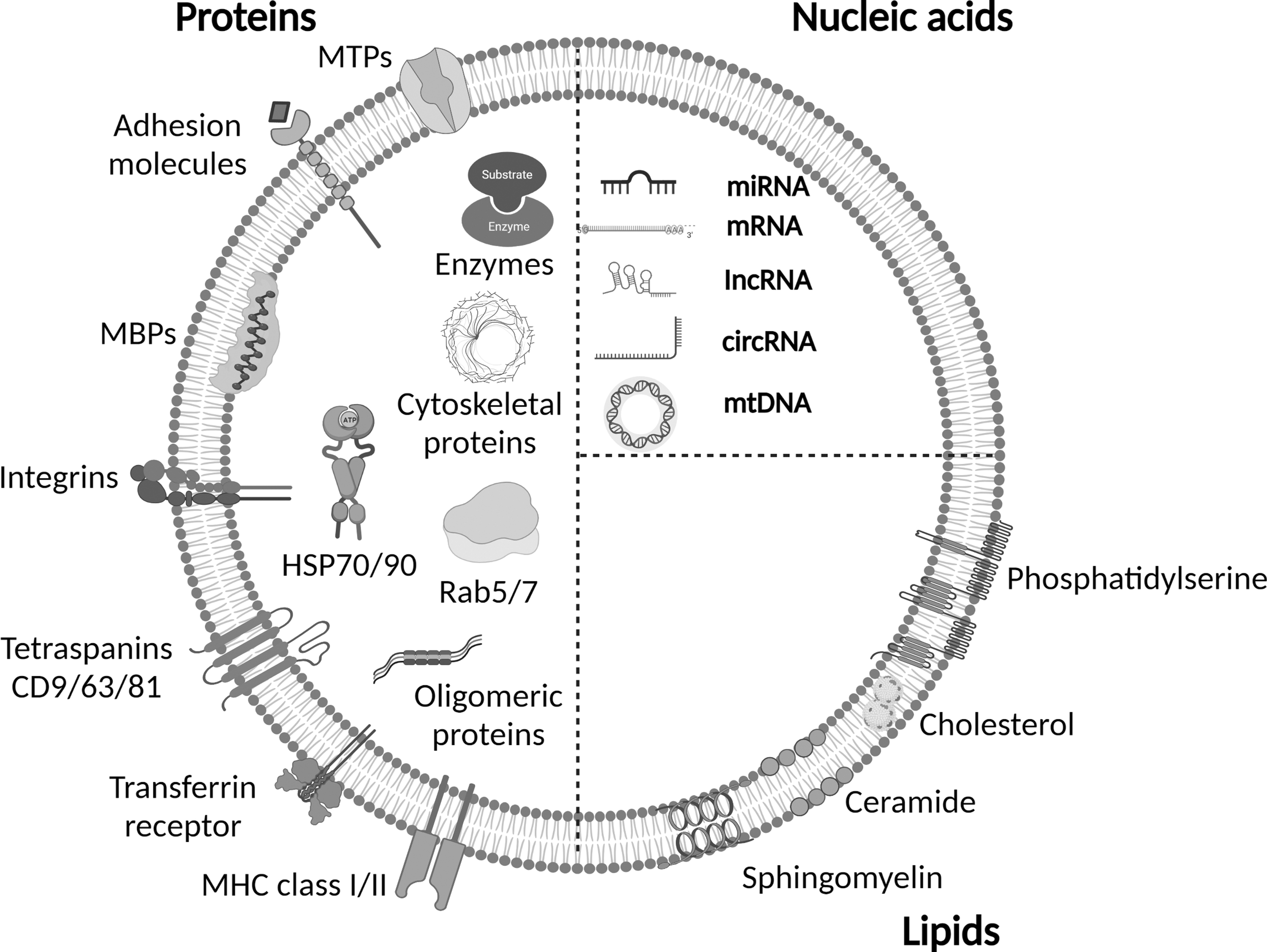
Exosomes: Structure and composition and molecules present as nucleic acid (miRNA, mRNA, IncRNA, circRNA, and mtDNA), soluble and MBPs (MTPs, adhesion molecules, MBPs, integrins, tetraspanins CD9/63/81, transferrin receptors, MHC class I/II, etc.), and lipids (phosphatidylserine, cholesterol, ceramide, and sphingomyelin). circRNA, circular RNA; MBP, membrane-bound protein; MHC, major histocompatibility complex; miRNA, microRNA; mRNA, messenger RNA; mtDNA, mitochondrial DNA; MTPs, metal tolerance proteins.
The most commonly characterized proteins in exosomes are Hsp70 and CD63. In addition, exosome formation and protein loading were improved by the tetraspanins CD9, CD63, and CD81. 16
FATE OF EXOSOMES
The efficacy and potential of exosomes are determined by the in vivo biodistribution method. They can deliver target-specific cells as therapeutic tools to a particular site. More specifically, the size, as well as the surface, of nanoparticles affects systemic stability, distribution, and intertissue biodistribution, for example, in cancer. Exosomes can be a promising controlled-release drug delivery pathway in the treatment of cancer due to their dimensions, lipid bilayer involvement, surface integrity, and the cargo they transport. Once the in vitro specificity of modified targeting exosomes is determined, the next crucial step is to assess their in vivo biodistribution characteristics. 17
Researchers looked at many studies on the biodistribution of EVs in people. 18 In addition, the biodistribution of DiR-labeled EVs from various cell types was examined in different research. Furthermore, they observed that 24 h after intravenous (i.v.) injection into mice, the liver, spleen, lung, and gastrointestinal system release the highest amount of EVs. EV parent cells may impact the biodistribution. Researchers have investigated the bioluminescent signals of Gaussia luciferase (Gluc)-labeled EVs in the spleen and liver, used for EV collection after i.v. therapy in athymic nude mice for 6 h. 19 A study recently reported that marked EVs, synthetic liposomes, and liposomes generated from EV lipid extracts all had identical biodistribution patterns and clearance rates. 20
According to recent research, exosomes were more likely to remain in the bloodstream because CD47-mediated protection against the microphysiological system (MPS) successfully facilitated tumor targeting in mice. Furthermore, regarding durability and tumor-targeting specificity, the benefits of overproduced exosomes are yet to be thoroughly exploited. Therefore, bioengineering and exosome modifications are required to get tumor-targeting exosomes. In addition to this, it effectively targets specific tissues and malignancies. Furthermore, further research is needed to create EVs with prolonged half-lives and improved vascular mobility. Finally, the exosomes' cellular origin, surface modifications, and transportation method are all critical factors. 21
REGULATION OF EXOSOMES BASED ON IMMUNE RESPONSE
The term immune response refers to how the body defends itself against potentially harmful substances, whether they originate from outside or are produced in the body. It is possible to divide the immune response into innate and adaptive immune responses. Several immune cell types are included in the above-discussed general and specialized immune responses. Phagocytes (monocytes, macrophages, and DCs) and natural killer (NK) cells act as the first line of defense against infection and engage in innate immunity. Moreover, T and B cells, cytokines, and immunoglobulins synergistically contribute to the adaptive acquired immune response against invading microbes. 22 –24
Consequently, exosomes may drive or decrease immunological responses depending on their source. The components govern the actions of exosomes' surface and intracellular constituents. Diverse, innovative concepts for developing exosomes as competent are summarized in Table 1. 25 The characterization of EVs is according to their size, shape, origin, and markers. Exosomes, also known as microvesicles, are apoptotic organs that are 30–150, 100–1,000, and 50–5,000 nm in size. Apoptosis is caused by TSG101 (tumor susceptibility gene 101), CD63, CD9, CD81, Alix, and CD63 integrin, heat shock, and cytoskeletal proteins. There are no damaged chromatins, organelles, or histones. Early-phase clinical studies used immunological medicine therapeutic transportation mechanisms as tumor immunizations demonstrated encouraging outcomes. 24,25
Exosome Regulation Based on Immune Responses
circUHRF1, circular ubiquitin like with PHD and ring finger domains 1; DC, dendritic cell; FasL, Fas ligand; HMGB1, high mobility group box 1; KRAS, Kirsten rat sarcoma; MHC, major histocompatibility complex; NK, natural killer; PD-L1, programmed cell death ligand-1; TGF, transforming growth factor; TRAIL, TNF-related apoptosis inducing ligand; Treg, regulatory T.
EXOSOME FORMATION
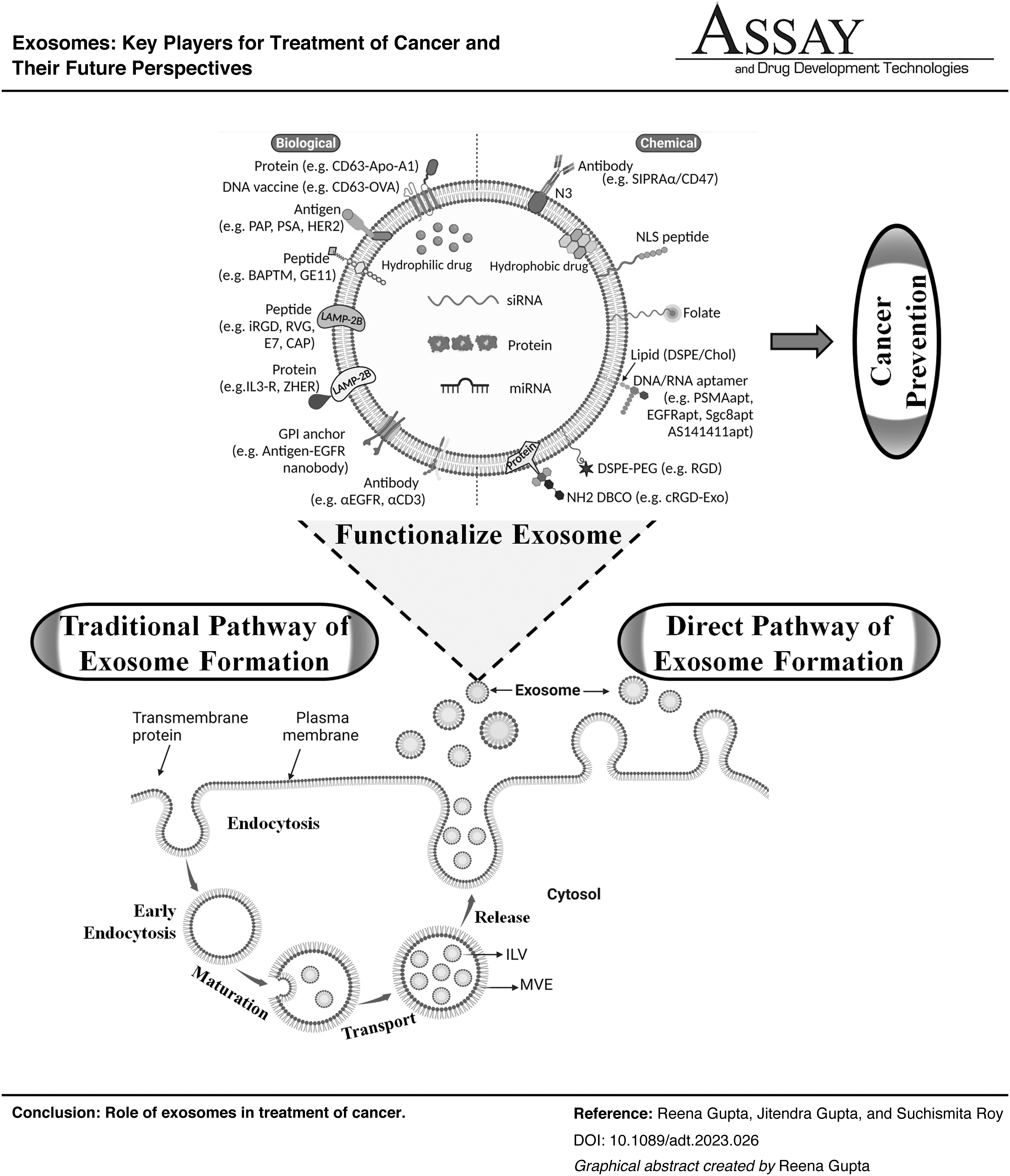
Exosome formation is directly associated with endocytosis origination. 26 The invasion of the plasma membrane creates the early endosome. It is accompanied by the appearance of vesicles and the invading of the endosomes' limiting membrane. A formation in the endosome known as a multivesicular body (MVB) comprises intraluminal vesicles (ILVs). 27 Several proteins and cytosolic components are encapsulated during this process into vesicles. 28 Following that, MVBs might have a choice between the two possible scenarios. The first one led to the destruction of the cell's contents when MVBs and lysosomes accumulated. Second, merging MVBs with the plasma membrane culminates in the liberation of exosomes and ILVs into the extrinsic environment (Fig. 2). 29 Exosome genesis and synthesis have been linked to endosomal sorting complexes required for transport (ESCRT) proteins, Rab-GTPase family, phospholipids, and lipid molecules.

Mechanistic path of exosome formation. (I) The traditional pathway creates ILVs inside MEVs. ILVs are then released by fusing plasma membrane with the MVE membrane and secreted as exosomes. (II) Alternatively, the direct pathway is responsible for exosome vesicle release from the plasma membrane resembling exosomes. ILVs, intraluminal vesicles; MEVs, medium extracellular Vesicles; MVE, multivesicular endosome.
Furthermore, other cells are SNARE (soluble NSF attachment protein receptors), TSG101, syndecan-1, and tetraspanins. 30,31 However, the procedures of these regulators throughout this system are yet unknown and need more exploration. In addition, environmental and genetic cell states may impact changes in the range of regulators involved in exosome formation. Due to their manufacturing mechanism, exosomes are believed to be bioactive cargo carriers. 32
Steps of Exosome Formation
Endocytic vesicle implementation from the plasma membrane.
Endosomal vesicle membrane inward budding produces MVBs having ILVs.
MVB fusion with the plasma membrane is needed for transferring the vesicular contents. 33
In the first step, endocytic vesicles comprised the plasma membrane and are transformed into late endosomes from early endosomes. Next, the limiting membrane undergoes inward budding in these late endosomes, producing vesicles inside the lumen. So, these ILVs gather within the early endosomes to form MVBs. MVBs are produced by one of two known techniques. The two routes use ESCRT. To release ILVs into the extracellular environment, these MVBs fuse either with the cell's plasma membrane or the lysosome, and then destroy them. Exosomes constitute the released ILVs. 34 The ESCRT is organized into four soluble multiprotein complexes called ESCRT-0, ESCRT-I, ESCRT-II, and ESCRT-III. The complex is called upon to sort specific proteins into ILVs. The ubiquitination of endocytosis receptors depends on cargo clustering carried out by ESCRT-0. 35
These payloads are proteins that will be incorporated into ILVs and ultimately become a component of the released exosomes. TSG101, a part of ESCRT-I, forms a complex with the ubiquitinated cargo protein and contributes to activating the ESCRT-II complex, promoting its initiation. This complex binds MVB proteins and recruits the deubiquitinating enzyme before sorting cargo proteins into ILVs. In the last phase, vascular protein sorting-associated protein 4 (VPS4) adenosine triphosphates (ATPase) disassemble ESCRT-III. 36
In addition, several investigations demonstrated exosome production even when ESCRT was inhibited, indicating the existence of a separate ESCRT process. This distinct process includes substances such as lipids, tetraspanins, or HSPs. 37 The creation of ILVs is caused by lipids such as ceramides twisting the restricting membrane of MVBs inward. ESCRT-independent mechanisms also connect phosphatidic acid and cholesterol to the generation of exosomes. Tetraspanins, a protein unrelated to lipids, are linked to integrating melanosome proteins into ILVs. It helps in sorting exosome cargo in an ESCRT-independent way. 38
Exosome Synthesis Pathway
According to recent research, the traditional pathways of exosome synthesis and internal multivesicular endosome (MVE) may not constantly be the origins of exosomes, making identifying them challenging. 39
Traditional pathway
The most considerable intensive research on exosome development mechanism is the traditional method. It results in MVE formation inside ILVs. The plasma membrane or lysosomes merge with MVEs, release ILVs, and expel as exosomes. Several intracellular sorting mechanisms are used to get proteins to ILVs targeted for either destruction or secretion, providing many MVEs. First, repetitive transmembrane receptors are segregated to ILVs for lysosomal degradation after ubiquitination. Second, ESCRT carries out a post-translational modification. Finally, exosomes include many ESCRT and ubiquitinated proteins, suggesting that ubiquitination occurs in the cytosol. 39 –47 Similarly, ESCRT is not required for sorting the proteolipid protein into ILVs. However, ESCRT releases exosomes on sphingolipid ceramide. 48 Furthermore, evidence suggests that lysophosphatidic acid stimulates the creation of ILVs, causing lysosomal breakdown. However, lysosomal breakdown has no impact on the production of exosomes.
Studies utilizing the cholesterol-labeling compound perfringolysin O revealed that only the MVEs carry cholesterol fused with the plasma membrane and release exosomes. Moreover, the other MVE perfringolysin remains O-negative. 49 As a consequence, the lipids bis(monoacylglycero)phosphate (BMP)/lysobisphosphatidic acid (LBPA) are not essential for an epidermal growth factor (EGF) and its receptor (EGFR), causing migration to exosomes using MVEs. 50,51
In contrast, lysosomes degrade the vesicles containing these lipids because the two intracellular MVE sorting pathways are present. The transmembrane protein sorting toward ILVs involved cytosolic lipid and protein domains. They are abundant in the tetraspanins CD63 and CD9. Rab families of tiny guanosine triphosphatases (GTPases) are also implicated, in addition to cytosolic calcium levels. They are involved in the transport of intracellular MVEs to the plasma membrane. 52 –57
Direct pathway
In addition to the traditional route, exosome biogenesis is done by the direct pathway. Therefore, it is a more appropriate technique for exosome development. Furthermore, erythroleukemia cell lines and T cells spontaneously liberate exosomes from the plasma membrane. Exosomes are released in response to human immunodeficiency virus (HIV) synthesis during surface receptor (Gag, Nef) crosslinking. These vesicles can be distinguished from exosomes and generated by the standard endosomal route. Furthermore, significant concentrations of exosome markers (CD81/63) have the same breadth and density, as discussed in Figure 2. 29,58 –60
EXOSOME ISOLATION METHODS
Numerous methods for isolating exosomes of high purity and quantity have beendeveloped due to the rapid development of technology and science (Fig. 3). Each methodology utilizes a distinct exosome characteristic, such as surface proteins, size, shape, and density. Furthermore, it helps to separate exosomes quickly. 38 Table 2 summarizes several isolation procedures, including their mechanisms and the benefits and drawbacks of each method. 9,61 –66 Unfortunately, there are no effective strategies for separating exosomes. The present methods are either economically impossible because they need specialized equipment, or they failed because of interference by proteins and nonexosomal vesicles. 67 Purified exosomes are produced using affinity-based methods, but their high prices and low productivity have restricted their use. Researchers use two techniques to improve isolated exosomes' effectiveness and purity. 68 The field flow fractionation methods have significant interest for their adaptability and capacity. They can isolate specific exosome subpopulations. 69
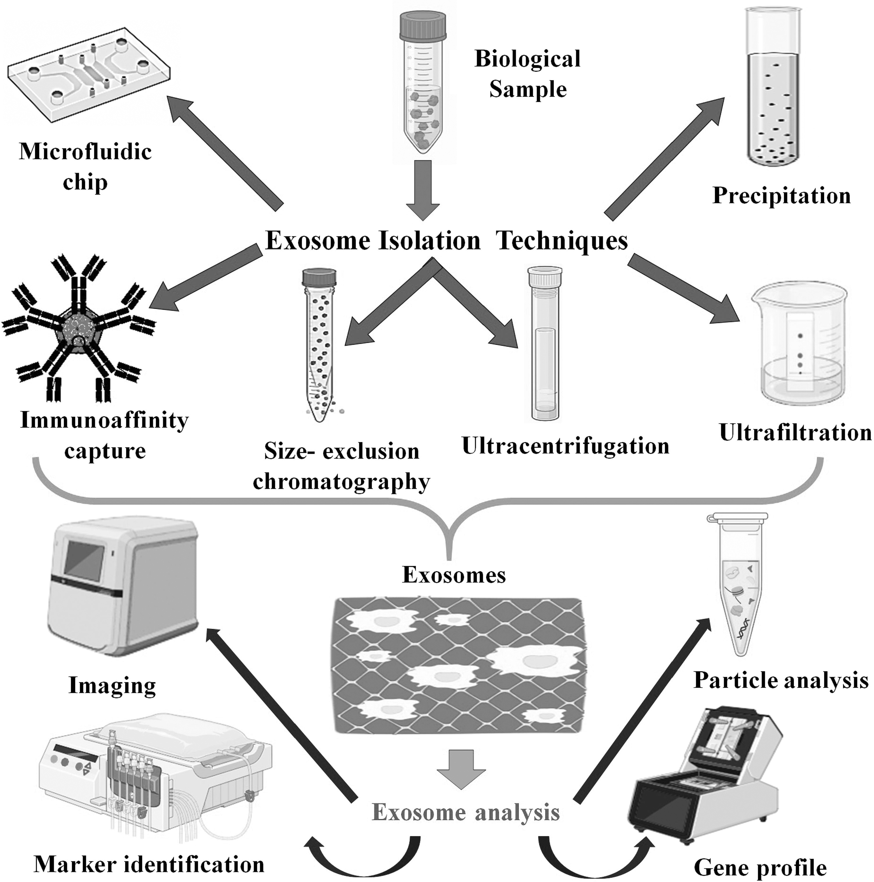
Isolation techniques of exosomes by using precipitation, ultrafiltration, centrifugation, size exclusion chromatography, microfluidic chip, and immunoaffinity capture; and their analysis such as gene profile, marker identification, particle analysis, and imaging.
Isolation Techniques of Exosomes and the Limitations and Purpose
The methodologies include immunoaffinity and size exclusion chromatography (SEC), differential centrifugation, ultrafiltration, microfluidics, and polymer-based precipitation. In addition, exosomes are extracted from physiological fluids or cell culture media. 70 These include gold-standard techniques such as density gradient centrifugation and differential ultracentrifugation. 9 Each method has significant advantages, disadvantages, and applications. However, combining many tactics may maximize benefits while minimizing negatives, compared with using a single process. For example, exosomes enhance the bioavailability of natural products in cancer management. It is due to their significant biodistribution, biocompatibility, and low immunogenicity. 71
Size Exclusion Chromatography
Applying SEC, a heterogeneous solution is separated into parts by classifying the portions by size. Significant smaller components (such as exosomes or other tiny sacs) may flow through these holes more effectively, extending their retention period inside the column. SEC is accomplished using a column of porous beads such as a filtering device. By contrast, more critical components are eluted from the column more rapidly because they cannot pass through these pores. Because SEC relies mainly on gravity flow, which puts significantly less stress on the membranes of these vesicles, it is more successful than ultracentrifugation in maintaining the biological activity of EVs. In addition, this technique produces very accurate and repeatable exosome collection findings. Furthermore, ultracentrifugation is typically the method used to concentrate the final exosome sample since, in part, gravity flow separation makes this separation process time-consuming. 18,72,73
Ultracentrifugation
Ultracentrifugation is one of the methods most often used in isolating exosomes. The modern gold standard for centrifugation-based exosome isolation would be this after being exempted from the cell and superfluous deposits from the effluent at lower rates. The residual supernatant churned faster to generate the exosomal pellets. Even though this method is widely used for separating exosomes, there is a chance that sample loss might occur. According to data, repetitive centrifugation may damage vesicles and enhance the probability. Immunogenic protein clumps would cosediment with them. 74 –76
Immunoaffinity Capture
Immunoaffinity capture isolated exosomes and identified individual surface proteins that distinguish exosomes from other particles. It primarily utilizes antibodies that associate with specific protein surfaces of exosomes to a filter and then eluted for further use. Medication delivery uses naturally occurring nanocarriers called exosomes. Magnetic bead immunization was a prominent variation of this technique used to extract exosomes quickly. Anti-EpCAM-coated magnetic beads made from LIM1863 colon cancer cell exosomes showed this approach. It is more convenient than density gradient separation and ultracentrifugation. 72,76
Microfluidic-Based Isolation
Accurately, exosomes were obtained using lab-on-a-chip or microfluidic-based technologies, such as antibody-coated surfaces. According to the original study researchers, implementing a microfluidic approach was faster, less costly, and required less space and chemicals. In addition, it could isolate exosomes with a specific cell origin. They further said that, in contrast to current exosome separation criteria, this method is congruent with clinical laboratory procedures. Furthermore, microfluidic devices enable the one-step isolation of exosomes. It is isolated from serum compared with the magnetic bead-based approach. Furthermore, microfluidic devices, as opposed to magnetic bead-based techniques, separate exosomes from serum in a single step. 77
Ultrafiltration
In ultrafiltration, exosomes are separated from other macromolecules and proteins using a collection of membranes. Exosomes accumulate on the membrane, while proteins, and other large molecules, are rinsed aside. This method produces more exosomes than ultracentrifugation. 78 Nonetheless, it has some potential issues. Exosomes and exosomal proteins may attach to the membrane, blocking the extraction for further investigation. Utilizing a nanomembrane ultrafiltration concentrator, rapid separation of urine exosomal biomarkers. 79 Exosomes become damaged during the operation due to the increased force necessary to move the liquid under investigation through the membranes. In research, the authors were worried that critical exosomal proteins may be difficult to discover and characterize if some proteins are not appropriately filtered out. 80
Polymer-Based Precipitation
Compared with ultracentrifugation, which is considered the gold standard, a precipitation procedure might be able to get pure RNA and protein from exosomes and also get more of them. However, this has only been proven by isolating exosomes from ascites. Another, utilizing urinary exosomes as a target, a commercially available exosome is prepared. It is prepared by a precipitation reagent combined with a modified precipitation protocol to determine which isolation method showed the highest yield. The greatest exosome, miRNA, and mRNA yields are obtained through precipitation compared with other widely used isolation methods. However, the precipitation approach was rated as a simple, quick, and scalable option for separating and identifying exosomes. The authors admitted that other ultracentrifugation processes were preferable for isolating exosomes. 80,81
Sonication
Exosomes and drugs use a probe sonicator to accelerate the growth rate of cells and act as therapeutic cargo. Drug diffusion is enhanced into exosomes through considerable membrane distortion by sonication. 82 Numerous research has shown the efficacy of this approach. Consequently, the drug is occasionally encapsulated in many layers confined under the exosome and rests inside the membrane. Sonication has resulted in two different drug releases, with the internalized medicine released over a longer time and the membrane-bound medication released more rapidly. In addition, sonication did not affect the stability of the drug and had a high drug loading efficiency. 64 However, this approach is more troublesome for nucleic acids since it produces aggregation and degradation. However, it may be advantageous for certain medications. 83
EXOSOMES: FUNCTION AND TRANSPORT
Exosomes transport functional molecules present in their lumen and surface from one cell to another. These include mtDNA, mRNA, lipid, proteins, and multiple noncoding RNAs. As a possible consequence, exosomes may mediate prevalent physiological processes via cell–cell interactions. 64,84 Direct contact between the nanoscale drug delivery device and the surface of the receiving cell-to-cell communication through the device's surface. As a result, it performs the earliest and most significant contribution to cellular identification and internalization for in vitro and in vivo studies. In addition, exosomes are naturally occurring products compatible with biological systems rather than artificially created nanoparticles. Furthermore, the delivery of specific miRNAs and mRNAs by exosomes affects the expression of genes in recipient cells. 84,85 For example, exosomes of colorectal cancer patients contain mRNAs that induce tumor angiogenesis, promoting endothelial proliferation. Consequently, it also enhanced the rate of endothelial cell division. 86
In addition, glioblastoma-derived exosomes contain proteins, mRNA, and sphingomyelin. These enhance angiogenesis, tumor invasiveness, and vascular lumen development. Furthermore, it is essential to figure out which sources make the ideal exosomes based on the outcome of the medication administration. Furthermore, it prevents negative consequences that encourage tumor development. 87
Moreover, the best technique to integrate therapeutic drugs into exosomes is to insert them into a solution with an elevated density of the required medication. Moreover, the medicine enters the vesicles via a concentration gradient. They used curcumin (CUR)-loaded exosomes that allowed to demonstrate exosomes incubated with therapeutic cargo. Then samples were centrifuged for 1.5 h at 36,000 rpm in a sucrose gradient. Finally, high performance liquid chromatography (HPLC) was performed to determine the concentration of CUR. Depending upon the results of this in vivo study, the researchers use a loading strategy for investigating brain inflammatory diseases. 88
EXOSOMES AND THEIR CORRELATION WITH TUMOR MICROENVIRONMENT
The tumor microenvironment (TME) comprises an ECM, stromal cells, immune cells, blood, and the lymphatic network. It contains fibroblasts, mesenchymal stem cells (MSCs), NK cells, pericytes, adipocytes, T and B lymphocytes, and macrophages. 89 In addition, the TME is implicated in tumor development, proliferation, and resistance to treatment and plays an essential part in the biology of tumors. Exosomes are an integral component of the TME. They are efficient communication osteosarcomas depending on exosomes for transporting genetic information. The following sections emphasize the TME's essential cells and their communications with exosomes. This communication promotes cell proliferation and invasion while also establishing a premetastatic niche that anticipates the arrival of cancer cells with the appropriate exosomal integrin variation (Fig. 4) as a consequence of these CDEs and their illicit trafficking. 90,91
Systematic representation: effects of exosomes on the tumor microenvironment.
Exosomes with Integrated Therapeutic Benefits
The researchers could not predict exosomes' pharmacological and biological potential in 1980. 38,92 Furthermore, these membrane vesicles allow the cell to interact by transmitting its constituents, especially proteins, mRNAs, and miRNAs, used as target cells, with or without direct contact between the cells. Exosomes also affect both pathological and healthy processes. Exosomes have essential properties that allow them to improve motor and neurological functions in the nervous system. They consider giving repeated i.v. doses with no nasty side effects, passing the BBB, and reducing inflammation. These membranous vesicles are crucial in detecting and predicting many disorders, particularly multiple cardiac problems, kidney and liver disease, neurological diseases, and tumors. 93
Cancer-Associated Fibroblast and Exosomes
Cancer-associated fibroblast (CAF) is the fundamental cellular component of the TME in most solid tumors. 94 CAF-derived exosomes (CAFDEs) are a crucial component of oncogenic transformation. 95 CAFDEs increase tumor development, treatment resistance, and tumor metastasis. 96 In addition, CAFDEs may promote the growth of tumors and neoplastic angiogenesis in colorectal cancer. They may also produce cancerous cell dedifferentiation by the Wnt pathway, enhancing disease resistance to chemical therapies. 97 Furthermore, TGF-β1 is a protein that may stimulate the metastatic ability of cancer cells by controlling the production of lncRNA. CAF induces epithelial–mesenchymal transition (EMT) in breast cancer by the TGF-β1 signaling pathway. 98 –100
Cancer Stem Cells and Exosomes
Cancer stem cells (CSCs) and cancer-initiating cells are present as a tiny group of diverse cells in tumor tissue. Cancer formation, recurrence, metastasis, and treatment resistance depend on CSCs, which have an inexhaustible capacity for self-renewal and variety. 101 Exosomes play an essential role in converting non-CSCs into CSCs, restoring CSC homeostasis, and the technology that makes all of these functions happen. 102 First, exosomes generated from CSCs transmitted miR-19b-3p to clear cells, renal cells, and carcinoma cells. Then, exosomes activate the EMT to enhance tumor cell metastasis. 103
MSCs and Exosomes
MSCs are among the most beneficial progenitor cells in tissue engineering due to pluripotency. They convert into adipocytes, osteoblasts, cardiomyocytes, and neurons. 104 Exosomes produced through MSCs in the TME facilitate non-CSCs to become CSCs. Upregulating ERK1/2 (extracellular signals to regulate kinase 1/2) increases the frequency of CSCs and activates the Wnt signaling pathway. The exosomes produced by MSCs provide colorectal stem cell properties responsible for in vitro and in vivo activity. 105 Exosomes secreted by MSCs are essential for the inflammatory process and the regeneration of damaged tissue. 106
Immune Cells in the TME and Exosomes
The predominant immune cells present in the TME are myeloid cells, DCs, myeloid-derived suppressor cells, and other myeloid-derived cells and lymphocytes (T and B cells). 107 Tumor-derived exosomes (TEXs’) ability to effectively communicate antigens to an immune system and excite the immune system enables them to stimulate antitumor responses and contribute to the recruitment and rebuilding of microenvironmental tumor components. 108,109 Exosomes generated from tumor cells are a rich source of immune-stimulating biological components. Furthermore, transmembrane proteins such as CD9, CD63, and CD81, HSPs, and tumor-associated antigens are also present. Exosomes are produced by TME immune cells. Treg cells hindered the maturation of antigen-presenting cells by expressing CTLA-4 or contributed to tumor formation by generating inhibitory cytokines and adenosine. 110,111
Exosomes Affect Epigenetic Mechanisms of Transformation, Invasion, and Migration
Epithelial-mesenchymal transition (EMT) and exosome signaling might increase the generation of cancer. 112 The mechanism in biology where epithelial cells might change into cells with mesenchymal phenotypes by using specific strategies is known as EMT. The complex transcription system required for EMT regulation comprises three different developmental transcription factors. TWIST1/TWIST2 family, ZEB1/ZEB2 ZEB (zinc-finger e-box binding homogenous box) family, and SNAIL/SLUG zinc-finger transcription factor family. 113 Exosomes originating from hepatocellular carcinoma cell (HCC) are susceptible to mediating EMT by stimulating the TGF-β/Smad signaling pathway. Exosomes inhibit the expression of E-cadherin while concurrently increasing the expression of vimentin, enhancing target cell migration and invasion. 114
Exosomes: Cancer Therapy as Bioactive Cargo
Exosomes have garnered much attention in cancer treatment since they can deliver their bioactive cargo to cancer cells. As a result, a diversity of chemotherapeutics has already been established. 115
The application of exosomes originating from immune cells inhibits cancer cells; the administration of cytokines induces apoptosis in cancer cells. 115
Preventing the release of CDEs. 116
Utilizing exosomes as gene transporters. 117
Using exosomes as carriers for anticancer drugs. 118
However, there is a significant probability that exosomes are utilized to treat cancer. Furthermore, it is challenging to use natural exosomes for the therapeutic result. On the contrary, according to current research, exosomes are genetically altered to transport specific proteins, RNAs, or medications that may prove to be extraordinarily effective weapons in the battle against cancer. Furthermore, exosomes play a crucial role in the pathogenesis of cancer and their biological compatibilities. Therefore, it may be possible to target exosomes as cancer progenitors as therapeutic devices. Furthermore, it helps find new biomarkers for early detection and uncover molecular targets. Cargo-loaded exosomes are selectively advantageous for detecting cancer as a biomarker. Researchers utilized exosomes to discover chemicals for targeting cancers. However, this approach is more effective for tailored identification, diagnostic, and prognostic methodologies (Fig. 5). 119

Exosomes: Captured from cultured cell homogenate or a patient's biological fluids, used as analytic and predictive indicators for cancer and synthesis and release from the producer cell or exosome absorption in the targeting of cancer.
Exosome-Mediated RNA Cancer Therapeutics
RNAs are significant macromolecules that are difficult to distribute in vivo. Some delivery strategies have been proposed, including cationic polymer-based particles, dendrimers, and cationic liposomes. 120 –122 Conversely, these transporters are inadequate for therapeutic intervention because of the off-target and insufficient sustainability and efficacy. 123 Exosomes are lipid bilayers-encapsulated nano EVs that have the potential to transport nucleic acids because of their innate capacity to shuttle DNA and RNA between cells and natural attraction toward destination cells. 124,125 Exosome-based nanocarriers conveying diverse RNAs, including mRNA, small interfering RNA (siRNA), and miRNA, have been developed to treat various illnesses and malignancies. Consequently, cancer cells were suppressed by exosomes synthesized through donor cells implanted with the miRNA. 126,127
Exosomes as miRNA Carriers for Cancer Treatment
Endogenous miRNAs are short noncoding RNAs that control gene expression by binding to target mRNAs. As a consequence, miRNAs may be a proper cancer therapy method. miRNAs are degraded in vivo, and distribution to specific target cells, tissues, and organs is challenging. Exosomes are tiny, persistent vesicles capable of transporting functional bioactive compounds across long distances with high selectivity. 128 –132 They have suggested a potential miRNA carrier for use in cancer treatment. Furthermore, pancreatic ductal adenocarcinoma cell growth and invasion were suppressed by exosomes. It is transfected with miR-145-5p through TGF-/Smad3 pathways. 133 In contrast, the utilization of inhibitors of exosomal miRNAs is an additional effective cancer treatment strategy. Through exosomes, these miRNAs play pivotal roles in the development of cancer.
In contrast, exosomal miR-25-3p is a miRNA related to colorectal cancer that increases metastasis dissemination. Exosomes containing miR-25-3p enhanced the vascular permeability and metastasis of colorectal cancer in the liver and lung of mice. However, these effects reverse by inhibiting the standard action of exosomal miR-25-3p using an miR-25-3p inhibitor. 134 –137
Protein-Containing Exosomes for Chemoprevention
Membrane proteins on the surface of the exosomes are involved in signaling. These immunogenic proteins have antitumor effects. In addition, they act by triggering immune cells. 138 As a result, exosomes can contribute to developing innovative anticancer vaccinations. For example, exosomes from DCs include MHC-I, which binds to tumor-derived peptides and activates immune cells to fight cancer. 139 Furthermore, protein antagonists are transported via exosomes. For example, macrophage phagocytosis is blocked when the signal-regulatory protein alpha (SIRPα) and CD47 on the surface of tumor cells interact. However, recombinant SIRPα is ineffective as a competitive antagonist in promoting macrophage phagocytosis. SIRPα-containing exosomes bind to CD47 on tumor cells and block the release. This further encourages macrophage phagocytosis and suppresses tumor growth. 114
Exosomes: Therapeutic Carriers for Cancers
Chemotherapeutic drugs effectively eradicate rapidly growing tumor cells. However, these drugs may also damage healthy, normal cells that are dividing quickly, having very negative consequences. In addition, certain hydrophobic medications have a hard time explicitly targeting tumor cells. Therefore, the need for an effective transporter for these drugs is essential. 140 –143
Furthermore, exosomes may transport chemotherapeutic medicines to cancer cells by altering their surface proteins. Lysosomal associated membrane protein-2 isoform B (LAMP2B)-internalizing Arg-Gly-Asp peptide (iRGD peptide; v integrin-specific) expressing immature mouse DCs (imDCs), extracted their exosomes. It is further utilized to transport doxorubicin (Dox). They showed that by using this technique, iRGD-exosomes could efficiently target and deliver Dox to v-integrin-positive breast cancer cells in vitro and to tumor tissues in a particular way, which prevents tumor growth in vivo. 119
CHEMOTHERAPEUTIC DELIVERY FOR CANCERS
Exosomes as a carrier help transport chemotherapeutics and treat prostate, lung, and pancreatic malignancies. For instance, paclitaxel (PTX)-loaded exosomes might be utilized. 144 Exosomes and Dox also demonstrated remarkable efficacy in treating cancerous mammary cells. 119 However, exosomes generated from different donor cells perform various physiological functions depending on their origin. For example, tumor-derived exosomes may contribute to antitumor immunity by delivering tumor-specific antigens, proteins, and miRNAs. 145 However, the same exosomes cause the death of T cells and impede monocyte differentiation. Exosomes released by MSCs can regulate the immune system and support tissue repair. Still, they also have the potential to stimulate the formation of tumors by activating proteins related to tumor angiogenesis. 146
Exosomes released by immune cells can evade elimination by the immune system and increase the amount of time spent circulating in the body's periphery. 147 Exosomes generated from milk do not cause the immune system to reject or cause inflammation. They can boost the bioavailability of medications when taken orally. When choosing exosomes for drug administration, picking exosomes generated from appropriate donor cells is vital. 148
Paclitaxel
PTX
American researchers' PTX anticancer effects on cancer cells resistant to the therapy are enhanced by macrophage-derived exosomes. 64 The researchers also showed the macrophage-produced exosomes with aminoethylanisamide-polyethylene glycol vectorization. It has a high PTX loading capacity, an enhanced tendency to cluster in cancer cells after systemic dispersion, and better therapeutic effects. 124 Exosomes produced by cancer cells could deliver PTX to the corresponding mother cells. Cancerous mother cells use the endocytic pathway to tailor the drug to the target cells. Furthermore, it helps to achieve a high degree of cytotoxicity. 144
Patients resistant to cisplatin are frequently treated with PTX to overcome their medication resistance. The fact that PTX has a dose-dependent toxic effect and a limited bioavailability is a significant barrier to its use in clinical settings. In addition, researchers found that PTX could not cross the BBB. 154,155 Nevertheless, pretreated PTX mesenchymal stromal cells generate PTX-loaded exosomes that exhibit a potent anticancer effect on human duodenal carcinogens. 156 Exosomes produced from cancer contain PTX. It helps target drug-resistant CSCs and increase their cytotoxicity toward autologous cancer cells. 157 One of the most significant barriers to effective cancer treatment is multiple drug resistance (MDR). Exosomes are an effective means of overcoming MDR in tumors. Exosomes synthesized from PTX-loaded macrophages avoided degradation by the P-glycoprotein drug efflux facilitator, showed significant cellular uptake in Madin–Darby canine kidney (MDCK) MDR1 cells, and exhibited a lower IC50 than free PTX. 32
Exosomes originating from U-87 MG cells might overcome MDR, penetrate the BBB, and distribute PTX, resulting in a significant therapeutic effect on GBM. 158

Curcumin
As a naturally occurring polyphenol, CUR (II) inhibits the development and spread of several malignancies, including pancreatic, colon, breast, and oral cancers. 159,160 CUR has been the subject of several human clinical trials that have investigated its efficacy, as well as its safety and pharmacokinetics, in the treatment of various malignancies. 161 CUR is appropriate for clinical applications because of its dominating characteristics, which include its low cost and low toxicity. 162 However, the utility of CUR is restricted in patients because of its low bioavailability, low solubility in water, short half-life in plasma, and low stability. Therefore, the researchers used exosomes to incorporate CUR. Furthermore, it helps to administer the exosome-CUR complex to a GL26 brain tumor model. However, this substantially slowed the progression of the brain tumor, accompanied by a reduction in inflammation. It also helped mitigate a catastrophic failure of the endothelial cells in the cerebral. 145,163
Furthermore, CUR-loaded exosomes trigger apoptosis in pancreatic cancer cells. However, exosomes originating from cancer cells stimulate the growth of pancreatic cancer cells by increasing their proliferation. 164
A recent study found exosomes originating from cow's milk and intestinal epithelial cells. It improves the cellular absorption and intestinal permeability of CUR. This finding demonstrates whether exosome-based delivery may boost the bioavailability of buccal therapy. 165 In addition, exosomes containing CUR reduce the toxicity caused by homocysteine. 166

Doxorubicin
Dox
Dox-loaded exosomes reduce the availability of Dox to cardiac endothelial cells and minimize cardiotoxicity. 168 In addition, researchers obtained exosomes from MSCs. It boosted the effectiveness and cellular absorption rate of Dox in osteosarcoma. However, MSCs tend to move toward tumor tissues, establishing the importance of cautiously choosing exosome origins. 169

Celastrol
Celastrol (CEL)

Beta-Elemene
Beta (β)-elemene
EXOSOMES: APPLICATIONS FOR CANCER PREVENTION
Exosome Bioengineering Through Surface Molecules
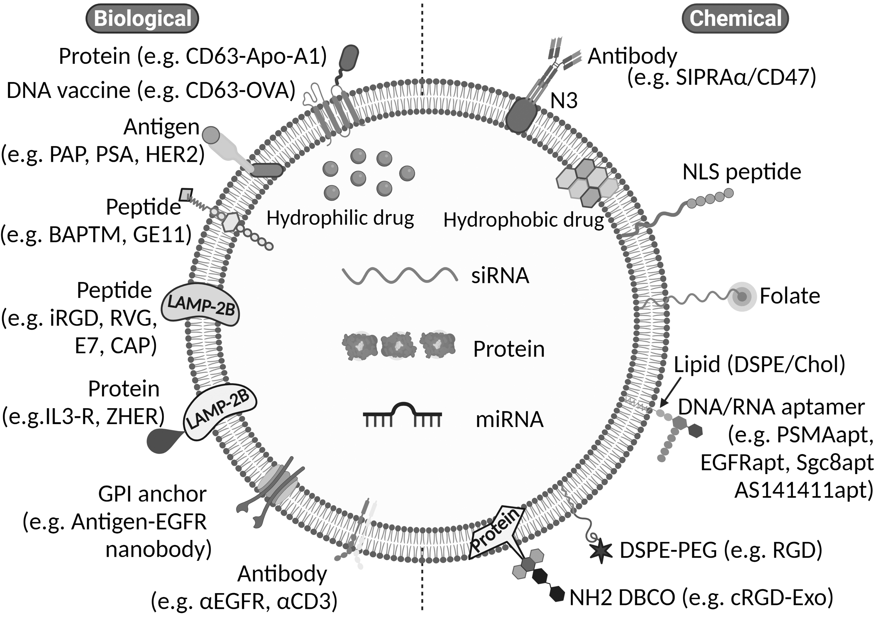
Exosome bioengineering aims to make specific exosomes utilized to treat cancer. For this technique to work, a particular moiety of segmentation (peptides or other ligands) must be attached to the surface of exosomes to interact with and taken inside the chosen cell type. Numerous uncharacterized cellular components and surface molecules regulate their internalization upon uptake and produce exosomes. This further results in posing the greatest obstacle to this strategy discussed diagrammatically in Figure 6. 185

Engineering exosomes through surface molecules
Genetic engineering
The induction of new qualities in exosomes is achieved using genetic engineering, a practical method. Ligands or homing peptides are initially coupled to the surface of exosome-expressed transmembrane proteins. Next, donor cells are induced with plasmids expressing fusion proteins, and exosomes containing targeting ligands are secreted. 186 The phage displays technique screens out and selects cell-specific binding peptides for targeting specific organs/tissues. The N-terminus of these peptides can then be genetically changed to accomplish the targeting effects detailed in Table 3. 186 –190 Finally, synthetic peptide ligands transfected with the plasmid exosomes are produced by donor cells. For example, the rabies virus glycoprotein (RVG) peptide generates neurospecific exosomes that help transport medicines to the central nervous system. 191
Exosomes Involved in Genetic Engineering for Delivery of Anticancer Drugs
ApoA-1, apolipoprotein A-1; Dox, doxorubicin; HCT-116, human colorectal tumor; HepG2, liver hepatocellular carcinoma; iRGD peptide, internalizing Arg-Gly-Asp peptide; LAMP-2B, lysosomal associated membrane protein-2 isoform B; miRNA, microRNA; OVA, ovalbumin; siRNA, small interfering RNA.
Surface engineering
Exosome surfaces are modified easily despite being vehicles of natural origin. Surface engineering primarily aims to elevate cell-type targeting specificity. The proteins on the surface of exosomes are covalently linked and permanently altered through conjugation processes. However, the complexity of the exosome surface interferes with their effectiveness. For example, they often lack the control needed to be site-specific. In addition, covalent modification may damage the design and function of vesicles. Furthermore, the lipids and amphipathic molecules are integrated into the lipid bilayer of the exosome. This mechanism, mediated by lipid self-assembly, makes exosomes more hazardous. 11
Exosome surface engineering was by chemical, genetic, chemical/biological modification through chemical reactions of the lipid/membrane-bound protein and lipid–lipid interactions. Furthermore, chemical modifications install a variety of moieties, including lipids, peptides, aptamers, polymers, and small molecules, as discussed in Table 4. 192 –196 Surface engineering is the process that introduces particular molecules to the surface of exosomes. Surface engineers frequently perform the following operations on exosomes. Biological interference methodologies, such as genetic fusion of membrane-bound proteins, consist of targeting motifs (peptides and proteins). 11
Exosome-Based Chemical Modification for Delivery of Anticancer Drugs
PTX, paclitaxel.
Tumor-associated endothelial cells (TAEs) are linked with tumors. Exosomes perform critical functions in DCs that are engaged in immune responses against cancer. Cancer immunotherapy aims to stimulate intracellular cytotoxic T lymphocyte (CTL) activity, aid in the generation of tumor-specific CTLs in lymphoid organs, and generate efficient and long-lasting anticancer immunity; consequently, CD8+ T cells are essential to tumor management. 197
DCs are essential targets in cancer immune therapy because they can take in and express antigens linked to tumors. In addition, DCs play a vital role in the immune system response to tumors. It is due to propensity and making them imperative targets. 198 An immunotherapy approach that genetically alters T cells to demonstrate the chimeric antigen receptor (CAR) is quickly emerging as a potentially beneficial novel medicine. 199
T cells are divided into CD8+ CTLs and CD4+ helper T cells based on their phenotypes and receptors expressed on their cell surfaces. Furthermore, the antigens are the most specific. 200 Due to their different activities and surface antigens, CD4+ helper T cells may split into several types, including Th17 cells, Treg cells, and follicular helper T cells (Tfhs). 201 They play a crucial role for NK cells in the innate immune system. Furthermore, they are essential for immune surveillance and act as the body's first line of defense against various diseases, such as bacterial infections and cancer. In addition, the direct recognition and eradication of tumor-transformed cells lacking class I MHC antigen affirmation by NK cells result in anticancer immunity. 202
In addition, the exosomes are recognized as adjuvants and antigenic elements of antitumor vaccines. It improves the immune response against a tumor cell and promotes immunosuppression. Consequently, exosomes act as additives and antigenic components of antitumor vaccinations (Table 5). 64,203 –217
Exosome Molecules for Cancer Progression
BBB, blood–brain barrier; CEL, celastrol; CUR, curcumin; ECM, extracellular matrix; GC, gastric cancer; HCC, hepatocellular carcinoma cell; HR-MM, hypoxia-resistant multiple myeloma; HSP, heat shock protein; HUVEC, human umbilical vein endothelial cell; lncRNAs, long noncoding RNA; MBC, metastatic breast cancer; MSC, mesenchymal stem cell; NPC, nasopharyngeal carcinoma; SIRP, signal-regulatory protein.
Dual Roles of Exosomes in Tumorigenesis
The ExoCarta database lists 9,769 proteins, 3,408 mRNAs, 2,838 miRNAs, and 1,116 lipids among the exosomes recovered from various animals and body fluids. In addition, exosomes carry miRNA and mRNA. 218 Exosomes play a vital role in the cell–cell communication by transferring bioactive molecules between the donor and recipient cells. 131,219,220 Several studies have shown that malignant cells release exosomes that interact with nearby and distant cells. Furthermore, by transferring their bioactive contents, cancer cell-derived exosomes promote tumor growth and progression. 221 Exosomes from functional cells, including DCs, B cells, and T cells, are crucial in preventing the growth of tumors. lncRNAs, proteins, and miRNAs are implicated in cancer development and progression. It also acts as antiapoptotic signaling, angiogenesis, immune evasion, and drug resistance. Consequently, exosomes slow down or speed up cancer development depending on the type of cell they produce and their bioactive compounds (Fig. 7). 222,223
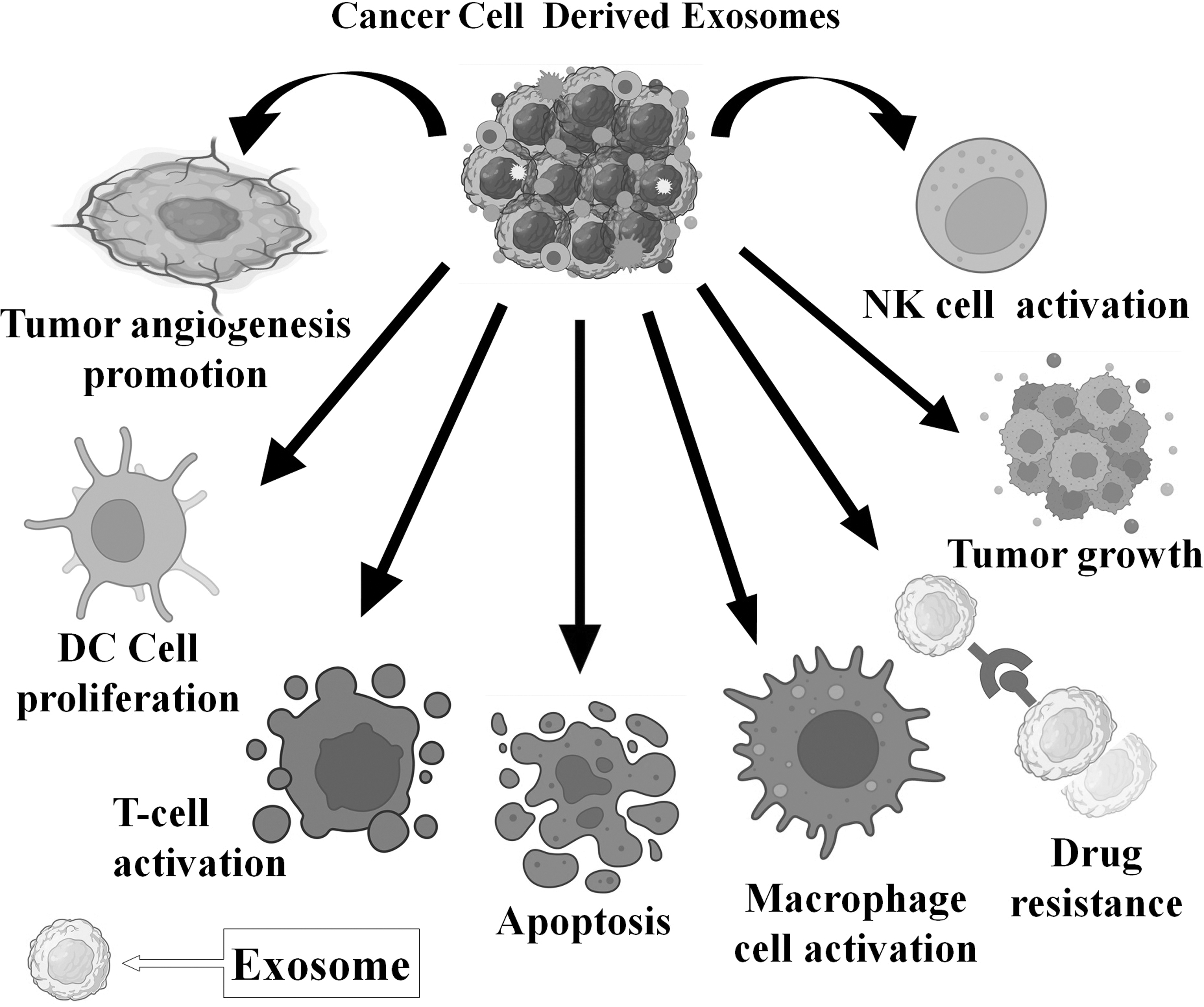
Dual role (tumor angiogenesis promotion, DC proliferation, T cell activation, apoptosis, macrophage cell activation, drug resistance, tumor growth, and activation of NK cell) of exosomes and its significance in tumorigenesis. DC, dendritic cell; NK, natural killer.
Exosome-Mimetic for Cancers
Naturally released exosomes are a model for creating nanovesicle mirror exosomes (NVMEs). Exosome secretion is believed to be efficient. Even single-cell explorations indicate that around 100 exosome particles are secreted per cell each hour. 224 –226 A typical yield (<0.1 exosomes per cell per hour) is obtained using culture conditions to isolate exosomes. 227 Researchers have overcome large-scale manufacturing by adopting a method known as exosome mimetic nanovesicle (EMNV) creation. Cells are extruded serially through filters of different micron diameters in this method. EMNVs derived from a broad range of cell lines are often used to address various illnesses. For example, EMNVs produce from primitive hepatocytes. It enhances liver regeneration in animals undergoing a partial hepatectomy. 228 However, they encourage BM cells to specialize in insulin-producing cells. EMNVs produced through pancreatic beta cells might be exploited to develop possible diabetes therapeutics. 229
Furthermore, anticancer medications such as Dox might be incorporated simultaneously with cell extrusion to enhance cancer therapy. 215 On the contrary, the membrane components of EMNVs are obtained both from the plasma membrane and intracellular organelles. In contrast, the exosome membrane is mostly generated from ILVs through endosomal pathways. 230 Furthermore, exosomes include more sphingolipids, cholesterol, and phospholipid phosphatidylserine than their parent cells. 231 Exosome–liposome hybrid nanoparticles are being initiated to solve the problem of large nucleic acids, such as plasmids, not being well wrapped up. 232,233
FUTURE PERSPECTIVES AND CHALLENGES
In the existing article, researchers focused on creating exosomes and their usage for cancer detection and treatment. We propose the notion of exosomes as a viable alternative to synthetic liposomes as medication carriers or delivery vehicles by highlighting results from different investigations. Exosomes naturally formed vesicles used as medication delivery vehicles or carriers. Furthermore, the simultaneous packing of several payloads in vesicles is characteristic, making them appropriate as drug delivery vehicles. 234
Despite the various benefits, using exosomes as drug carriers presents several obstacles. Exosomes enhance cellular interaction by transmitting biomacromolecules from the host to the receiving cell. However, the particular chemicals conveyed their functions, and the variety of exosomes remains unknown. 235 In addition, it is vital to choose suitable donor cells to avoid activating an immunological reaction. 236 However, if the originating cells are recognized, producing a sufficient amount of vesicles for therapeutic intervention represents an impending obstacle. Exosomes derived from wide-scale cell cultures also suffer from heterogeneity. So, research is necessary on the composition of exosomes and their effectiveness as a human therapeutic carrier. 237
In complement to exosomes, cells have the capacity of ectosomes (30–100 nm) and apoptotic bodies (50–500 nm), which are directly generated and ejected from the plasma membrane during apoptosis. 238 –240 Since some of these EVs replicate exosomes in the aspects of their physical features, including size and density, it might be challenging to distinguish them from other EVs. 241 The fundamental distinction between the different EVs is assumed to be their manner of biogenesis, which dictates cargo contents and functions. Exosomes are formed by the inward budding of endosomes into MVBs, whereas nonexosomal EVs form by direct budding of plasma membranes. Some MVBs are guided into the lysosomal compartment for breakdown and recycling. It forms ILVs and is released into bodily fluids as exosomes outside cells.
Throughout this approach, parent cell data become packed into exosomes in the form of lipids, proteins, and nucleic acids, which can subsequently affect the functioning of destination cells upon arrival. 242
Furthermore, messages conveyed from the parent cell may be sent to cells in the vicinity even without direct cell-to-cell contact. Regardless of the parent cell, exosomes have several properties. These characteristics include tetraspanins (CD9, CD63, and CD81), HSPs (Hsp 60, Hsp 70, and Hsp 90), biogenesis-related proteins (Alix and TSG101), membrane transport and fusion proteins (GTPases, annexins, and Rab proteins), nuclear acids (mRNA, miRNA, and lncRNAs), and lipids (cholesterol and ceramide) for cancer diagnosis and drug delivery of therapeutics. The distinctive properties of exosomes provide opportunities for novel medicinal and diagnostic strategies. Exosome composition, biogenesis, and release mechanism studies will advance our understanding of certain diseases. In addition, it also improves the therapeutic targeting of exosomes. Researchers may also use exosomes as natural drug delivery systems to enhance targeting accuracy while reducing the minimum dosage and side effects. 243,244
EXOSOME: CLINICAL FINDINGS FOR CARCINOMA THERAPY
Several preclinical studies involve cancer immunotherapy and its significance in detecting rehabilitative targets. Given the function of tumor-associated endothelial cells (TAEs) in boosting cancer cell survival and progression, targeting TAE disruption mechanisms, such as the heparinase/syndecan-1 axis, is a novel strategy for treating cancer. 245,246 Exosomes are used as therapeutic indicators in immunotherapy. For example, cancerous glioblastoma patients reduced CD9+/GFAP+/SVN+/CD9+ and SVN+ exosomes. Exosomes cause the production of antisurviving immunotherapy resulting in progression-free survival. Recent research indicates that tumor-derived exosome DNA (ExoDNA) may modulate tumor immunity. In addition, ExoDNA can activate immune cells through STING/cGAS. 247,248
Further phase I study includes autologous dendritic cell-derived exosomes (DEX) for patients with metastatic melanoma. Since no particular CD4+/CD8+ T cell reactions are shown in the peripheral blood, further research into the mechanism of vaccination antigen dissemination is required. 249 Furthermore, the administration of DEX in clinical trials on nonsmall cell lung cancer (NSCLC) patients resulted in the activation of Melanoma antigen gene-specific T cell activation and boosted NK lysis effectiveness. DEX induced from cancer patients' blood cells has proven to be secure and advantageous for immunotherapy. A few minor clinical investigations have been used successfully. 250
A crucial example is a phase II clinical research of a DEX with T cell-dependent anticancer activity carried out in France. 251 DEX immunotherapy may be a helpful treatment for cerebral tumors since it was shown to be viable against glioma in rats, even in the brain, to impede the entrance of tumor-specific immune cells. 252 DEX immunotherapy creates a more targeted immune response against tumor cells. Furthermore, DEX immunotherapy offers superior bioavailability and biostability. It also provides outstanding production and affordable costs compared with other cell-based drugs. 253
Exosomes were researched for immunotherapeutic vaccines, prognostic indicators, and cancer recurrence and metastasis predictors. However, clinical trials still occur (Table 6). 254 –268 Exosomes are used in pancreatic cancer immunotherapy. It encompasses TAEs paired with an antisense molecule for glioma and DEX mixed with cyclophosphamide for NSCLC. In addition, KrasG12D siRNA was from mesenchymal stromal cell-derived exosomes (exosomes). Several clinical studies have examined the potential benefits of exosomes as diagnostic, predictive, and meditative markers for several cancers, such as lung, prostate, glomerular cell, gastric, breast, gallbladder, pancreatic, and rectal cancers. In addition, randomized trials have shown that using exosomes to carry CUR to treat colorectal cancer is safe and productive. 269 Exosome separation, preservation, cargo loading, quality monitoring, and effectiveness assessment techniques will need to be standardized for clinical studies and therapy. 270,271
Exosomes Involving Clinical Trials in Humans for Cancer Treatment
AUC, area under the curve; ccRCC, clear cell renal cell carcinoma; CDE, cancer-derived exosome; NSCLC, nonsmall cell lung cancer; PFS, progression-free survival; PSA, prostate specific antigen.
PATENTS: EXOSOMES FOR CANCER TREATMENT
The finding of preclinical studies involved in cancer management helps predict successful therapy that significantly suppresses cancer using exosomes, and their biomarkers, summarized in Table 7. 271 –281
Patents on Exosomes for Cancers
APC, antigen-presenting cell.
CONCLUSION
Exosomes play a significant role in the cancer premetastatic environment and tumorigenesis. It further improves the immune system, generating new blood vessels, blocking apoptosis from occurring, and attempting to make cancer resistant to treatment. It would be possible to load various payloads, including chemotherapeutic medicines, RNAs, DNAs, proteins, and certain hydrophobic pharmaceuticals. It is still necessary to validate the activities of such modified carriers, even though the complex purification processes and the limited effectiveness of drug transfer appear to be significant obstacles to further clinical use. Another was moving toward advancing the goal of precision medicine in cancer treatment.
Past research has shown that several molecular abnormalities are associated with cancer development. Exosomes are recognized as a unique model of intracellular communication. They transport RNAs, proteins, lipids, and various other biomolecules. In addition, the miRNAs contained within exosomes play an essential part in mediating treatment responses in cancer. Methods for the purification and mass manufacturing of exosomes need additional investigation to achieve targeted therapy. Furthermore, exosomes are used for drug administration and targeted treatment. Cancer treatment options based on exosomes are expected to develop shortly, satisfying many cancer patients substantially.
ETHICS APPROVAL AND CONSENT TO PARTICIPATE
This is a review article. No participation of animals and human beings.
HUMAN AND ANIMAL RIGHTS
This is a review article. No use of animals.
Footnotes
ACKNOWLEDGMENT
The authors acknowledge the efforts of GLA University, Mathura, India, for the assistance.
AUTHORs' CONTRIBUTIONS
R.G. was involved in writing—original draft and language. J.G. was in charge of conceptualization, figures, reviewing, and editing the article. S.R. carried out data collection and the making of tables.
DISCLOSURE STATEMENT
The authors declared no conflict of interest.
FUNDING INFORMATION
Authors have no source of funding.