




Abstract
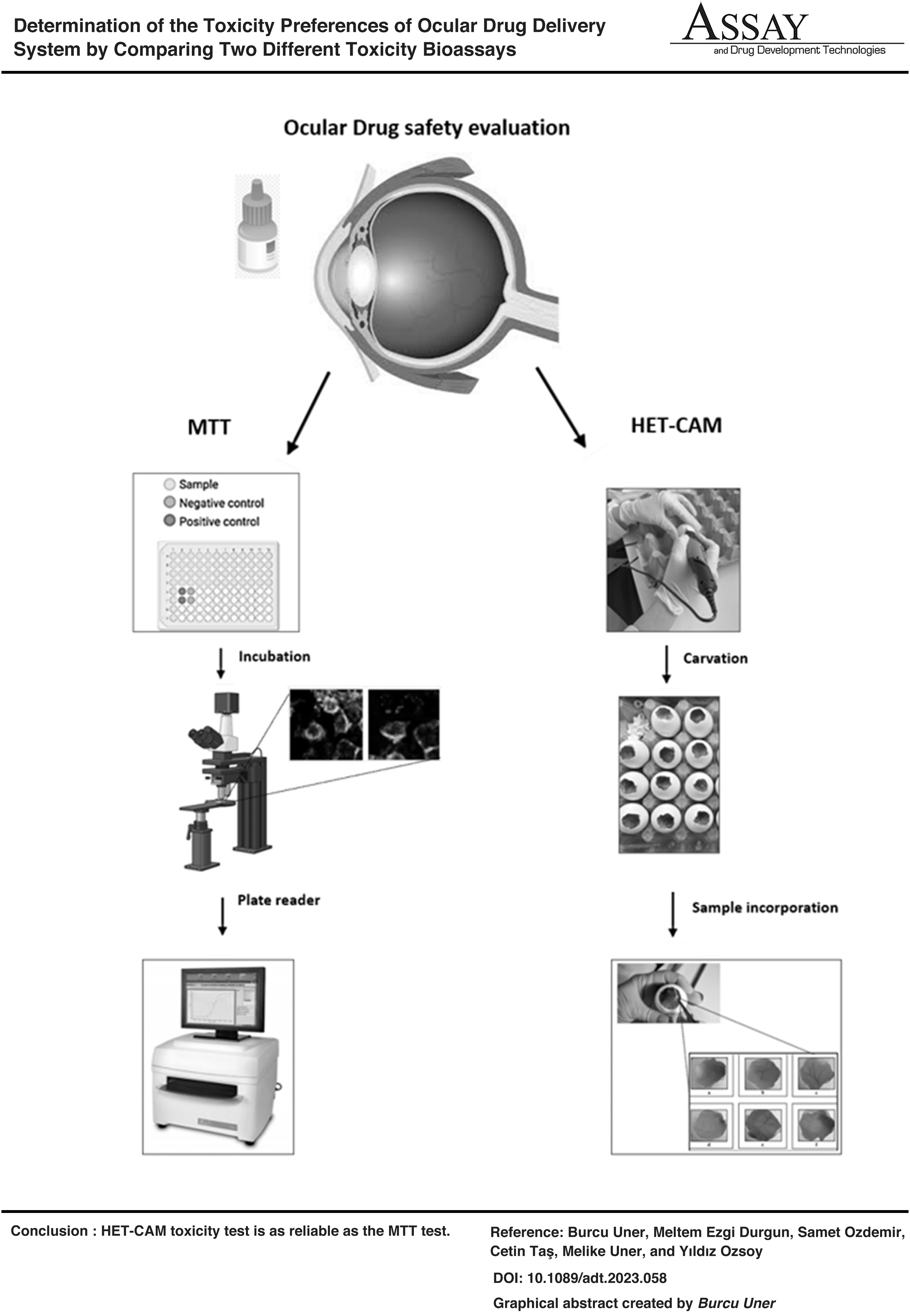
Ocular drug delivery methods are highly favored for boosting bioavailability, patient compliance, and lower adverse effects and dose frequency. In addition to preventing adverse effects from the active ingredient, the parts of drug delivery systems must be nontoxic and nonallergic as well. Mitochondrial toxicity test (MTT) and Hen's egg chorioallantois membrane (HET-CAM) assay are the most often utilized tests based on this dilemma. The toxicity of loteprednol etabonate loaded solid lipid nanoparticles, lipid nanostructured carriers, and nanoemulsion were compared. Oleic acid, Precirol®ATO5, and Pluronic® F68 were used in the preparation. Their toxicities were evaluated by using two different toxicity tests (MTT and HET-CAM). The results suggest that there are no significant differences between the HET-CAM and MTT assays. It is noteworthy that the HET-CAM assay offers a more cost-effective and environmentally friendly alternative to the MTT assay, as it does not require cell culture and generates less toxic waste. This information may be useful to consider when selecting between the two assays.
INTRODUCTION
In the past two decades, remarkable progress has been made in administering drugs to ocular tissues and maintaining effective drug levels in these tissues. 1,2 Inflammation of anterior ocular tissues occurs in two ways. First, these tissues are constantly exposed to the external environment which occurs in constant contact with various disease-causing factors such as dust, bacteria, viruses, fungi, and parasites. The other things are proteins and cells' penetration from the peripheral circulation. 3,4
Ocular inflammation is a crucial and ubiquitous problem in ophthalmology. Causes include primary ocular disorders such as keratitis, chronic superficial keratitis (degenerative pannus), uveitis, systemic disease processes such as mycoses, autoimmune disease, septicemia, and rickettsia infections, as well as secondary disorders. Topical corticosteroids (loteprednol etabonate [LE], fluorometholone, prednisolone, and dexamethasone) have been shown to play a major role by inhibiting inflammatory cell chemotaxis leading to vascular cell proliferation to reduce the inflammatory effect. 5 –7 Although steroid therapy is effective, it has many side effects such as glaucoma, cataract development, and risk of exceeding infection. Meanwhile, these side effects do not make long-term use possible.
LE existed in the group of novel generation corticosteroids. Besides, its anti-inflammatory effect is similar to other topical corticosteroids and lack of increasing intraocular pressure. Meantime, LE also has similar side effects such as other corticosteroids, for instance, blurred vision, tearing, itching, and sensitivity to light. 8 Liposomes, nanocapsules, nanosponges, polymeric nanoparticles, and biodegradable implants developed for ophthalmological applications have disadvantages such as limited physical and chemical stability, costly scale-up methods, rapid release of the drug from the carrier system, stability challenges, and polymer-induced toxicity. 9
Solid lipid nanoparticles (SLN), nanostructured lipid carriers (NLC), and nanoemulsion (NE) are invented especially preferred for transport against hydrophobic drugs. In addition to increasing the stability of the drug, they also increase the bioavailability by transporting the drug to the targeted area. Meantime, they provide controlled drug release by increasing drug retention period in tissue. 10
Ocular epithelial cell line (regular human cell; PCS-100-010; ATCC, Houston, TX) was exposed to mitochondrial toxicity test (MTT) with marketed product (5 mg/mL ocular suspension) (MP), SLN, NLC, and NE (LE-loaded). Placebo formulations (UL) of SLN, NLC, and NE were also analyzed to observe the ocular safety of polymers (Pluronic® F68-PF68; Sigma Aldrich, St. Louis, MO) and lipids (Sigma Aldrich) (Oleic acid-OA and Precirol® ATO5-PATO5) used in the preparation of nanocarriers SLN, NLC, and NE formulations, and available in MP. After careful consideration and analysis of preliminary data, we have decided to use Pluronic F68 (PF68) as the surface-active agent. 11
Our findings indicate that the use of PF68 has resulted in improved particle size distribution and stability. In contrast, our previous studies have shown that the addition of oleic acid to the drug delivery system leads to a decrease in crystallization ratio %. 11,12 When it comes to the toxicity studies we conducted on our drug delivery systems, the agar medium was supplemented with Dulbecco's modified Eagle's medium to culture PCS-100-010 cells: Nutrient mixture (DMEM/F-12)(Sigma Aldrich) with 10% (v/v) fetal bovine serum and 1% (v/v) penicillin–streptomycin. 13 Subsequently, Hen's egg chorioallantois membrane (HET-CAM) assay study was performed using the Leghorn chicken eggs (Has Tavuk, sivrihisar, Eskisehir, Turkey) relevant with ICCVAM guidelines. 0.3 mL (∼10 μM) of formulations were applied onto CAM of eggs.
CURRENT EVIDENCE
Fabrication and Evaluation of the Formulations
The formulations were fabricated using ultrasonication. LE (0.5%) and PATO5 were dissolved in 5 mL of methanol before being added to the mixture. After the evaporation of methanol in a rotary evaporator (Hei-Vac, Heidolph, Germany), the lipid layer loaded with LE was gently melted by heating it up to 80°C. The molten lipid layer containing LE was then carefully dispersed in the aqueous surfactant solution using a water bath at the same temperature. The o/w emulsion was sonicated at 75% amplitude for 7 min using a Bandelin sonicator. The final formulation was left to cool at room temperature for about 30 min for the formation of SLN.
The NLC formulations were prepared consistently with the same method and conditions. PATO5 and OA were utilized to create appropriate NLC while maintaining the total lipid content of the design area. To produce NLC, a reduced solid lipid ratio in the formulation was used along with 1.3% of OA. NE was created by replacing all solid lipids in the SLN formulations with OA in the same manner. 11,13 While the entrapment efficiencies of SLN, NLC, and NE find 91.07%, 92.23%, and 89.17%, the average sizes and polydispersity index (PI)s of nanoparticles were collected 86.2 nm (PI: 0.14), 82.4 nm (PI: 0.17), and 126.4 nm (PI: 0.17), respectively.
Mitochondrial Toxicity Test
In the MTT experiment, mitochondrial dehydrogenase enzyme activity is enhanced in proliferating cells, allowing for the reduction of yellow 3-(4,5-dimethylthiazol-2-yl)-2,5-diphenyltetrazolium-bromide into purple water-insoluble formazan crystals. 14 First, the cytotoxic effects of SLN, NLC, and NE formulations on PCS-100-010 cells were assessed using the MTT assay to verify if the study's aims would be met. PCS-100-010 cells have 10% phenol, making them suitable for MTT testing. Five thousand cells/well were seeded in the red-tinged media in each well of the 96-well plate.
The cells were incubated at 37°C, with 5% CO2 for 24 h, the medium was discarded. Diluted formulations were added (10 μM-5 mg/mL), then incubated until 24 h. Over the 24 h, the formulations were removed with 100 μL of fresh medium and 20 μL of MTT solution in each well. The viability (%) was determined by measuring absorbance at 570 nm with a LogPhase 600 Microbiology Reader (Agilent Technologies, Atlantic City, NJ). 13
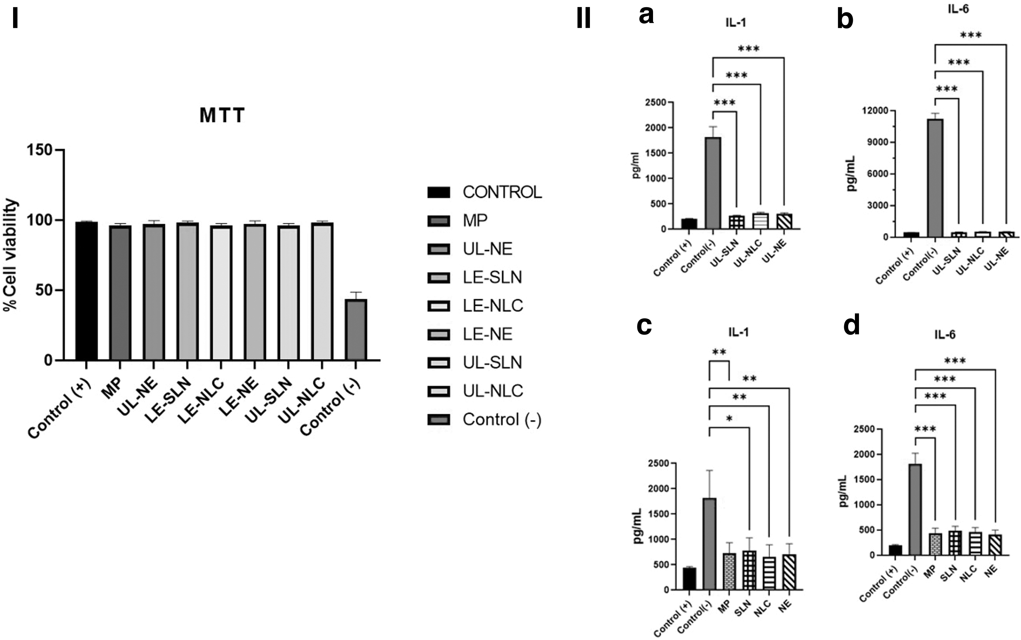
The MTT experiment demonstrated that LE was harmless to PCS-100-010 cells and that UL-SLN, UL-NLC, and UL-NE had no discernible impact on cell viability compared with the + control. Despite the fact that the difference between the control (97.7%) and LE-NE (98.1%) formulations was not statistically significant (p > 0.05), the marketed formulation had the highest percentage of viable cells (Fig. 1I). IC50 values of the LE, LE-SLN, LE-NLC, and LE-NE were collected 5.32, 7.83, 8.06, and 7.55 mg/mL, respectively.
The results of the MTT and ELISAs (
In addition, interleukin (IL)-1 and IL-6 cytokines are proinflammatory cytokines that contribute to the development of inflammatory alterations and a quick immunological response. Consequently, Uner et al. also performed an enzyme-linked immunoassay (ELISA) on the LE-loaded lipid-based nanoparticles. 13 In this context, IL-1 and IL-6 markers were analyzed. In the context of inflammation, levels of both IL-1 and IL-6 raised. According to graphs and micrographs (Fig. 1II), the markers of the SLN, NLC, and NE groups were lowered compared with the control (−) group (p < 0.05; p < 0.01; p < 0.001), and there was no significant difference between commercially available products containing lipid nanoformulations (p > 0.05).
HET-CAM Test
Hereby, aim of our study covered to toxicological assessment of SLN, NLC, and NE formulations. HET-CAM toxicity test was also performed on the chorioallantoic membranes (CAM) of eggs and the reactions occurring in this membrane are followed in terms of lysis, hemorrhage, and coagulation. 15 CAM is similar to rabbit conjunctival tissue in terms of vascular and inflammatory processes. 16 Although the HET-CAM test seems to be based on the examination of vascular toxicity, collected data illuminate general pathological phenomena. Therefore, the HET-CAM test is an alternative nonocular organotypic model to the Draize Rabbit Eye Test.
In this circumstance, the identical approach that applied to the MTT was used to formulation types (LE-SLN, LE-NLC, LE-NE, UL-SLN, UL-NLC, UL-NE, MP, 0.1 N NaOH (−control group) and 0.9% isotonic solution was used (+) control group). At the end of the incubation, eggs were examined as so define nonviable or defective eggs were discarded or appropriate eggs' air cell marking. Last step before the initiated administration, marked section of egg was carved by shaft rotary (Dremel® Stylo, Germany), as well as taken off by facilitating with forceps. 17
Besides, CAM's reaction was observed within 300 s, being recorded the time for the appearance of each noted endpoints. Depending on the endpoints of formulations, toxicity was scored in the level of formation of the venous appearance (lysis: 30 s—5 point; 120 s—3 point; 300 s—1 point/hemorrhage: 30 s—7 point; 120 s—5 point; 300 s—3 point/angiogenesis: 30 s—9 point; 120 s—7 point; 300 s—5 point) regarding with their appearing time. Subsequently, formulations' toxicity was assessed according to the cumulatively rate of this score: score ≤0.9: NA 1 ≤ score ≤4.9: Negligible 5 ≤ score ≤8.9: Severe 9 ≤ score: Robust.
LE has been used for years and its ocular safety is known. 4 As anticipated, no CAM reaction occurred for 300 s in MP applied samples (Fig. 2).

SS and reaction evaluation of marketed product 5 mg/mL ocular suspension, SLN, NLC, and NE with the HET-CAM assay.
Pluronic F68, oleic acid, and PATO5, which are used in the preparation of the SLN, NLC, and NE formulations, are frequently preferred to develop ocular drug delivery systems. Ocular safety has been demonstrated in many previous studies. 18 –20 However, we also analyzed UL-SLN, UL-NLC, and UL-NE in toxicity tests. No CAM reaction was observed in any of the test groups after 300 s (Fig. 2). Collected data are in line with backward studies, which follow that the ocular use of both polymers and lipids in nanocarrier formulation is safe. Neither lysis, hemorrhage nor coagulation reactions were observed by 300 s with LE-loaded SLN, NLC, and NE formulations. According to these results, it can be stated that both optimized LE-loaded and blank formulations are nontoxic, practically.
CONCLUSION
It has been demonstrated that the HET-CAM toxicity test is reliable as much as the MTT assay. In the results compared according to the UN Globally Harmonized System of Classification and Labeling of Chemicals applied in the European Union and the “Dangerous Substances Directive” criteria of the European Union, HET-CAM test results were found in line with 80%–90% reliable compared with the other pathological cases. In this context, it can be anticipated that the ocular toxicity of the analyzed substances could be achieved by in vitro study design.
So, the obtained both MTT and HET-CAM data confirmed the safety of LE nanocarriers on ocular cells and CAM, respectively. SLN, NLC, and NE have been developed both to increase ocular bioavailability and safety profile of LE, since they can be used in the treatment of ocular inflammation. In addition, these advantages of HET-CAM may be supplied for future studies, including time savings, protection of the environment from chemical waste, and cost savings.
Footnotes
ACKNOWLEDGMENT
The authors extend sincere thanks to Yeditepe University Faculty of Pharmacy for supporting throughout the project period.
AUTHORs' CONTRIBUTIONS
Introduction by B.U., M.E.D., and S.O. Methods by B.U. and M.E.D. Conclusion by B.U., M.E.D., S.O., C.T., M.U., and Y.O. Proof reading by C.T., M.U., and Y.O.
CONSENT FOR PUBLICATION
The authors could be given the permission to publish research findings after approval.
DATA AVAILABILITY
The data could be shared upon request.
DISCLOSURE STATEMENT
No competing financial interests exist.
FUNDING INFORMATION