




Abstract
Diabetes mellitus (DM) is the most common endocrine disorder characterized by increased blood glucose levels resulting from defective insulin secretion, resistance to insulin action, or both. DM is often associated with severe complications, and there is an increasing appreciation that cognitive function declines in DM. The aim of this research work was to evaluate Kigelia pinnata root bark extract in Streptozotocin (STZ)-induced type-2 diabetes. Experimental diabetes was induced by a single administration of STZ (60 mg/kg, intraperitoneal [i.p.]), immediately after the STZ administration, and all animals were fed with normal food and water. Nicotinamide was administered (120 mg/kg, i.p.) 15 min before STZ. The development of hyperglycemia was confirmed by the elevated blood glucose levels determined at fixed intervals, which was confirmed by measuring fasting blood glucose levels in rats' blood taken from the tail vein. Supplementation with ethanolic extract of K. pinnata root bark (EEKP) significantly reduced the elevated blood glucose in STZ-induced hyperglycemia in rats. EEKP significantly restored the biochemical and antioxidant defense system. On the final day of the protocol, the extract also reduced inflammatory cytokines in the blood serum.
INTRODUCTION
Diabetes mellitus (DM) is an endocrine disorder, which further interrupts metabolic functionality and characterized by hyperglycemia caused by an inherited or acquired deficiency in the production of insulin by the pancreas, or by the ineffectiveness of the insulin produced. 1 Clinically, symptoms of marked hyperglycemia include thirst (polydipsia), large volume of urine (polyuria), frequent feeling of hunger (polyphagia), feeling of tiredness, blurred vision, and weight gain or weight loss. As per published scientific reports, ∼463 million individuals globally suffered from DM and in India 74–145 million people until 2021 were affected due to DM. 2 Moreover, DM in the aspect of mortality and morbidity is ranked 3rd after cancer and cardiovascular disease. 3
Reactive oxygen species (ROS) is known to play a significant role in the pathogenesis of DM, where increased ROS production and oxidative stress lead to cell membrane damage, enzyme inactivation, apoptosis, and altered gene expression of endogenous antioxidants. 4 Hyperglycemia encourages oxidative stress and the subsequent generation of ROS. Recent research has shown that activating intracellular antioxidant genes like NAD(P)H quinone oxidoreductase, glutathione S-transferase-α, and heme oxygenase-1 inhibits the production of ROS and metabolic dysfunction brought on by hyperglycemia in human microvascular endothelial cells. 5 Moreover, uncontrolled diabetics were found in both human and animal studies to have considerably lower levels of the blood enzyme catalase (CAT), which removes hydrogen peroxide, superoxide dismutase, which neutralizes superoxide radicals, and glutathione (GSH), which is an endogenous antioxidant. 6,7
According to experimental research, tumor necrosis factor alpha (TNF-α) mRNA and protein levels rose in glomerular and proximal tubule cells from diabetic rats, supporting the idea that DM is a chronic inflammatory state linked to the production of proinflammatory cytokines and chemokine genes. 8 –10 Moreover, it has been noted that patients with type 2 DM (T2DM) had serum levels of interleukin (IL-6) that were considerably greater than those seen in diabetic patients who did not have nephropathy. 11,12 These studies showed that TNF-α and IL-6 played a substantial impact on the onset of diabetic neuropathy and renal impairment.
In T2DM there is a disturbance in the physiology of the beta cell of the pancreas commonly seen in animals and human individuals as well. To understand a detailed study about T2DM, animals contribute their lives for the betterment of mankind. Several chemical compounds are being utilized to create animal models for T2DM, but Streptozotocin (STZ) is the most reliable and widely used animal model for T2DM. 13 Thus, STZ is glucosamine–nitrosourea compound responsible for the destruction of beta cells of the pancreas that affects the physiology of insulin.
In the modern era several pharmaceutical drugs are available in the market for the management of the DM, but these chemical entities have side effects as well, which originates different types of the complication as well. Numerous side effects are linked to the synthetic medications. For example, plant-based medications have less side effects than conventional antidiabetic medications, such as decreased risk of cardiovascular disease, hepatotoxicity, weight gain, and hypoglycemia. Due to the side effects of synthetic compounds, researchers gave great attention toward organic and herbal formulations. Several natural compounds are currently used for maintaining glucose levels and insulin release, action, and metabolism in diabetic patients. 14
In this study, we investigated the pharmacological potential of Kigelia pinnata in STZ-induced T2DM. K. pinnata (Balam Kheera) belongs to the family of Bignoniaceae and commonly is called the Sausage tree because of its huge fruits. 15 K. pinnata has a diverse range of medicinal values with many attributes and considerable potential. The plant has traditional uses, which include anticancer, 16 antimicrobial, 17 antiaging, 18 antioxidant, 17 anti-inflammatory, 19 and antimalarial 18 properties. It is also widely applied in the treatment of genital infections, 20 gynecological disorders, 20 renal ailments, fainting, epilepsy, 20 and so on.
In our study, we showed the therapeutic potential of K. pinnata root bark at a dose of 200, 400, and 600 mg/kg in T2DM induced by a single administration of STZ (60 mg/kg, i.p.) and administration of nicotinamide (120 mg/kg, intraperitoneal [i.p.]) 15 min before STZ administration. Furthermore, all the parameters were performed through blood collection from the tail vein and orbital plexuses under mild anesthesia. Lastly, our aim was to analyze the pharmacological potential of ethanolic extract of K. pinnata root bark (EEKP) in STZ-induced T2DM in experimental animals.
MATERIALS AND METHODS
Plant Collection and Authentication
K. pinnata root bark was collected from a botanical garden in January 2022 of Gurukula Kangri (Deemed to be University), Haridwar, Uttarakhand, India, and the botanical authentication was carried out by the National Institute of Science Communication and Policy Research (CSIR-NIScPR), Dr. K.S. Krishnan Marg, Pusa Campus, New Delhi, India, by Dr. Sunita Garg, Principal Scientist and Officer in Charge. A voucher specimen of the plant was deposited in the Raw Material Herbarium and Museum (RHMD), CSIR-NIScPR, Delhi, under the number NIScPR/RHMD/Consult/2021/3901-02.
Preparation of K. pinnata Root Bark Extract
K. pinnata root barks were dried under shade and then powdered with a mechanical grinder. The powder was then passed through sieve No. 40 and was stored in an airtight container for further use. Dried and coarsely powdered K. pinnata root bark (290 g) was extracted with petroleum ether (40°C–60°C), chloroform (60°C), and ethanol (78°C), respectively, using the Soxhlet apparatus. The percentage yield (ethanolic extract) was found to be 22.7%.
Animal Ethics Approval and Experimental Protocol
The present experimental study was conducted using Wistar rats of 11–12 weeks weighing about 220–250 g procured from the Central Animal House facility of ISF College of Pharmacy, Moga, Punjab (India). All the animals were kept in clean polyacrylic cages and maintained in an air conditioned animal house under standard laboratory conditions (room temperature 25 ± 2°C and relative humidity of 55%–60%) with a 12-h light/12-h dark cycle (lights turned on at 7 AM). The animals were maintained with food (dry pellets) and water ad libitum. The standard diet was procured from local vendors and given to mice with following ingredients (g/kg) as: amido (397.5), corn starch dextrinized (132), sucrose (100), carbohydrate (629.5), casein (200), L-cysteine (3), choline bitartrate (2.5), protein (205.5), soyabean oil (70), lard (saturated fat), total fats (70), cellulose microfine (50), fiber (50), mineral wax (35), vitamin wax (10), and energy content (3.97 kcal/g). The experimental protocol was approved by the Institutional Animal Ethics Committee (IAEC) under protocol number (ISFCP/IAEC/CPCSEA/2022/P16).
All the experiments were carried out in accordance with the Committee for the Purpose of Control and Supervision of Experiments on Animal (CPCSEA) guidelines for the use and care of experimental animals. All experiments were performed using age-matched animals in an attempt to avoid variability between experimental groups.
Rats weighing 220–250 g were selected and fasted for 12 h before experiments and allowed an access to water ad libitum. Experimental diabetes was induced by a single administration of STZ (60 mg/kg, i.p.) dissolved in freshly prepared cold citrate buffer (pH 4.5, 0.1 M). Immediately, after the STZ administration, all animals were fed with normal food and water. Nicotinamide was dissolved in normal saline and administered (120 mg/kg, i.p.) 15 min before STZ, 21 after which, EEKP was administered daily for 3 weeks as respective doses. Standard drug (metformin 500 mg/kg) was also used in STZ-treated rats. 22 Moreover, animals were divided into six groups (Table 1). The development of hyperglycemia was confirmed by the elevated blood glucose levels determined on days 3, 7, 14, and 21, which was confirmed by measuring fasting blood glucose levels in rats blood taken from the tail vein. The rats were considered as diabetic, whose blood glucose levels were above 226 mg/dL on day 3 after STZ injection. Glucose levels were measured by using a glucometer (ACCU-CHEK active). On the 21st day under mild anesthesia, blood was withdrawn from the retro-orbital plexus for further biochemical investigations.
Experimental Animal Grouping
EEKP, ethanolic extract of Kigelia pinnata root bark; Met, metformin; STZ, Streptozotocin.
Drugs and Chemicals
STZ was obtained from Sigma-Aldrich, St. Louis, MO, USA, and was used in the study to induce diabetes in rats. Metformin was obtained from Sigma-Aldrich and was used as a standard drug. ELISA Kits of TNF-α, IL-6, and IL-1β (ELK Biotechnology, China) were purchased from Ana Bioenergy, Haryana, India. All other chemicals and solvents used in this study were of analytical grade and purchased from commercial sources.
Evaluation Parameters for Diabetes
Measurement of body weight
The body weight of all the animals was measured during the treatment follow-up at days 1, 7, 14, and 21, respectively, and marked change in body weight was recorded. 23
Estimation of fasting sugar test
Fasting sugar test was performed weekly. After overnight fasting, blood samples were collected at 0-, 30-, 60-, and 120-min intervals and then blood glucose levels were measured by using a glucometer. 24
Estimation of blood glucose test
Blood glucose test was performed on a weekly basis. Blood samples were collected at 0, 30, 60, and 120 min from the tail vein and blood glucose levels were measured by using a glucometer. 25
Estimation of biochemical parameters
At the end of the experiment, blood sample was collected from the orbital plexus under mild anesthesia. Then, the blood sample was centrifuged at 3,000 g for 10 min and then the levels for serum creatinine (SCr), blood urea nitrogen (BUN), uric acid (UA), calcium (Ca2+) and nitric oxide were determined by using commercial kits (Thermo Fisher Scientific) 25 and UV spectroscopy (UV-1700; Shimadzu, Kyoto, Japan). Furthermore, few antioxidants' signs, such as lipid peroxidation (LPO), GSH, and CAT, were also measured according to the method described by Wills 26 , Hu, 27 and Aebi. 28
Estimation of inflammatory markers
All the inflammatory markers
Measurement of serum insulin
Rats were partially anesthetized with ketamine and blood was taken in heparinized tubes. Samples were centrifuged at 3,000 rpm for 12 min and serum was separated. Serum insulin was measured using an ELISA Kit.
Statistical analysis
The observations are expressed as a mean ± standard deviation. The results were analyzed by using GraphPad Prism 9.0.2 and were observed in different time intervals, and the same parameter was examined by two-way ANOVA followed by Bonferroni's post hoc test for several comparisons and those parameters were observed only once during protocol and calculated by one-way ANOVA followed by Tukey's post hoc test.
Ethics Approval
The present study was conducted using animals, which were duly approved by IAEC with protocol number (ISFCP/IAEC/CPCSEA/2022/P16) and experiments were carried out in accordance with CPCSEA and Indian National Science Academy guidelines for the use and care of experimental animals.
RESULTS
Effect of K. pinnata on Body Weight in STZ-Induced Diabetic Rat
The present 21-day study demonstrated that there is no significant difference seen in day 1. Furthermore, on days 7, 14, and 21 STZ-treated rats showed a significant decrease (a p < 0.001) in body weight as compared with normal control rats. After the administration of EEKP at doses of 200, 400, and 600 mg/kg, body weight significantly increased (b p < 0.001) dose dependently as compared with STZ rats. Furthermore, body weight significantly increased (c p < 0.05) in the STZ+EEKP 600 mg group as compared with the STZ+EEKP 400 mg. Remarkably, significant increase (d p < 0.05) in body weight was also observed in the STZ+Met 500 mg group as compared with the STZ+EEKP 600 mg group (Fig. 1).

Effects of EEKP on body weight in STZ-induced diabetes in rats. Data are expressed as mean ± standard deviation represented by columns and bars. a p < 0.001 vs. NC; b p < 0.001 vs. STZ; c p < 0.05 vs. STZ+EEKP 400 mg; d p < 0.05 vs. STZ+EEKP 600 mg. Statistical analysis performed by two-way ANOVA followed by Bonferroni's multiple comparison. The significant test was confirmed with p < 0.05. EEKP, ethanolic extract of K. pinnata root bark; Met, metformin; NC, normal control; STZ, Streptozotocin.
Effect of K. pinnata on Fasting Blood Glucose Test in STZ-Induced Diabetic Rat
The present 21-day study demonstrated that there is no significant difference seen in day 1. Furthermore, on days 7, 14, and 21 STZ-treated rats showed a significant increase (a p < 0.001) in fasting blood glucose levels as compared with normal control rats. After the administration of EEKP at doses of 200, 400, and 600 mg/kg, fasting blood glucose significantly decreased (b p < 0.001) dose dependently as compared with STZ rats. Furthermore, fasting blood glucose significantly decreased (c p < 0.05) in the STZ+EEKP 600 mg group as compared with the STZ+EEKP 400 mg. Remarkably, a significant decrease (d p < 0.05) in fasting blood glucose was also observed in the STZ+Met 500 mg group as compared with the STZ+EEKP 600 mg group (Fig. 2).
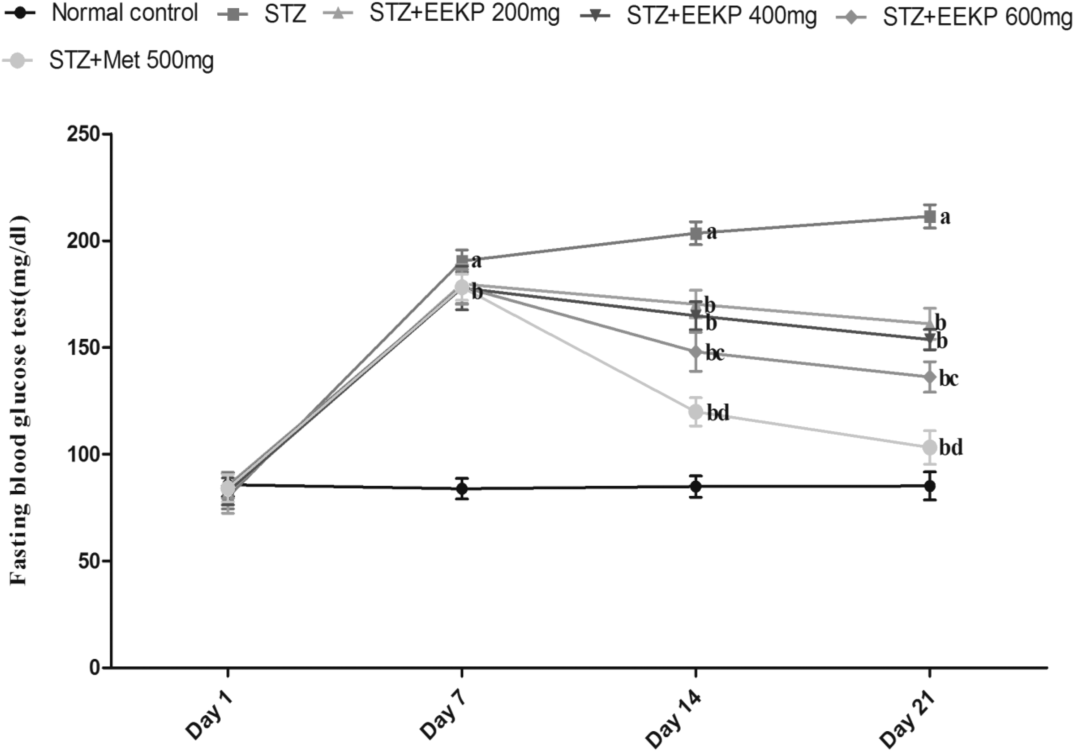
Effects of EEKP on fasting blood glucose levels in STZ-induced diabetes in rats. Data are expressed as mean ± standard deviation represented by columns and bars. a p < 0.001 vs. NC; b p < 0.001 vs. STZ; c p < 0.05 vs. STZ+EEKP 400 mg; d p < 0.05 vs. STZ+EEKP 600 mg. Statistical analysis performed by two-way ANOVA followed by Bonferroni's multiple comparison. The significant test was confirmed with p < 0.05.
Effect of K. pinnata on Blood Glucose Test in STZ-Induced Diabetic Rat
The present 21-day study demonstrated that there is no significant difference seen in day 1. Furthermore, on days 7, 14, and 21, STZ-treated rats showed a significant increase (a p < 0.001) in blood glucose levels as compared with normal control rats. After the administration of EEKP at doses of 200, 400, and 600 mg/kg, blood glucose significantly decreased (b p < 0.001) dose dependently as compared with STZ rats. Furthermore, blood glucose significantly decreased (c p < 0.05) in the STZ+EEKP 600 mg group as compared with the STZ+EEKP 400 mg. Remarkably, a significant decrease (d p < 0.05) in blood glucose was also observed in the STZ+Met 500 mg group as compared with the STZ+EEKP 600 mg group (Fig. 3).

Effects of EEKP on blood glucose levels in STZ-induced diabetes in rats. Data are expressed as mean ± standard deviation represented by columns and bars. a p < 0.001 vs. NC; b p < 0.001 vs. STZ; c p < 0.05 vs. STZ+EEKP 400 mg; d p < 0.05 vs. STZ+EEKP 600 mg. Statistical analysis performed by two-way ANOVA followed by Bonferroni's multiple comparison. The significant test was confirmed with p < 0.05.
Effects of K. pinnata on Biochemical Parameters in STZ-Induced Diabetic Rats
Effect of K. pinnata on SCr level
In SCr estimation, STZ-treated rats showed a significant increase (a p < 0.001) in SCr levels as compared with normal control rats. After the administration of EEKP at doses of 200, 400, and 600 mg/kg, levels of SCr significantly decreased (b p < 0.05) dose dependently as compared with STZ rats. Furthermore, SCr level significantly decreased (c p < 0.05) in the STZ+EEKP 600 mg group as compared with the STZ+EEKP 400 mg. Remarkably, a significant decrease (d p < 0.05) in SCr levels was also observed in the STZ+Met 500 mg group as compared with the STZ+EEKP 600 mg (Table 2).
Data Are Expressed as Mean ± Standard Deviation Represented by Columns and Bars
Statistical analysis performed by one-way ANOVA followed by Tukey's post hoc test.
p < 0.001 vs. NC; b p < 0.001 vs. STZ; c p < 0.05 vs. STZ+EEKP 400 mg; d p < 0.05 vs. STZ+EEKP 600 mg.
BUN, blood urea nitrogen; Ca2+, calcium; NC, normal control; NO, nitric oxide; SCr, serum creatinine.
Effect of K. pinnata on BUN level
In serum BUN estimation, STZ-treated rats showed a significant increase (a p < 0.001) in BUN levels as compared with normal control rats. After the administration of EEKP at doses of 200, 400, and 600 mg/kg, levels of BUN significantly decreased (b p < 0.001) dose dependently as compared with STZ rats. Furthermore, BUN level significantly decreased (c p < 0.05) in the STZ+EEKP 600 mg group as compared with the STZ+EEKP 400 mg. Remarkably, a significant decrease (d p < 0.05) in BUN levels was also observed in the STZ+Met 500 mg group as compared with the STZ+EEKP 600 mg (Table 2).
Effect of K. pinnata on Ca2+ level
In serum Ca2+ estimation, STZ-treated rats showed a significant increase (a p < 0.001) in Ca2+ levels as compared with normal control rats. After the administration of EEKP at doses of 200, 400, and 600 mg/kg, levels of Ca2+ significantly decreased (b p < 0.001) dose dependently as compared with STZ rats. Furthermore, Ca2+ level significantly decreased (c p < 0.05) in the STZ+EEKP 600 mg group as compared with the STZ+EEKP 400 mg. Remarkably, a significant decrease (d p < 0.01) in Ca2+ levels was also observed in the STZ+Met 500 mg group as compared with the STZ+EEKP 600 mg (Table 2).
Effect of K. pinnata on UA level
In serum UA estimation, STZ-treated rats showed a significant increase (a p < 0.001) in UA levels as compared with normal control rats. After the administration of EEKP at doses of 200, 400, and 600 mg/kg, levels of UA significantly decreased (b p < 0.001) dose dependently as compared with STZ rats. Furthermore, UA level significantly decreased (c p < 0.05) in the STZ+EEKP 600 mg group as compared with the STZ+EEKP 400 mg. Remarkably, a significant decrease (d p < 0.001) in UA levels was also observed in the STZ+Met 500 mg group as compared with the STZ+EEKP 600 mg (Table 2).
Effect of K. pinnata on nitric oxide level
In serum nitric oxide estimation, STZ-treated rats showed a significant increase (a p < 0.001) in nitric oxide levels as compared with normal control rats. After the administration of EEKP at doses of 200, 400, and 600 mg/kg, levels of nitric oxide significantly decreased (b p < 0.001) dose dependently as compared with STZ rats. Furthermore, nitric oxide level significantly decreased (c p < 0.05) in the STZ+EEKP 600 mg group as compared with the STZ+EEKP 400 mg. Remarkably, a significant decrease (d p < 0.001) in nitric oxide levels was also observed in the STZ+Met 500 mg group as compared with the STZ+EEKP 600 mg (Table 2).
Effects of K. pinnata on Antioxidants' Level in STZ-Induced Diabetic Rats
Effect of K. pinnata on LPO
In LPO estimation, STZ-treated rats showed a significant increase (a p < 0.001) in serum LPO levels as compared with normal control rats. After the administration of EEKP at doses of 200, 400, and 600 mg/kg, levels of LPO significantly decreased (b p < 0.001) dose dependently as compared with STZ rats. Furthermore, LPO level significantly decreased (c p < 0.01) in the STZ+EEKP 600 mg group levels as compared with the STZ+EEKP 400 mg. Remarkably, a significant decrease (d p < 0.01) in LPO levels was also observed in the STZ+Met 500 mg group as compared with the STZ+EEKP 600 mg group (Table 3).
Data Are Expressed as Mean ± Standard Deviation Represented by Columns and Bars
Statistical analysis performed by one-way ANOVA followed by Tukey's post hoc test.
p < 0.001 vs. NC; b p < 0.01 vs. STZ; c p < 0.05 vs. STZ+EEKP 400 mg; d p < 0.05 vs. STZ+EEKP 600 mg.
CAT, catalase; GSH, glutathione; LPO, lipid peroxidation.
Effect of K. pinnata on GSH
In GSH estimation, STZ-treated rats showed a significant increase (a p < 0.001) in serum GSH compared with normal control rats. After the administration of EEKP at doses of 200, 400, and 600 mg/kg, levels of GSH significantly decreased (b p < 0.001) dose dependently as compared with STZ rats. Furthermore, GSH level significantly decreased in (c p < 0.001) in the STZ+EEKP 600 mg group as compared with the STZ+EEKP 400 mg. Remarkably, a significant decrease (d p < 0.01) in GSH levels was also observed in the STZ+Met 500 mg group as compared with the STZ+EEKP 600 mg group (Table 3).
Effect of K. pinnata on CAT
In CAT estimation, STZ-treated rats showed a significant increase (a p < 0.001) in serum CAT levels as compared with normal control rats. After the administration of EEKP at doses of 200, 400, and 600 mg/kg, levels of CAT significantly decreased (b p < 0.01) dose dependently as compared with STZ rats. Remarkably, a significant decrease (d p < 0.05) in CAT level was also observed in the STZ+Met 500 mg group as compared with the STZ+EEKP 600 mg group (Table 3).
Measurement of serum insulin
In serum insulin estimation, STZ-treated rats showed a significant increase (a p < 0.001) in serum insulin levels as compared with normal control rats. After the administration of EEKP at doses of 200, 400, and 600 mg/kg, levels of serum insulin significantly decreased (b p < 0.01) dose dependently as compared with STZ rats. Remarkably, a significant decrease (d p < 0.05) in serum insulin level was also observed in the STZ+Met 500 mg group as compared with the STZ+EEKP 600 mg group (Table 3).
Effects of K. pinnata on Inflammatory Markers in STZ-Induced Diabetic Rats
Effect of K. pinnata on TNF-α
In TNF-α estimation, STZ-treated rats showed a significant increase (a p < 0.001) in serum TNF-α levels as compared with normal control rats. After the administration of EEKP at doses of 200, 400, and 600 mg/kg, levels of TNF-α significantly decreased (b p < 0.01) dose dependently as compared with STZ rats. Furthermore, TNF-α level significantly decreased (c p < 0.05) in the STZ+EEKP 600 mg group as compared with the STZ+EEKP 400 mg. Remarkably, a significant decrease (d p < 0.05) in TNF-α levels was also observed in the STZ+Met 500 mg group as compared with the STZ+EEKP 600 mg group (Fig. 4).
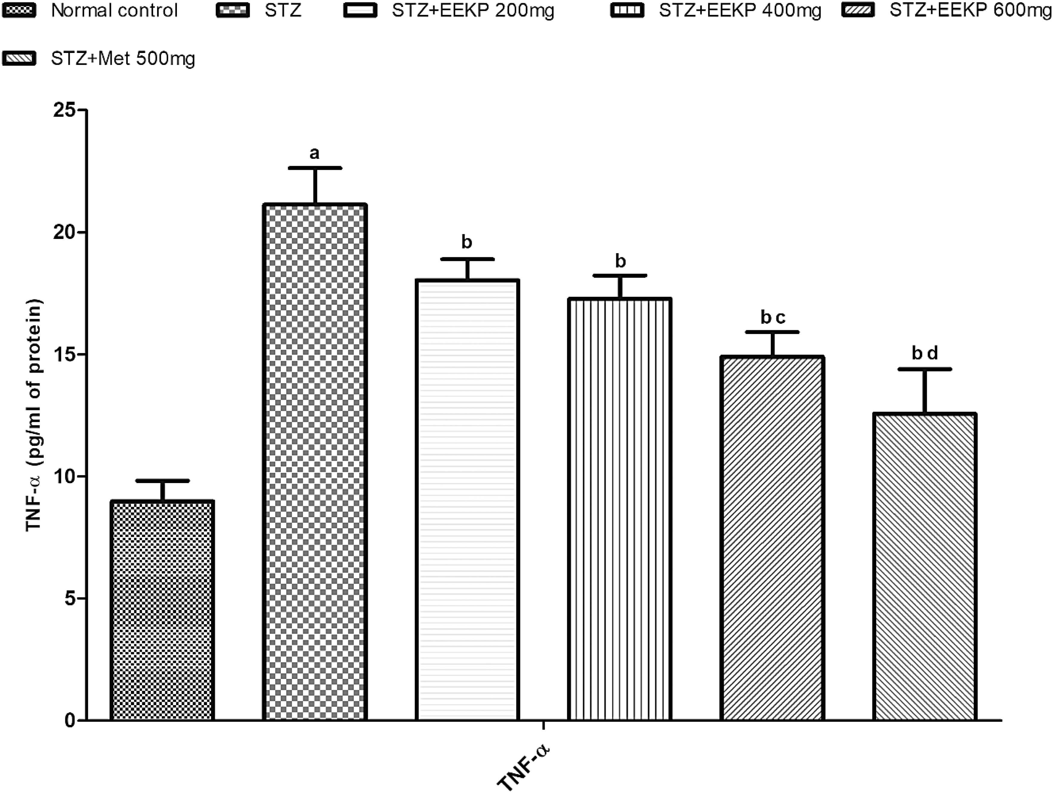
Effects of EEKP on TNF-α level in STZ-induced diabetes in rats. Data are expressed as mean ± standard deviation represented by columns and bars. a p < 0.001 vs. NC; b p < 0.01 vs. STZ; c p < 0.05 vs. STZ+EEKP 400 mg; d p < 0.05 vs. STZ+EEKP 600 mg. Statistical analysis performed by one-way ANOVA followed by Tukey's post hoc test. The significant test was confirmed with p < 0.05. TNF-α, tumor necrosis factor alpha.
Effect of K. pinnata on IL-6
In IL-6 estimation, STZ-treated rats showed a significant increase (a p < 0.001) in serum IL-6 levels as compared with normal control rats. After the administration of EEKP at doses of 200, 400, and 600 mg/kg, levels of IL-6 significantly decreased (b p < 0.001) dose dependently as compared with STZ rats. Furthermore, IL-6 level significantly decreased (c p < 0.001) in the STZ+EEKP 600 mg group as compared with the STZ+EEKP 400 mg. Remarkably, a significant decrease (d p < 0.01) in IL-6 levels was also observed in the STZ+Met 500 mg group as compared with the STZ+EEKP 600 mg group (Fig. 5).
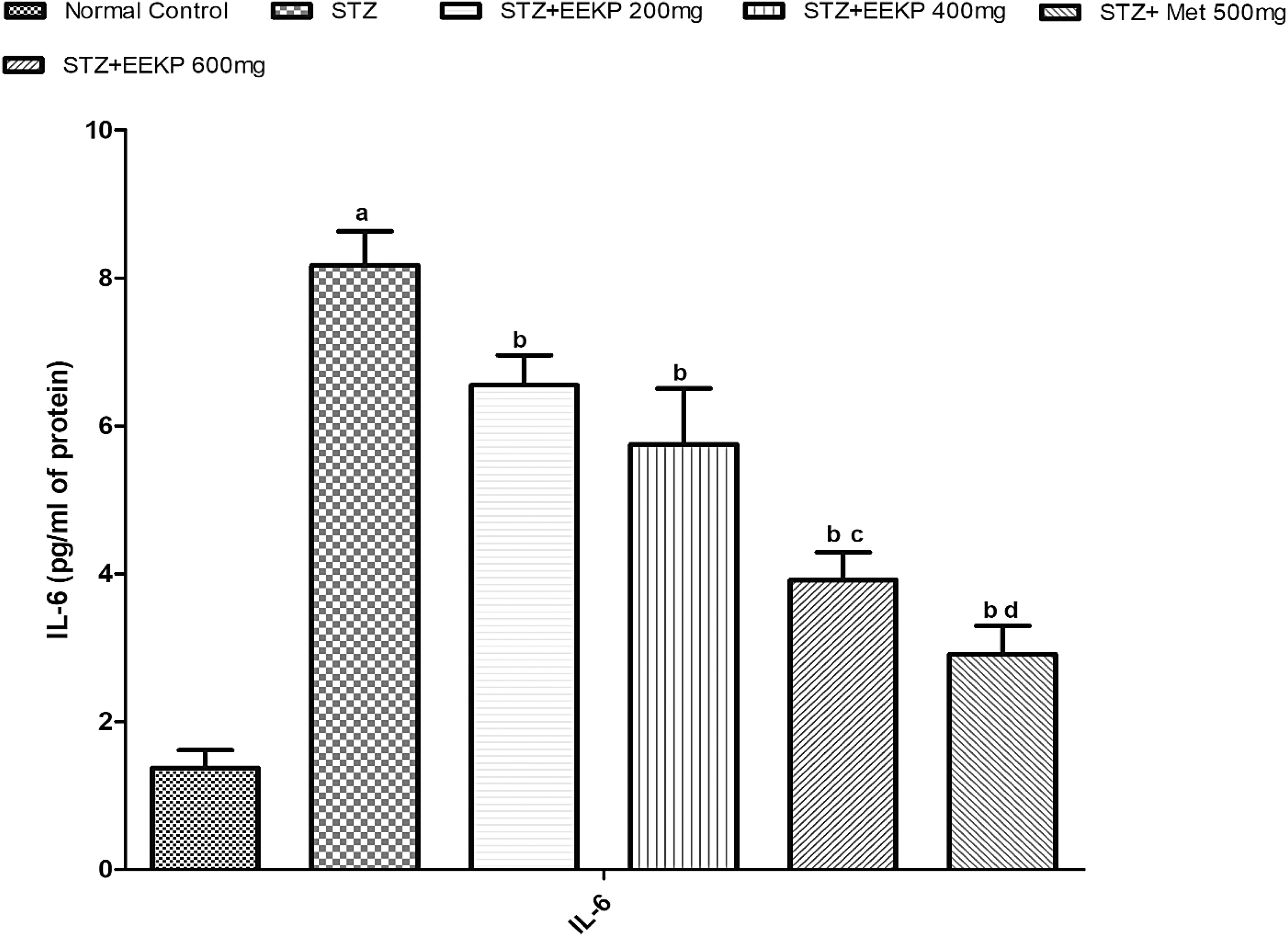
Effects of EEKP on IL-6 level in STZ-induced diabetes in rats. Data are expressed as mean ± standard deviation represented by columns and bars. a p < 0.001 vs. NC; b p < 0.001 vs. STZ; c p < 0.001 vs. STZ+EEKP 400 mg; d p < 0.01 vs. STZ+EEKP 600 mg. Statistical analysis performed by one-way ANOVA followed by Tukey's post hoc test. The significant test was confirmed with p < 0.05. IL-6, interleukin 6.
Effect of K. pinnata on IL-1β
In IL-1β estimation, STZ-treated rats showed a significant increase (a p < 0.001) in serum IL-1β levels as compared with normal control rats. After the administration of EEKP at doses of 200, 400, and 600 mg/kg, levels of IL-1β significantly decreased (b p < 0.01) dose dependently as compared with STZ rats. Furthermore, IL-1β level significantly decreased (c p < 0.01) in the STZ+EEKP 600 mg group as compared with the STZ+EEKP 400 mg. Remarkably, a significant decrease (d p < 0.01) in IL-1β levels was also observed in the STZ+Met 500 mg group as compared with the STZ+EEKP 600 mg group (Fig. 6).

Effects of EEKP on IL-1β level in STZ-induced diabetes in rats. Data are expressed as mean ± standard deviation represented by columns and bars. a p < 0.001 vs. NC; b p < 0.01 vs. STZ; c p < 0.01 vs. STZ+EEKP 400 mg; d p < 0.01 vs. STZ+EEKP 600 mg. Statistical analysis performed by one-way ANOVA followed by Tukey's post hoc test. The significant test was confirmed with p < 0.05.
DISCUSSION
In the present study, we demonstrated the protective effect of K. pinnata at different doses in STZ-induced T2DM in the experimental animals, in which a single dose of STZ compromises the release of insulin from pancreatic beta cell leading to T2DM, as confirmed by enhanced levels of blood glucose in fasting as well as random condition. Furthermore, T2DM is evidenced by a significant increase in SCr, BUN, serum UA, serum Ca2+, nitric oxide, and oxidative stress that further promotes the disease condition. The present investigation reveals the first evidence that EEKP alleviated the fasting and random blood glucose levels along with restoration of body weight and biochemical parameters. Lastly, K. pinnata also shows its impressive modulatory effect on the levels of proinflammatory mediators in STZ-induced T2DM.
K. pinnata belongs to the family Bignoniaceae [syn Kigelia africana (Lam.) Benth], which is a multipurpose medicinal plant with many attributes and considerable potential. 29 The present study has been proposed for the pharmacological evaluation of root bark extracts of K. pinnata for antidiabetic activity. Previously, it has been reported that they have in-vitro cytotoxic potential against G361 melanoma cell lines and the leaves of K. pinnata also have the capacity to eradicate tumors in repair deficiency human cancer cell line. 16,30 In 2020, Nabatanzi et al., demonstrated that K. pinnata has an inhibitory effect on cytokines (IL-1β, TNF-α, and IL-6) in different in-vitro assays, which has the potential against inflammatory mediators. 31 Furthermore, several studies reveal that K. pinnata shows a protective effect against oxidative stress, microbial infection, and cancer studies.
We hypothesized that the EEKP has a protective potential against STZ-induced T2DM. In this regard, treatment drugs have dose-dependent action on different parameters, and increase in body weight every week is observed at all doses. Moreover, elevated levels of fasting blood glucose and blood glucose are reduced but the effect on metformin has a lesser significant difference compared with EEKP 600 mg. With hyperglycemia, alleviation in the functionality of the antioxidant defense system that further facilitates ROS production results. The primary source of cellular ROS production is mitochondria, and diabetes has been linked to a decline in mitochondrial activity. However, STZ modulates the biochemical as well as inflammatory cytokine balance in the body that contributes to the severity of diseases. For this instance, K. pinnata restored the elevated levels of biochemical parameters and inflammatory cytokines after 3 weeks of treatment.
The production of endogenous TNF-α in microvascular and neural tissues was also found to be elevated in chronic hyperglycemia, which may increase microvascular permeability, hypercoagulability, and nerve damage, as well as start and promote the development of the typical diabetic microangiopathic polyneuropathy and encephalopathy. 32 Our observations indicated the nuclear factor kappa B's translocation into the nucleus and the raised TNF-α and IL-6 transcript levels strongly suggested that the inflammatory cascade was started in the diabetic rats. Intriguingly, K. pinnata treatment significantly reduced TNF-α, IL-1β, and IL-6 in diabetic rats, pointing to an anti-inflammatory activity of K. pinnata in the serum. Research on the anti-inflammatory properties of K. pinnata has been conducted in numerous additional properties, including renal melanoma and epidermis cancer cell lines. 33 Although our study results are primarily descriptive, we feel that based on our work, further trials may be devised for mechanistic exploration of the mechanisms underlying the benefits of K. pinnata on DM.
CONCLUSION
The aim of the study was to assess the antidiabetic activity of EEKP in STZ-induced diabetes in rats. The present study reveals that K. pinnata significantly attenuated the glucose levels and suppressed the overactivation of inflammatory cytokines in diabetic rats. In addition, it attenuates ROS production and promotes the antioxidant defense fate. Taken together, our findings suggested that K. pinnata might be a novel therapeutic option for those with type 2 diabetes.
Footnotes
ACKNOWLEDGMENT
Authors are highly thankful to Mr. Parveen Garg, Chairman, ISF College of Pharmacy, for providing excellent research platform with advanced instruments.
AUTHORs' CONTRIBUTIONS
R.K. and S.M. performed the research and analyzed the data. N.K., S.K.R., A.K., and S.S. designed the research study. B.S.R. and N.K.R. contributed to essential reagents and tools. All authors wrote and reviewed the article.
DISCLOSURE STATEMENT
The authors declare that they have not conflicts of interest.
FUNDING INFORMATION
No funding was received for this work.