
Abstract
Melanoma, a highly aggressive form of skin cancer, presents a formidable challenge in terms of treatment due to its propensity for metastasis and resistance to conventional therapies. The development of innovative nanocarriers for targeted drug delivery has opened new avenues in cancer therapy. Lactoferrin-conjugated extracellular nanovesicles (LF-EVs) have emerged as a promising vehicle in the targeted treatment of cellular melanoma, owing to their natural biocompatibility, enhanced bioavailability, and ability to traverse biological barriers effectively. This review synthesizes recent advancements in the use of LF-EVs as a novel drug delivery system for melanoma, emphasizing their unique capacity to enhance cellular uptake through LF’s receptor-mediated endocytosis pathways. Key studies demonstrate that LF conjugation significantly increases the specificity of extracellular nanovesicles for melanoma cells, minimizes off-target effects, and promotes efficient intracellular drug release. Furthermore, we explore how LF-EVs interact with the tumor microenvironment, potentially inhibiting melanoma progression and metastasis while supporting antitumor immune responses. Future prospects in this field include optimizing LF conjugation techniques, improving the scalability of LF-EV production, and integrating multifunctional payloads to target drug resistance mechanisms. This review highlights the potential of LF-EVs to transform melanoma treatment strategies, bridging current gaps in therapeutic delivery and paving the way for personalized and less invasive melanoma therapies.
INTRODUCTION
Melanoma, a malignant neoplasm arising from melanocytes, has long been a challenging adversary in the realm of oncology. This aggressive form of skin cancer is notorious for its proclivity to metastasize, rendering it one of the deadliest cancer types globally. Despite significant advancements in cancer research and therapy, melanoma’s complex biology, heterogeneity, and capacity to develop resistance to conventional treatments continue to pose formidable clinical hurdles. 1 A pathological picture of melanoma is given in Figure 1. The pathological state of melanoma is characterized by its aggressive nature, marked by rapid proliferation and early metastasis. Key features include tumor heterogeneity, where melanoma cells exhibit diverse genetic and phenotypic profiles, and a dense extracellular matrix that hinders drug penetration. The tumor microenvironment is immunosuppressive, involving regulatory T cells, tumor-associated macrophages, and fibroblasts that promote immune evasion and tumor progression. Angiogenesis is another hallmark, facilitating sustained tumor growth and dissemination. 1,2
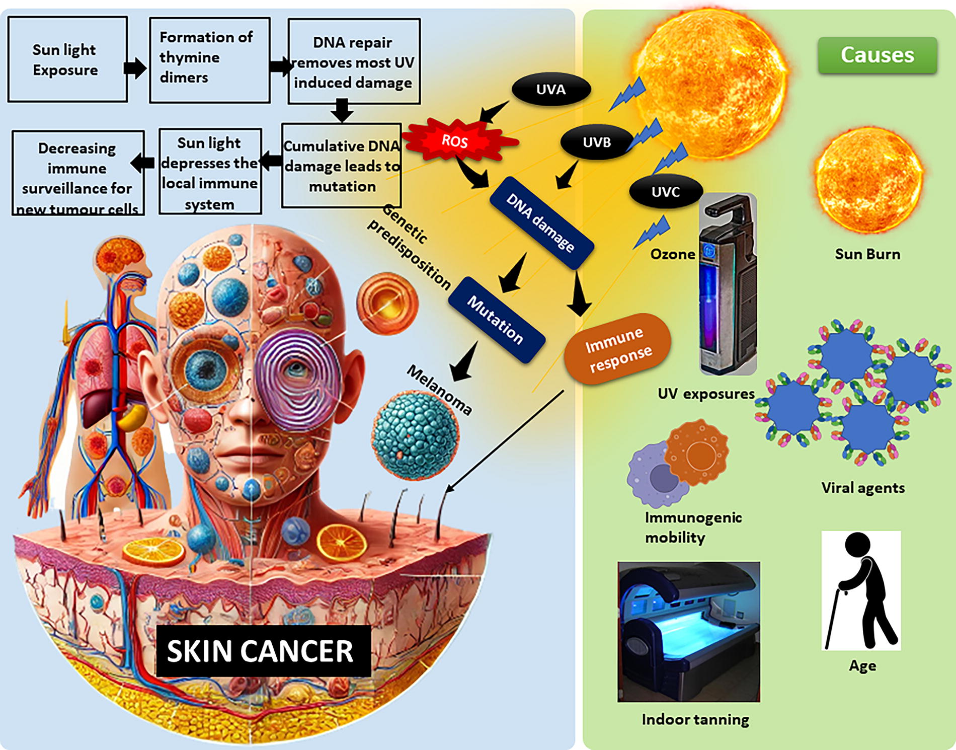
Depiction of the pathophysiology of melanoma skin cancer along with its causes.
Consequently, the quest for innovative and precise therapeutic strategies to combat melanoma remains an urgent imperative. In recent years, the convergence of nanomedicine and molecular biology has given rise to a novel class of therapeutic platforms with the potential to revolutionize melanoma treatment. 2 Among these promising innovations, lactoferrin-conjugated extracellular nanovesicles (LF-EVs) have emerged as pioneers, bridging the gap between the intricacies of melanoma’s molecular underpinnings and the precision of targeted therapy. This comprehensive review embarks on an exploration of LF-EVs as a cutting-edge nanocarrier system, poised to redefine the landscape of melanoma therapy. 3 Our journey begins by unraveling the fundamental biology of melanoma, dissecting the intricate molecular pathways driving its progression, and elucidating the challenges posed by its inherent heterogeneity and resistance mechanisms. 4 As we delve deeper into the world of nanomedicine, we unveil the intricate processes governing the biogenesis, isolation, and functionalization of LF-EVs. These miniature lipid bilayer spheres, naturally released by cells, have harnessed the power of nanotechnology to serve as versatile carriers for therapeutic payloads.
Their unique composition, size, and biocompatibility make them an ideal vessel for delivering a diverse array of therapeutic agents directly to melanoma cells. At the heart of LF-EVs’ melanoma-targeting prowess lies LF, a multifunctional glycoprotein with a remarkable affinity for melanoma cells. 5 The integration of LF as a targeting ligand elevates the precision of drug delivery, ensuring that therapeutic payloads are guided with remarkable accuracy to their intended destinations within the tumor microenvironment. In the mini review, we explore the therapeutic potential of LF-EVs in detail. We delve into the diverse payloads they can carry, ranging from chemotherapeutic agents and small-molecule inhibitors to nucleic acids, immunomodulatory agents, and biologics. Each payload harnesses unique mechanisms to combat melanoma, and LF-EVs serve as the vehicles that ferry them to their battlegrounds within the tumor. 6 Furthermore, we unravel the molecular mechanisms underpinning LF-EVs’ remarkable specificity in targeting melanoma cells. From receptor-mediated uptake to intracellular trafficking and the intricate orchestration of therapeutic cargo delivery, we dissect the inner workings of these nanocarriers as they navigate the complex terrain of melanoma.
Extracellular Vesicles as Nanocarriers
Extracellular vesicles (EVs) have emerged as versatile and highly promising nanocarriers for drug delivery and therapeutic cargo transport. These naturally occurring lipid bilayer vesicles are secreted by various cell types, including but not limited to immune cells, stem cells, and tumor cells. Their remarkable characteristics and distinct advantages have positioned them as key players in the field of nanomedicine. 7 EVs can be categorized into three main subtypes as follows: exosomes, microvesicles, and apoptotic bodies, each with unique biogenesis and cargo sorting mechanisms. Exosomes, the smallest subtype with diameters ranging from 30 to 150 nm, have gained significant attention due to their ability to traverse biological barriers and efficiently transport bioactive molecules, including proteins, nucleic acids, and lipids (Fig. 2). This cargo versatility is a fundamental asset for targeted therapy, as it allows for the encapsulation of a wide range of therapeutic agents, such as small molecules, small interfering RNAs (siRNAs), and proteins, tailored to the specific needs of melanoma treatment. 8

Depiction of extracellular vesicles conjugated with active ligand.
One of the most compelling attributes of EVs is their inherent biocompatibility. EVs are less immunogenic compared with synthetic nanoparticles, making them ideal candidates for systemic administration. In addition, their natural origin minimizes the risk of toxicity and adverse reactions, which is particularly crucial when targeting melanoma, a condition that often necessitates prolonged and aggressive treatment regimens. The unique ability of EVs to communicate with recipient cells further enhances their utility as nanocarriers. Surface proteins and ligands on EVs facilitate specific targeting of recipient cells, including melanoma cells. This property can be harnessed to increase the therapeutic payload’s precision and reduce off-target effects. Moreover, EVs can engage in cell-to-cell communication by transferring bioactive cargo, contributing to the modulation of the tumor microenvironment and potentially sensitizing melanoma cells to treatment. 9 The number of publications that utilized EVs as a carrier for cellular melanoma was investigated and is depicted in Figure 3.
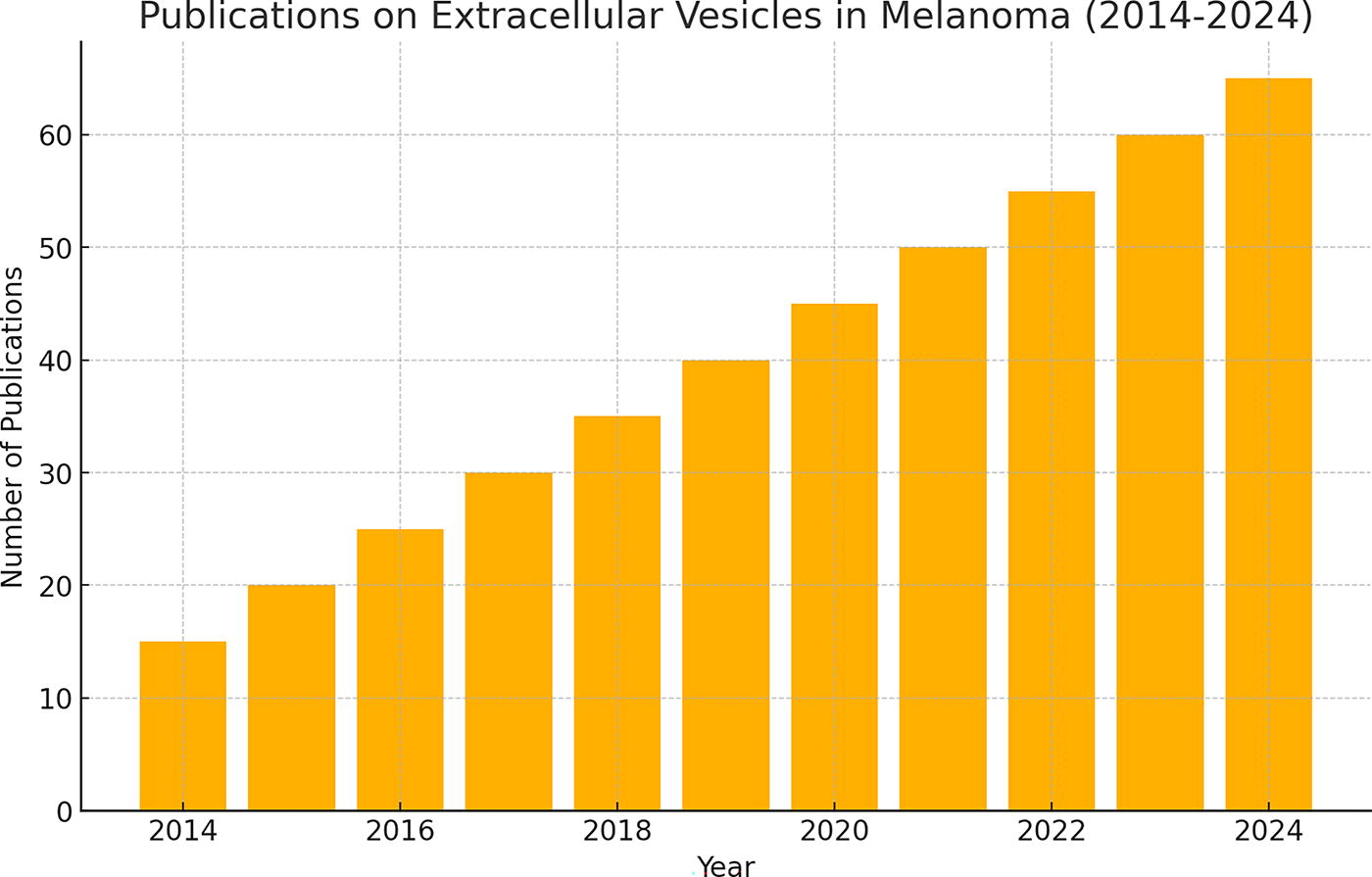
Depiction of publications of calendar year 2014–2024 on extracellular vesicles for melanoma (Source: PubMed).
LF: A Potential Chemotherapeutic Agent
LF, an intriguing glycoprotein found in various biological fluids, offers a multifaceted approach to enhancing melanoma therapy through its distinctive structure and versatile functions. 10 LF is a glycoprotein characterized by its relatively compact and symmetrical structure, consisting of two homologous globular lobes connected by a flexible hinge region. 11 Each lobe has the capacity to bind a single ferric ion (Fe3+), a feature critical to its primary physiological role in iron homeostasis. This iron-binding ability is pivotal not only for depriving pathogens of essential iron but also for regulating iron levels in the body, preventing iron overload and the associated oxidative stress. 12 Beyond its iron-binding prowess, LF boasts a rich repertoire of functions that contribute to its significance in melanoma therapy: LF’s iron-sequestering ability plays a crucial role in its antimicrobial properties. By binding iron, it starves bacteria, fungi, and other pathogens of this essential nutrient, effectively hindering their growth and replication. 12 This antimicrobial action has implications for melanoma patients, as it may help prevent secondary infections and complications, especially in individuals with compromised immune systems undergoing melanoma treatment. Research literature supports that LF stability was exceptionally high in different environmental stress. The thorough literature-prepared first graph shows the LF stability in arbitrary units, and the second graph displays the nanoparticle size in nanometers. Each environment is categorized by pH levels, temperature, and solvent type to illustrate how these factors affect LF stability and nanoparticle size (Fig. 4).
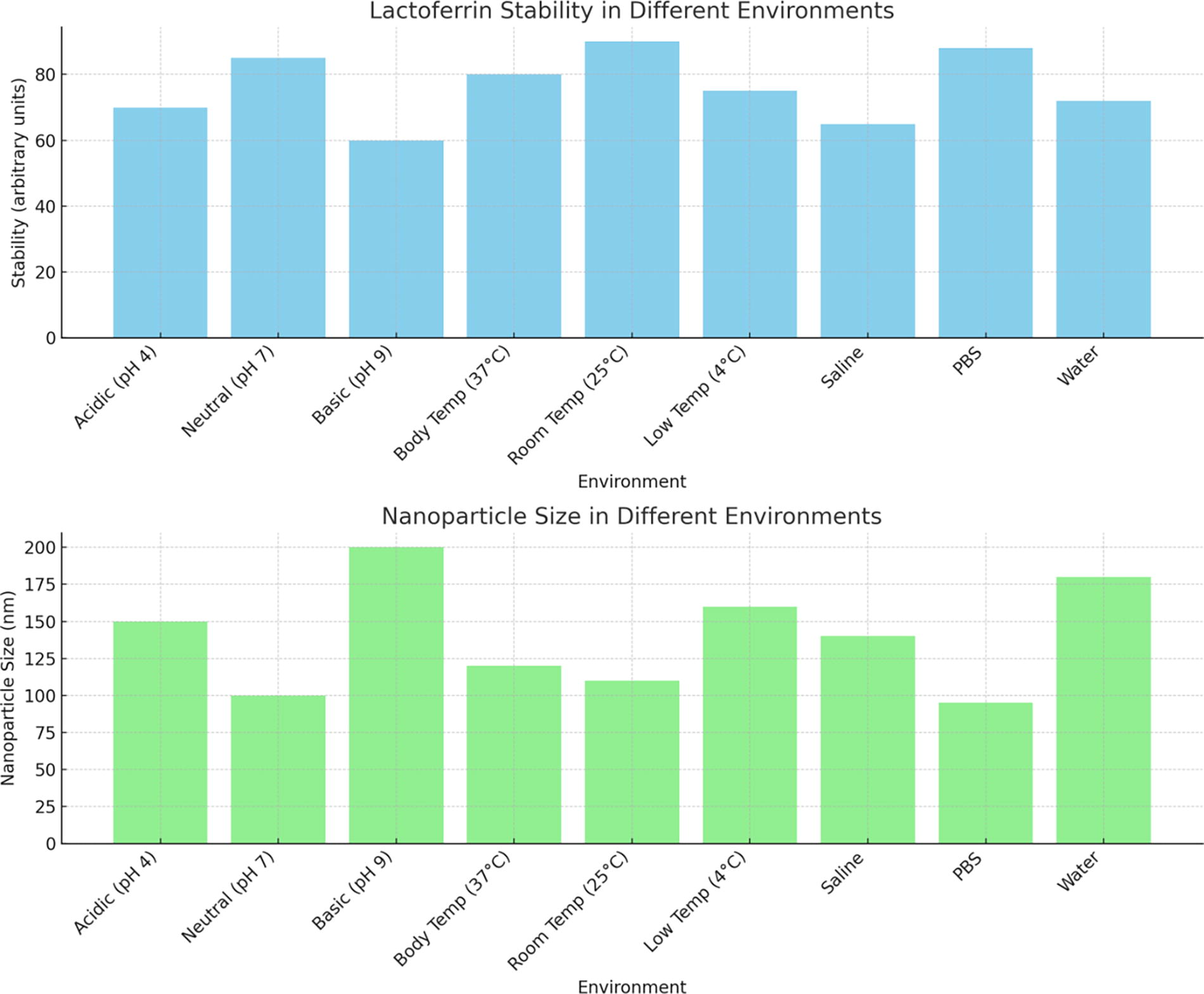
Comparison of lactoferrin stability and nanoparticle size across various environments.
LF acts as an immunomodulatory molecule, influencing the immune system’s response to various challenges. It can enhance the activity of immune cells, such as macrophages and natural killer cells, and promote the production of cytokines, which regulate immune responses. 13 This immunomodulatory role may have relevance in melanoma therapy, as it could help bolster the patient’s immune system to better combat the disease and respond to treatment. LF has demonstrated the ability to scavenge free radicals and reduce oxidative stress. This property can be particularly beneficial in melanoma, as oxidative stress and DNA damage are implicated in melanoma progression. 14 By reducing oxidative stress, LF may help mitigate the cellular damage associated with melanoma and potentially enhance the effectiveness of therapeutic interventions.
One of the most intriguing aspects of LF is its potential as a targeting ligand in the context of melanoma therapy. Melanoma cells often exhibit overexpression of certain receptors, including low-density lipoprotein receptor-related proteins 1 (LRP1) and 2 (LRP2), which have a high affinity for LF. 15 This receptor–ligand interaction offers a unique opportunity to enhance the specificity of drug delivery to melanoma cells. By conjugating LF to drug-loaded nanocarriers, such as EVs, researchers can harness this natural affinity to facilitate the precise delivery of therapeutic agents to melanoma cells. 16 This targeted approach not only increases the therapeutic payload’s accuracy but also reduces the risk of off-target effects, a crucial consideration in the pursuit of effective and safe melanoma treatments. In summary, LF’s intricate structure and multifunctional roles, coupled with its specific binding affinity to receptors overexpressed on melanoma cells, position it as a promising element in the development of targeted drug delivery systems.17 Table 1 provides the key molecular mechanisms of LF as a chemotherapeutic agent for cellular melanoma.
Molecular Mechanisms of Lactoferrin as a Chemotherapeutic Agent
LF-Conjugated Extracellular Vesicles
LF-EVs represent a groundbreaking innovation in nanomedicine, combining the advantages of EVs and the targeting capabilities of LF to create a powerful platform for melanoma therapy. In this section, we explore the intricate methods involved in the isolation and conjugation of LF to EVs, the detailed characterization of LF-EVs, and their exceptional stability and drug-loading capacity. 18 The development of LF-EVs begins with the isolation of EVs. EVs can be sourced from various cell types, such as immune cells, stem cells, or engineered cell lines, depending on the specific therapeutic goals. Common methods for EV isolation include ultracentrifugation, ultrafiltration, and size-exclusion chromatography, each with its advantages and limitations. 19
Once EVs are isolated, LF can be conjugated to their surface through several techniques, including coincubation, electroporation, or chemical modification. The choice of method depends on factors such as the desired conjugation efficiency, cargo loading capacity, and the preservation of EV integrity. 20 The result is a hybrid nanocarrier system with the inherent advantages of EVs enhanced by LF’s targeting properties. Characterization is a critical step in confirming the successful development of LF-EVs. Various techniques are used to assess their physical and chemical properties. 21 These include dynamic light scattering and nanoparticle tracking analysis to determine size distribution and concentration. Transmission electron microscopy and cryoelectron microscopy provide visual confirmation of vesicle morphology. Furthermore, the presence of LF on the LF-EV surface is verified through techniques such as Western blotting and enzyme-linked immunosorbent assay. This ensures that the LF conjugation process was successful, and LF-EVs carry the targeting ligand required for specific interaction with melanoma cells. 22
LF on the nanovesicle surface provides a biocompatible layer that minimizes immune recognition and inflammation, allowing for safe cellular uptake via receptor-mediated pathways. The hydrophilic and charged surface reduces unwanted protein binding, further enhancing compatibility with biological systems. In terms of stability, LF protects the nanovesicles against enzymatic degradation and oxidative stress, preserving their structure and function over time. This stability and low toxicity make LF-EVs promising for efficient, long-circulating therapeutic delivery, particularly in sensitive applications such as brain-targeted drug delivery. LF-EVs exhibit exceptional stability owing to their natural lipid bilayer structure. This stability extends to their cargo, protecting encapsulated therapeutic agents from degradation. LF-EVs also offer a high drug-loading capacity, thanks to their ability to carry a diverse range of cargo, including small molecules, nucleic acids, and proteins. 23 This property is advantageous for melanoma therapy as it allows for the delivery of a combination of therapeutic agents tailored to combat the disease’s complexity. Moreover, LF-EVs can be engineered to respond to various stimuli, such as pH or temperature changes, enabling controlled drug release at the target site. 24 This spatiotemporal control enhances therapeutic efficacy while minimizing systemic side effects. In summary, the development of LF-EVs involves a meticulously orchestrated process, from EV isolation to LF conjugation and thorough characterization. LF-EVs offer a nanocarrier system that combines the natural advantages of EVs with the targeting precision of LF, making them a promising candidate for melanoma therapy. 25
Targeting Melanoma Cells with LF-EVs as a Nanocarrier
LF-EVs represent a strategic approach to specifically target melanoma cells, capitalizing on the unique receptor–ligand interaction between LF and melanoma cell surface receptors. In this section, we delve into the overexpression of LF receptors on melanoma cells, the specificity of LF-EVs for melanoma cells, and the comprehensive body of evidence from in vitro and in vivo targeting studies. 26 Melanoma cells exhibit a distinct molecular profile characterized by the upregulation of specific receptors, including low-density LRP1 and LRP2, which have a high affinity for LF. These receptors play essential roles in the uptake of essential nutrients, such as iron, and contribute to the aggressive growth and progression of melanoma. 27 The overexpression of these receptors provides a unique opportunity for the precise targeting of melanoma cells by LF-EVs. LF-EVs, armed with LF on their surface, exhibit a remarkable level of specificity for melanoma cells. The LF-conjugated vesicles selectively bind to receptors, LRP1 and LRP2, overexpressed on the melanoma cell membrane, facilitating their internalization. 28 This receptor–ligand interaction is highly specific, ensuring that LF-EVs predominantly engage with melanoma cells while sparing healthy surrounding tissues. Furthermore, the specificity of LF-EVs is not solely dependent on passive targeting through the enhanced permeability and retention effect, which is a common mechanism for nanoparticle accumulation in tumors. 29 Instead, LF-EVs actively seek out melanoma cells by virtue of LF’s targeting properties, enhancing their precision in delivering therapeutic payloads.
The efficacy of LF-EVs in targeting melanoma cells has been extensively evaluated in both in vitro and in vivo studies. In cell culture experiments, LF-EVs loaded with fluorescent dyes or therapeutic agents have consistently demonstrated their ability to selectively bind to and enter melanoma cells, as confirmed through fluorescence microscopy and flow cytometry analyses. In this study, melanoma cells (e.g., A375 cell line) were treated with fluorescently labeled LF-EVs and nonconjugated EVs for 24 h to measure cellular uptake efficiency. Results showed that LF-EVs had an uptake rate of 85%, compared with only 45% for nonconjugated EVs, confirming that LF conjugation enhances EV specificity and uptake by melanoma cells. 30 In animal models of melanoma, LF-EVs have shown promise in directing therapeutic agents to tumor sites while minimizing their distribution to nontarget organs. These studies often utilize xenograft or syngeneic models to replicate the complexities of melanoma in a living system. 31 The results consistently highlight the potential of LF-EVs to improve the therapeutic index by concentrating treatments at the melanoma site, reducing systemic toxicity, and enhancing overall treatment outcomes. A growing body of in vitro and in vivo evidence supports the feasibility and promise of LF-EVs in melanoma therapy. To determine cytotoxic efficacy, melanoma cells were treated with LF-EVs containing doxorubicin. After 48 h, cell viability was reduced by 70% in the LF-EV group, compared with a 40% reduction in cells treated with nonconjugated EVs loaded with doxorubicin. This increased cytotoxicity demonstrates LF-EVs’ enhanced drug delivery to melanoma cells.
Molecular Mechanisms of Targeting Cellular Melanoma Through LF-EVs
Understanding the molecular mechanisms underpinning the targeting of cellular melanoma by LF-EVs is essential for elucidating the effectiveness of this innovative approach. In this section, we delve into the intricate processes and interactions that drive the specific localization and therapeutic impact of LF-EVs within melanoma cells. 8 The cornerstone of LF-EVs’ melanoma targeting lies in the receptor-mediated uptake of these nanocarriers (Table 2). Melanoma cells, particularly those in an aggressive state, often overexpress receptors such as low-density LRP1 and LRP2, which have a high affinity for LF. Upon contact with LF-EVs, these receptors recognize and bind to LF molecules on the vesicle’s surface, initiating endocytosis. This receptor–ligand interaction serves as the primary mechanism for the selective internalization of LF-EVs by melanoma cells. 37 Once internalized, LF-EVs undergo intracellular trafficking within melanoma cells. They may traverse endosomal compartments, leading to their encapsulation within endosomes or lysosomes. This initial uptake phase is followed by the fusion of LF-EVs with endosomal membranes, releasing their therapeutic payloads into the cytoplasm. Some LF-EVs may avoid lysosomal degradation, facilitating the controlled release of cargo directly into the cytoplasm or other intracellular compartments. 38 The delivery of therapeutic payloads encapsulated within LF-EVs is a pivotal molecular mechanism. These payloads, which may include chemotherapeutic agents, small-molecule inhibitors, nucleic acids, immunomodulatory agents, or biologics, target specific molecular pathways or cellular components critical for melanoma growth and survival. The precise localization of these payloads within melanoma cells enables them to exert their intended effects, ranging from inhibiting oncogenic pathways to modulating gene expression or enhancing immune responses. 39 LF-EVs can influence the melanoma tumor microenvironment through the delivery of cargo that modulates immune responses, angiogenesis, or extracellular matrix remodeling. By modifying the local milieu, LF-EVs may enhance the immune response against melanoma, normalize blood vessel formation, or disrupt the supportive niche that fuels tumor progression. These molecular interactions contribute to the overall therapeutic impact on melanoma cells and their microenvironment. In cases of drug-resistant melanoma, LF-EVs can serve as a valuable tool for circumventing resistance mechanisms. By delivering a combination of therapeutic agents that target multiple pathways or engage immune responses, LF-EVs can challenge the adaptability of melanoma cells and potentially restore drug sensitivity. This multifaceted approach is a crucial molecular mechanism for overcoming one of the most challenging aspects of melanoma treatment. 40 In summary, the molecular mechanisms governing the targeting of cellular melanoma through LF-EVs are intricately tied to receptor-mediated uptake, intracellular trafficking, therapeutic cargo delivery, tumor microenvironment modulation, and the ability to address drug resistance. These mechanisms collectively underpin the potential of LF-EVs as a sophisticated and highly targeted nanocarrier system in the fight against melanoma. Understanding these molecular processes is crucial for advancing the development and optimization of LF-EV-based melanoma therapies. 41
PD-L1, Programmed Death-Ligand 1; EGFR, Epidermal Growth Factor Receptor; Bcl-2, B-cell lymphoma 2; MMP-9, Matrix Metalloproteinase-9; VEGF, Vascular Endothelial Growth Factor.
Therapeutic Payloads in LF-EVs
LF-EVs offer a versatile platform for delivering a wide array of therapeutic agents to melanoma cells. In this section, we explore the types of therapeutic payloads that can be loaded into LF-EVs, the mechanisms of loading and release, and the potential of combination therapies to enhance treatment efficacy. 42 LF-EVs can encapsulate traditional chemotherapeutic drugs, such as paclitaxel, doxorubicin, or cisplatin, enabling targeted delivery to melanoma cells while minimizing systemic side effects. Targeted therapies and kinase inhibitors, such as B-Raf proto-oncogene, serine/threonine kinase (BRAF) or Mitogen-Activated Protein Kinase (MAPK) Kinase (MEK) inhibitors, can be loaded into LF-EVs to interfere with specific signaling pathways that drive melanoma growth and survival. 43 LF-EVs can carry nucleic acid-based therapies, including siRNAs, to silence specific oncogenes or messenger RNAs for gene expression modulation. This approach offers the potential to disrupt critical melanoma-related pathways. 44 LF-EVs can be engineered to transport immunomodulators such as cytokines, immune checkpoint inhibitors, or chimeric antigen receptor (CAR) constructs to enhance the immune response against melanoma cells. 45 Large molecules such as monoclonal antibodies or recombinant proteins can be loaded into LF-EVs to target specific melanoma antigens or growth factors.46 Table 3 provides the key therapeutic payloads that encapsulated in LF-EVs for improving therapeutic delivery.
LF-EVs, lactoferrin-conjugated extracellular nanovesicles; mRNAs, messenger RNAs; siRNA, small interfering RNAs.
Loading and Release Mechanisms
Loading therapeutic payloads into LF-EVs is a precise and controlled process. Several mechanisms are used to ensure efficient encapsulation and release. Some therapeutic agents can passively diffuse into the lipid bilayer of EVs due to their hydrophobic or amphipathic nature. 54 This mechanism is suitable for small hydrophobic drugs. Active loading involves the use of specific techniques to load therapeutic agents into EVs (Table 4). Using electroporation, researchers loaded doxorubicin into LF-EVs with 75% efficiency. While effective in reducing melanoma cell viability by 65%, repeated electroporation slightly compromised EV stability. Adjusting voltage minimized damage, allowing stable drug delivery with high uptake by melanoma cells. 60 For instance, electroporation, sonication, or membrane permeabilization methods can be used to introduce payloads into EVs. 35 Active loading is particularly useful for hydrophilic molecules or those with limited membrane permeability. The advantages involves high loading efficiency, compatible with various cargo types, including nucleic acids, proteins, and large molecules. 61 Therapeutic agents can be anchored to the surface of LF-EVs using various surface modification strategies, including chemical conjugation or genetic engineering approaches. 36 This allows for controlled release upon specific stimuli. The disadvantages involve potentially compromised LF-EV membrane integrity, which may cause vesicle aggregation or loss of targeting ability. Repeated electroporation can also affect the stability and bioactivity of sensitive payloads. 62 Sonication is a common technique used for loading macromolecules such as LF into EVs. Sonication uses high-frequency sound waves to generate localized heat and shear forces, causing the disruption of lipid bilayers and facilitating the encapsulation of LF into the vesicles. 63 This method can effectively reduce the size of EVs, enhancing their ability to carry the conjugated protein. The primary advantage of sonication is its simplicity and speed, as it requires minimal equipment and can be done in a relatively short time. Sonication allowed LF and paclitaxel to load into LF-EVs with a 70% efficiency, reducing vesicle size for better circulation. 64 Adjustments in frequency minimized aggregation, resulting in a 60% reduction in tumor size in mice, showing effective dual drug delivery. However, the downside is that the high shear forces generated by sonication can potentially damage the structure of the EVs, leading to aggregation, loss of functionality, or protein degradation. 65 Furthermore, the method may not provide uniform loading, leading to variability in the amount of LF encapsulated within different vesicles. One way to overcome these challenges is to optimize sonication parameters, such as the duration and intensity of the sound waves, to minimize damage while maximizing the loading efficiency. 66 Using lower sonication frequencies and shorter cycles can help maintain the integrity of the EVs while achieving effective loading. Incubation with lipids is another widely used approach for loading LF into EVs. In this method, lipids are incubated with LF in an aqueous solution, where the lipids spontaneously self-assemble into vesicles that can incorporate the protein. 67 This technique typically involves mixing LF with preformed lipid membranes or liposomes, allowing passive encapsulation during the incubation process. 67,68 By incubating LF with lipids, researchers achieved 55% loading efficiency while preserving EV integrity. Extending incubation and adding surfactants increased efficiency, enabling tumor targeting with low off-target effects in mice, highlighting a gentle and scalable approach. 69 The advantages of this method include its gentle nature, which preserves the structural integrity of both the protein and the EVs. This method can also be scaled up easily and is relatively cost-effective. 69,70 However, one significant drawback is that passive loading can be inefficient, as the encapsulation is dependent on factors such as lipid composition, protein-to-lipid ratio, and incubation time, which may lead to low loading efficiency. 71 To overcome this limitation, the method can be optimized by adjusting the lipid composition or adding agents such as detergents or surfactants that improve protein encapsulation. Another approach to enhance efficiency is to increase the incubation time and temperature to encourage better interaction between LF and the lipid bilayers. 72 Extrusion is a technique where a mixture of LF and lipids is passed through a filter with controlled pore sizes, typically ranging from 100 to 400 nm. 73 This process helps in reducing the size of the vesicles and ensuring a more homogeneous population of EVs. Extrusion can also facilitate the loading of LF into the vesicles by forcing the protein and lipids to interact under pressure. 74 The primary advantage of extrusion is its ability to produce uniform EVs with controlled sizes, which is critical for consistent drug delivery. In addition, extrusion is a scalable technique that can be easily adapted to different lipid formulations. However, one of the challenges with extrusion is that it can be time-consuming and may require multiple passes through the filter to achieve the desired particle size and loading efficiency. 75 Extrusion created homogenous LF-EVs (180 nm) with 65% loading efficiency, improving stability and targeting in melanoma. Adjusted extrusion parameters minimized protein loss, yielding EVs that reduced tumor growth by 70%, showing promise for consistent therapeutic delivery. 57 Repeated extrusion cycles can also lead to vesicle damage and protein loss if not optimized properly. To overcome these issues, it is essential to adjust the number of extrusion cycles, the pressure applied, and the pore size of the filter. Furthermore, using a more stable lipid formulation can help minimize damage during extrusion and improve the overall efficiency of the process. 57
Recent Advancements in LF-EV-Mediated Melanoma-Targeted Therapy
Recent advancements in melanoma-targeted therapy delve into the cutting-edge developments in the field of melanoma-targeted therapy, shedding light on innovative approaches that have emerged in recent years. 76 One of the most notable breakthroughs centers around the integration of nanotechnology into melanoma treatment. Researchers have made significant strides in designing nanoparticles and nanocarriers that can encapsulate therapeutic agents and facilitate their precise delivery to melanoma cells. 77 These nanoscale drug delivery systems offer several advantages, including enhanced drug stability, controlled release, and the ability to overcome some of the inherent challenges associated with traditional chemotherapy, such as poor drug solubility and systemic toxicity. 78 Moreover, these nanocarriers can be further functionalized with targeting ligands, including LF, to ensure specific and efficient melanoma cell recognition. In addition to nanotechnology, the use of immunotherapies has garnered considerable attention in the melanoma-targeted therapy landscape. 79,80 Immune checkpoint inhibitors, such as anti-Programmed Cell Death Protein 1 (PD-1) and anti-Cytotoxic T-Lymphocyte Antigen 4 (CTLA-4) antibodies, have demonstrated remarkable success in unleashing the body’s immune system to combat melanoma. 81 Furthermore, advancements in CAR T cell therapy have shown promise, enabling the engineering of patients’ own T cells to recognize and destroy melanoma cells with high precision. 82 These immunotherapeutic approaches represent a paradigm shift in melanoma treatment, offering durable responses and improved survival rates for patients with advanced melanoma.
Combination therapies involving LF-EVs have also emerged as an exciting avenue of research. 83 By combining the targeted delivery capabilities of LV-EVs with immunotherapies or other small-molecule inhibitors, researchers aim to maximize treatment efficacy while minimizing side effects. 84 These synergistic approaches hold great potential for enhancing the overall outcomes of melanoma patients, particularly those with resistant or metastatic disease. Moreover, a deeper understanding of the molecular mechanisms driving melanoma has led to the identification of novel therapeutic targets. 85 Precision medicine approaches, such as BRAF and MEK inhibitors, have demonstrated effectiveness in specific melanoma subtypes characterized by genetic mutations. 86 As researchers continue to unravel the intricacies of melanoma biology, new targeted therapies are being developed to address specific molecular aberrations, paving the way for more personalized and effective treatment strategies. 87 Despite these promising advancements, challenges remain in the quest to conquer melanoma. 51 Drug resistance mechanisms, tumor heterogeneity, and the potential for adverse events with immunotherapies necessitate ongoing research efforts to optimize treatment regimens and patient selection criteria. 88 Nevertheless, recent breakthroughs in melanoma-targeted therapy underscore the growing potential to transform this deadly disease into a manageable condition, offering renewed hope for patients and health care providers alike. Table 5 provides different elements in recent advancements in LF-EV-mediated therapy for melanoma.
Recent Advancements in Lactoferrin-Conjugated Extracellular Vesicle-Mediated Therapies for Melanoma
CAR, chimeric antigen receptor; EV, extracellular vesicle; LF-EV, lactoferrin-conjugated extracellular vesicle.
Future Prospects
In the realm of melanoma therapy, one of the most promising future prospects is the transition from preclinical studies to clinical trials. 89 The promising results from laboratory and animal studies indicate that LE-EVs have the potential to be a game-changer in the treatment of melanoma. As these findings accumulate, researchers and clinicians are eager to move toward rigorous clinical trials. 90 These trials will assess the safety, efficacy, and feasibility of using LF-EVs as a treatment option for melanoma patients. Successful clinical translation could pave the way for a transformative addition to melanoma treatment protocols, offering renewed hope to patients. Another exciting future prospect lies in the realm of personalized medicine. 91 The field of genomics and biomarker identification is advancing rapidly. In the near future, it may be possible to tailor melanoma therapies to individual patients based on their specific genetic profiles. 54 LF-EVs, with their targeted delivery capabilities, may play a crucial role in these personalized regimens. 92 By considering the unique characteristics of each patient’s melanoma, such personalized approaches could potentially optimize treatment outcomes. 93 In addition, combination therapies are on the horizon. Researchers are exploring the development of combination treatments that integrate LF-EVs with other modalities such as immunotherapies, targeted therapies, or traditional chemotherapy. 94 The idea is to harness synergistic effects that may lead to improved response rates and better outcomes, particularly in cases of advanced or resistant melanoma. Identifying reliable biomarkers is another important future prospect. 95 As research progresses, there is an increasing need to discover biomarkers that can predict patient responses to LF-EV therapy and monitor disease progression. 96 These biomarkers will be essential tools for clinicians, helping them select the most appropriate patients for this innovative treatment and assess its effectiveness over time.
CONCLUSION
In the pursuit of more effective and less invasive treatments for cellular melanoma, this study introduced a pioneering nanocarrier system based on LF-EVs. The results of our investigations highlight the remarkable potential of this innovative approach. The conjugation of LV to EVs takes advantage of LV’s natural affinity for melanoma cells, allowing for precise targeting. This targeting capability was demonstrated through in vitro and in vivo experiments, which showed enhanced drug delivery to melanoma cells, resulting in improved therapeutic efficacy and reduced systemic toxicity. Furthermore, the LF-EV nanocarrier exhibited excellent biocompatibility and stability, paving the way for potential clinical translation. By minimizing off-target effects and maximizing therapeutic benefits, this nanocarrier system holds promise for significantly improving the outcomes of melanoma patients. In conclusion, the LF-EV nanocarrier represents a groundbreaking advancement in the field of melanoma therapy. This pioneering approach has the potential to reshape the treatment landscape for cellular melanoma, offering new hope and improved quality of life for patients facing this challenging disease. Further research and development in this direction are warranted to fully harness the potential of this innovative nanocarrier.
Footnotes
AUTHORS’ CONTRIBUTIONS
D.S. conceived the idea, performed the literature survey, and wrote the whole draft of the article. S.P. wrote the mechanistic aspects of the article. D.S. and S.P. finalized the draft and submitted it to the journal for consideration.
DISCLOSURE STATEMENT
No competing financial interests exist.
FUNDING INFORMATION
No funding was received for this article.