
Abstract
Skin is one of the largest organs in the human body. It acts as an outer protective cover and comprises the epidermis, dermis, and hypodermis. Liposomes are formed by phospholipids and have a vesicular character that improves the encapsulation of lipophilic, hydrophilic, and amphiphilic drugs. The invasome structure is flexible as opposed to regular liposomes; this is due to the presence of ethanol and terpene that increases lipid fluidity in the vesicle structure. Terpenes, ethanol, or terpene mixes are potential carriers that invasomes’ tiny liposomal vesicles used to improve skin penetration. Terpenes that are primarily derived from natural sources are the most efficient and secure kind of penetration enhancers (PEs). There are some methods for the preparation of invasomes, but mostly the techniques used for the preparation of invasomes are mechanical dispersion and film hydration methods. Although PEs are effective when applied topically, only a small number are clinically approved due to concerns about skin irritation and toxicity. Invasomes exhibit a higher rate of skin penetration than liposomes and ethosomes. This review examines the structure, components, preparation methods, and applications of invasomes in pharmaceutical formulations, focusing on their potential to treat skin disorders and improve therapeutic outcomes. The primary objective is to assess the future potential of invasome technologies in transdermal drug delivery, alongside an exploration of the regulatory challenges and pathways for their development and approval. Graphical abstract illustrating the composition, mechanism of action, and therapeutic applications of invasomes in transdermal drug delivery systems.

INTRODUCTION
Skin is the largest organ of the body, primarily serving to provide protection. It consists of the following three layers: the epidermis, dermis, and hypodermis, as shown in Figure 1. 1 It also acts as a barrier to prevent water loss, invasion by microorganisms, and damage from mechanical and chemical stress, as well as Ultraviolet (UV) radiation. Collagen and elastin fibers are located in the dermal papillary and reticular components of the skin. The dermal layer also has sweat and sebaceous glands present within it. 2 By the hypodermis connective tissue reticular structures, the dermis is related to the muscles and bones in the layer of adipose tissue at the base of the skin. 3 The epidermis consists of multiple layers, with the stratum basale as the deepest. This layer is separated from the dermis by a basement membrane that includes cells such as keratinocytes and melanocytes. The stratum spinosum consists of 8–10 irregular cell layers that resemble spines and are primarily composed of dendritic cells. The stratum granulosum is the third layer. 4 It has diamond-shaped cells with lamellar and keratohyalin granules and it is three to five cell layers thick. The stratum lucidum comes second and appears as two to three cell layers in thick skin, such as on the palms and soles. The stratum corneum (SC) is made up of 30–50 cell layers. The outermost layers, known as anucleate squamous cells because they lack a cell nucleus, are composed of horny scales. This layer is the thinnest of all of them and its thickness varies greatly, particularly in the callused skin. 5 Within this layer, the dead keratinocytes secrete defensins, which are part of our first immune defense. Skin disorders are conditions that affect the skin and are often characterized by inflammation, itching, and other symptoms. Skin disorders are the most prevalent disorders that affect people of all age groups. 6 Liposomes are the vesicular structure derived from phospholipids for effective drug encapsulation of lipophilic, hydrophilic, and amphiphilic drugs. 7 They are composed of cholesterol as well as anionic and cationic lipids. Lipophilic drugs are located in the inner part of the lipid bilayer, hydrophilic in the aqueous compartment, and amphiphilic in the vesicle intermediate compartment. 8 On the contrary, invasomes can be referred to as flexible liposomes formed from ethanol, phospholipids, and a single molecule of terpenes or more than one. By making the lipids in the vesicle structure more fluid than they would be in a conventional liposome, ethanol improves the liposome’s permeability through the skin. 9 It has been shown that terpenes increase penetration by rupturing the tight structure of the SC lipids. 10 Invasomes, which are less rigid than normal liposomes, have a flexible structure due to the combination of ethanol and terpene, which promotes lipid fluidity in the structure. 11 The combination of an edge activator, penetration enhancers (PEs), and a bilayer-forming agent provides a synergistic effect, enhancing the penetration ability of the drug or active ingredient and increasing the flexibility and fluidity of the invasome. 12 Invasomes offer several advantages over conventional methods, including enhanced patient comfort and adherence to treatment, as well as improved therapeutic efficacy. Soya phosphatidylcholine forms the invasomal bilayer matrix and lysophosphatidylcholine acts as an edge activator that enhances the flexibility of the phosphatidylcholine bilayer. 13 However, the inclusion of ethanol and terpenes improves the absorption of medications. In the case of ethanol, ethanol interacts with lipids in the SC polar group region causing keratinized/lipophilic regions to undergo structural changes, lowering the temperature at which lipids transition and disturbing and fluidizing the tightly packed SC lipids14. Figure 2 describes the structure and components of invasomes. The objective of this review is to provide an in-depth overview of the advancements in invasomal technologies for transdermal administration of drugs, highlighting the way they can improve therapeutic results. This article also covers the regulatory obstacles and procedures necessary for the effective creation and licensing of invasomal formulations, providing information on the potential future of this drug delivery method.
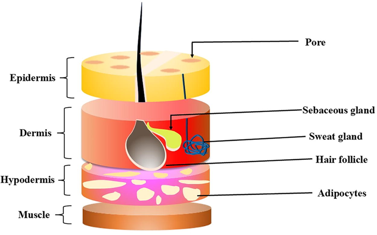
Structure of skin.
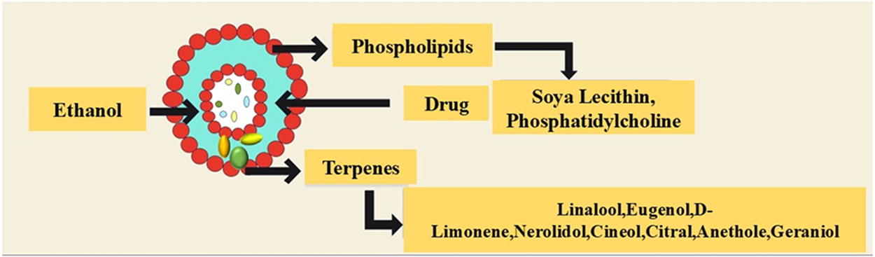
Structure and components of invasomes.
ADVANTAGES OF INVASOMES OVER LIPOSOMES IN TRANSDERMAL DELIVERY SYSTEMS
Liposomes and invasomes belong to the category of vesicle carriers for drug delivery, although they possess distinct properties. Invasomes are therefore formed by unsaturated soybean lecithin, ethanol, and a blend of terpenes and are nanoscale spheres. On the contrary, liposomes are made from phospholipids and cholesterol and this forms spheres that are minute in size. Both types form bilayer lipid vesicles; however, their permeation mechanisms vary: The invasomes penetrate through the SC via the fusion and disruption of both the SC and trans-appendageal permeation, while liposomes penetrate using diffusion, fusion, and lipolysis. Invasomes have higher deformability and elasticity due to the presence of ethanol and terpenes, compared with liposomes. In addition, invasomes have a negative zeta potential value, while liposomes have nearly zero value for the same.
The advantages of invasomes over liposomes include higher flexibility and elasticity, which make them to be better capsules in helping in skin penetration of the drugs. 15 Ethanol and terpenes in invasomes not only contribute to their higher deformability but also enhance their ability to disrupt the SC, thereby improving the drug’s permeation. Because of these characteristics, invasomes are a better option than liposomes for medication administration through the skin. The difference is shown in Figure 3 and Table 1.
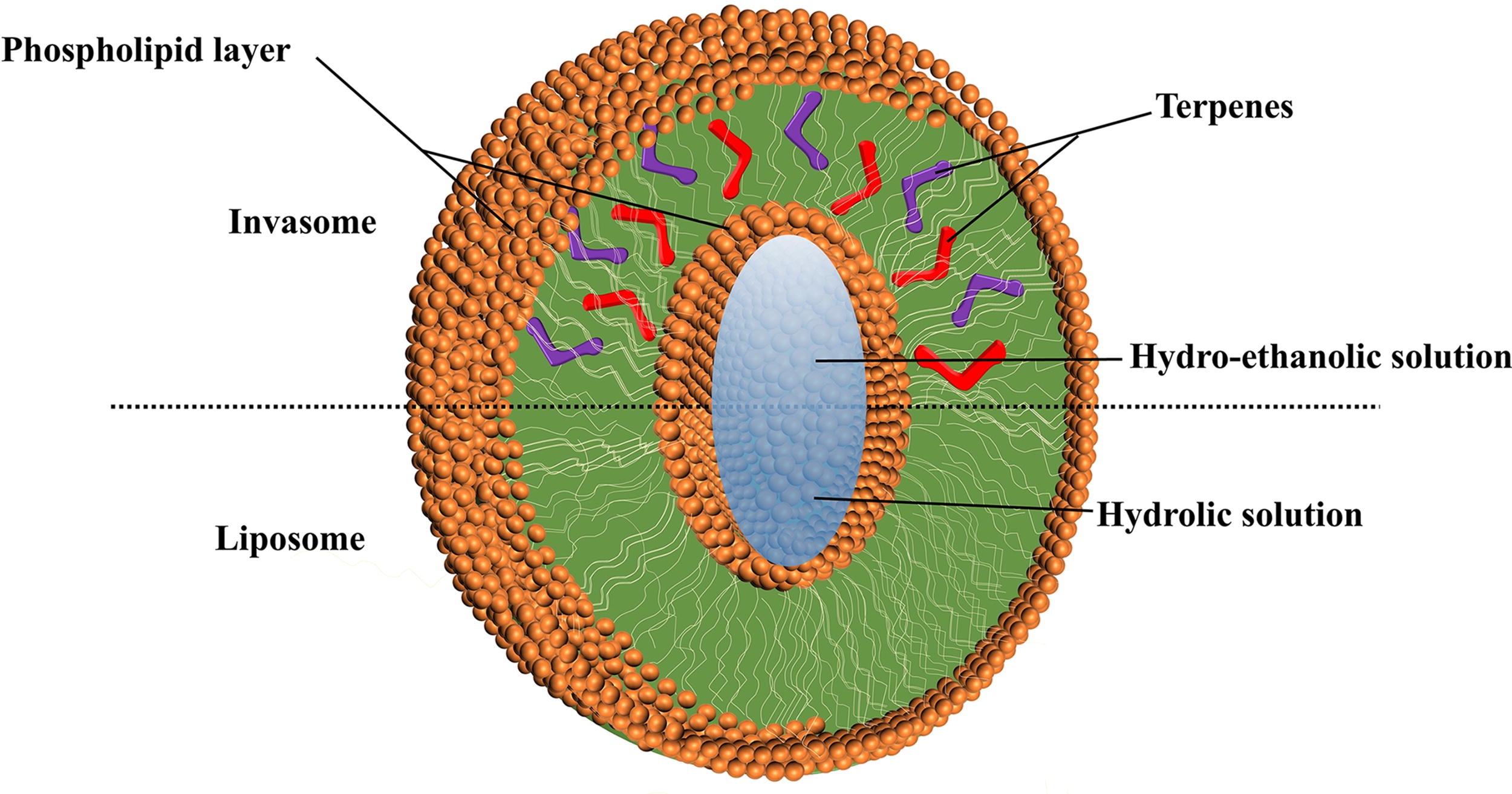
Structure showing invasomes versus liposomes.
Showing the Difference Between Invasomes Versus Liposomes
SC, stratum corneum.
Role of Terpenes and Ethanol in Enhancing Skin Penetration of Invasomes
A class of vesicles known as invasomes significantly improve the transdermal penetration of active pharmaceutical ingredients. 16 Terpenes, ethanol, or terpene mixes are potential carriers that invasomes’ tiny liposomal vesicles used to improve skin penetration. Invasomes exhibit a higher rate of skin penetration than liposomes and ethosomes. Invasomes are a promising alternative for many applications due to their enhanced skin penetration. 17 Invasomes are flexible phospholipid vesicles that include ethanol, phosphatidylcholine, and one or more terpene combinations. 18
Invasomes are a type of vesicular system that enhances the transdermal penetration of active pharmacological molecules. 19 The architecture of these vesicles is primarily composed of phospholipids, ethanol, and terpenes or a combination of various terpenes, which contribute to their structure and functionality. These substances functioned as appropriate transdermal penetrators with strong penetration abilities. 20 Electron spin resonance (ESR), differential scanning calorimetry (DSC), and cryo-electron microscopy have established the great fluidity of phospholipids in invasomes, which is thought to be a crucial dynamic strategy for their strong penetration-enhancing potential. 21 However, an invasome-enhanced skin penetration process involves additional phenomena in addition to fluidity. 22 Invasomes are a vesicular system that enhances the transdermal penetration of active pharmaceutical molecules. Invasomes are specialized, flexible liposomal vesicles that contain small amounts of ethanol and terpenes or mixtures of terpenes. 23 These constituents serve as efficacious transporters, augmenting the capacity for skin penetration. Phospholipids (such as phosphatidylcholine, phosphatidylserine, soya phospholipid, egg lecithin, phosphatidylinositol, phosphatidic acid, and phosphatidylglycerol), terpenes or a combination of terpenes (such as citral, cineole, limonene, and eugenol; 1%–5% v/v), and water are present in these lipid vesicles. 24 An important factor influencing the rigidity or fluidity of invasomes is the composition of their bilayer. Studies have shown that the flexibility and fluidity of a lipid bilayer are directly correlated with the enhanced penetration of invasomes. 25 Highly flexible lipid vesicles, significantly smaller than conventional liposomes, have been demonstrated in numerous studies to easily adapt to external stimuli and pass through skin pores. It takes a certain kind and quantity of edge activators to achieve the best possible vesicle membrane deformation. The most effective and safest terpenes for penetration enhancement are typically those derived from natural sources. Although penetration improvements are effective when applied topically, their skin irritation and toxicity are clinically authorized for only a small number of them. 26 Thus, it is imperative to remember that maintaining the right balance between the toxicity and potency of terpene or terpene combinations for humans is a critical regulatory requirement while developing invasomal formulations. 27
Methods to Enhance Transdermal Penetration
Optimizing transdermal drug distribution with chemical and physical approaches has been the focus of significant research in the last few decades. 28 Physical methods include iontophoresis, which involves applying an electrical charge to the skin to facilitate the penetration of charged particles; electroporation, where a voltage is applied to the skin to temporarily disrupt cell membranes and allow drug permeation; sonophoresis, which uses ultrasound to enhance skin penetration; microneedles, tiny needles that pierce the SC to deliver drugs while minimizing pain and improving compliance; and magnetophoresis, using a magnetic field to increase skin permeability. Chemical approaches include the use of salt formations, drug–ion pairs, eutectic mixtures, chemical PEs, and liposomal vesicles. 29 Liposomal vesicles, in particular, have shown promise when combined with physical methods, synergistically improving drug penetration. 30 In addition, strategies such as SC ablation, which involves removing the outer layer of the skin using techniques such as radiofrequency, laser, microwave, ultrasound, cryoablation, or chemical methods, are also explored for their potential in enhancing drug delivery. Figure 4 shows the process of penetration through SC. By targeting the skin’s barrier characteristics, these methods aim to increase the effectiveness and efficiency of transdermal medication delivery systems. 31

Process of penetration through stratum corneum (SC).
DEFORMABLE INVASOMES: KEY TO ADVANCING TRANSDERMAL DRUG DELIVERY
Terpenes are added to develop invasomes, deformable vesicles that enhance the penetration of active pharmaceutical compounds. 16 Due to their great membrane fluidity, these vesicles are soft. The presence of terpenes and ethanol makes invasomes distinctive. The presence of ethanol and terpenes in invasomes enhances vesicle flexibility, disrupts the bilayer structure of the SC, and promotes penetration. This combination improves the permeability of invasomes. 25 According to Dragicevic-Curic et al., during invasome penetration, terpenes, phospholipid segments, and individual phospholipid molecules are released from the vesicle, enhancing penetration and fluidizing the lipids in the SC. Smaller, nondivergent invasome vesicles remain unaffected throughout their travel through the SC. 20 Unsaturated phospholipids were selected because of their low melting temperature, which results in the development of liposomes in a liquid crystalline thermodynamic condition. 32
The objective of including terpenes is to improve the carrier’s deformability. 33 The decreasing transepidermal osmotic gradient in this application mode can cause deformable vesicles to lose their diffusion driving force. 34 These vesicles can function as PEs, with their lipid bilayers interacting with the SC and altering the intercellular lipid layers. This mechanism of enhanced penetration may be influenced by the drug’s lipophilicity. 35 The ethanol- and eugenol-containing vesicle increases diffusivity, which raises the rate at which invasomes penetrate. By disrupting the skin’s lipid matrix, intact invasome vesicles gradually access deeper dermal layers, eventually reaching the pilosebaceous region. 23 In addition, the wide pores around hair follicles act as channels that enable intact invasomes to localize effectively in the target region, with hair follicles serving as conduits for this targeted delivery. 36 The potential of smaller, more flexible invasomes as carriers for transdermal drug delivery is demonstrated by their capacity to penetrate deeper layers upon SC entry while remaining intact. On the contrary, after SC penetration, larger and more robust invasomes typically break down. 37
ROLE OF ETHANOL, TERPENES, AND PHOSPHOLIPIDS IN OPTIMIZING LIPID NANOVESICLES AND INVASOMES FOR TRANSDERMAL DELIVERY
The Role of Ethanol in Invasomes: Improving Stability and Skin Permeability
The fluidity of the skin’s lipid bilayer is successfully increased by adding ethanol to the formulation of lipid nanovesicles. 38 The enhancement of permeability by ethanol is possible. The specific size, zeta potential, entrapment efficiency, and skin permeability of vesicles in nanovesicular systems are important factors that determine their efficacy. 21 Recent studies show that the size and entrapment efficiency of vesicles decrease with increasing ethanol concentrations. Higher ethanol concentrations result in vesicle dissolution, reducing membrane thickness and consequently decreasing vesicular volume. 12 The net charge of the particles is altered as ethanol interpenetrates into the hydrocarbon chains, resulting in a decrease in the mean vesicle size. Ethanol alters the structure of lipophilic or keratinized domains and lowers the transition temperature of lipids. 20 Ethanol enhances lipid fluidity, making ethanol-based nanovesicles softer and less rigid than liposomes. 39 The negative surface charge of ethanol-containing nanovesicles and the resulting electrostatic repulsion can contribute to increased storage stability. 40
Terpenes in Invasomes: Improving Drug Penetration and Drug Delivery
The majority of transdermal drug delivery system exploratory researchers learned about how terpenes and mixtures of terpenes in very small amounts are also extensively found to be PEs that penetrate the skin to reversibly lower the barrier resistance. 31 Terpenes are considered “generally recognized as safe” due to their minimal risk of causing skin irritation. 23 Their effectiveness in penetrating the skin is attributed to their solubility, their ability to dissolve lipid and protein layers, and the depletion of skin microconstituents. 41 According to research using DSC and X-ray diffraction (XRD), terpenes increase drug penetration by breaking up the lipid structure and closely packed bilayers in the SC. 11
Dragicevic-Curic et al. discovered a synergistic effect of various terpene types on temoporfin penetration. Invasomes containing a 1% mixture of three distinct terpene types (citral, cineol, and limonene) demonstrated higher temoporfin permeability than invasomes carrying 1% citral alone. 42
Influence of Terpenes on the Size and Structure of Invasomes
According to Prasanthi et al., the molecular size and concentration of the added terpene impacted the size of invasomes loaded with finasteride. Nerolidol (molecular size: 222 g/mol)-containing invasomes were between 11 and 13 µm in size. Citral-, limonene-, and cineole-loaded nimesulide-loaded liposomes had vesicle diameters of 194, 216, and 244 nm, in that order. 20
The size of the invasomes and the concentration of terpenes were shown to be directly correlated, with the size of the invasomes increasing as terpene concentration increased, according to an examination of particle size. The size of vesicles containing 0.5% terpenes was 93.0 nm, but those containing 1% terpenes were 124 nm. 42
Terpenes and Invasome Shape: Structural Modifications and Their Implications
Terpenes directly affect the size and characteristics of the vesicles present in invasomes. 41 In the lipid bilayer, terpenes act as fluidizers increasing the membrane fluidity, reducing surface tension causing phase separation, and forming more stable vesicles of different shapes. 43 These changes help enhance drug encapsulation, release, and skin penetration, and therefore terpenes should be incorporated in the formulation of designs of invasomes. Cryo-transmission electron microscopy (TEM) results, consistent with DSC and ESR findings, demonstrated that terpenes affect the shape of invasomes. Besides spherical vesicles, the invasomal dispersions also contained irregularly shaped vesicles. 20
Effect of Phospholipids on Invasome Structure, Stability, and Functionality
Hydrophilic head groups and hydrophobic acyl chains joined by an alcohol backbone make up phospholipids. 44 A wide range of phospholipids are produced by differences in head groups, aliphatic chains, and alcohols. 45 Different sources of phospholipids, such as synthetic (polyethylene glycosylated phospholipids) and natural (soybean or egg phosphatidylcholine) varieties, contribute to this diversity even further. Nanovesicle formulations also involve the use of hydrogenated phosphatidylcholine. 20
Lakshmi et al. (2014) described the development of a transdermal gel containing curcumin. To enhance its solubility, curcumin was combined through cyclodextrin (CD) and hydroxypropyl-β-cyclodextrin. This mixture was then incorporated into invasomes, which were subsequently embedded in a hydroxypropyl methylcellulose gel. 21
INVASOME PREPARATION: METHODS AND APPROACHES
Preparation methods for invasomes typically involve lipid hydration techniques combined with edge activators to enhance membrane fluidity and penetration properties. 46 Commonly used methods include thin-film hydration, where lipid components are dissolved in organic solvents and subsequently hydrated with an aqueous phase containing the drug and edge activator. This process promotes the formation of multilamellar vesicles (MLVs) or large unilamellar vesicles, depending on the subsequent processing steps such as extrusion or sonication to achieve smaller and more uniform vesicle sizes. 47 The choice of lipids and edge activators is critical to controlling vesicle stability, drug encapsulation efficiency, and release characteristics. These preparation methods are versatile and adaptable, allowing for customization to optimize invasome properties for specific pharmaceutical applications in transdermal and systemic drug delivery. Although several methods exist for developing invasomes, the most commonly used are mechanical dispersion and film hydration. 48
Formulation and Preparation of Invasomes via Mechanical Dispersion
The active pharmaceuticals/biomolecule and terpene, or a combination of terpenes, are dissolved in a phospholipid that contains ethanol when using the mechanical dispersion approach. The mixture should be well mixed before being vortexed for 5 min and then sonicated for 5 min to yield a straightforward solution. Next, a syringe was used to add phosphate buffer (pH 7.4), phosphate-buffered saline (PBS), and an appropriate solvent to the mixture for vesicle hydration, continuously vortexing for 5 min. To extrude MLVs, the solution was finally sifted and this process was done multiple times using polycarbonate membranes with pore size ranges of 400, 200, 100, and 50 nm. Mechanical dispersion of invasome is depicted in Figure 5.

Mechanical dispersion method.
Invasome Preparation Using the Film Hydration Method
The conventional film process also can be applied to preparing invasomes. Here, methanol dissolves phospholipids in ethanol with the help of chloroform in the ratio 2:1 v/v. When using a rotary flash evaporator to dry the above-identified mixture to a thin film, the pressure is reduced from 500 mbar to 1 mbar at 50°C. The next process of the applied method is vacuuming the film to 1 mbar, followed by 2 h at room temperature and flushing with nitrogen. Subsequently, the deposited film is hydrated for 30 min at the lipid-phase transition using either PBS (pH 7.4) or a combination of phosphate buffer (pH 7.4) containing ethanol and terpenes, then adding ethanol and either one terpene or a combination of them to get invasomes. After these samples were vortexed, ultrasonicated, and extruded through polycarbonate membranes with 0.4, 1.0, and 3.0 µm pore sizes, the vesicles were sized. The film hydration technique of invasome is depicted in Figure 6.
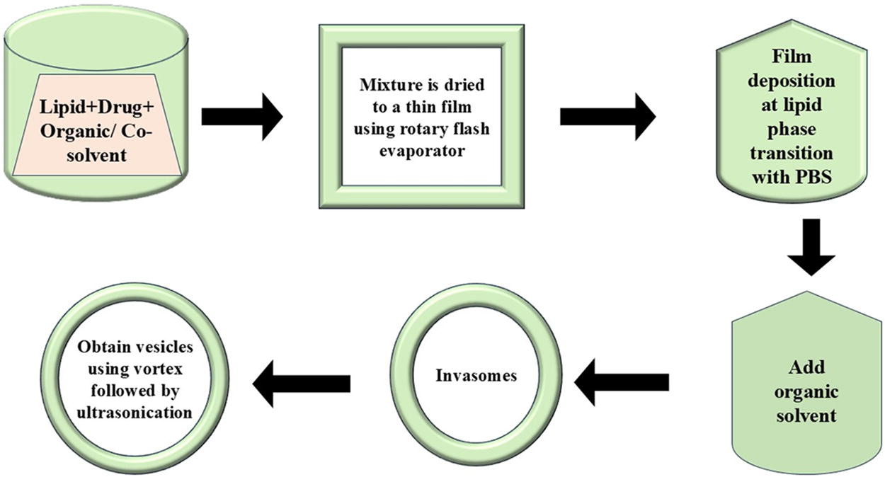
Film hydration technique.
INVASOME CHARACTERIZATION: KEY TECHNIQUES AND APPROACHES
The physical characterization of invasomes is crucial for understanding their structure and performance in drug delivery. Techniques such as dynamic light scattering (DLS) and nanoparticle tracking analysis are used to assess particle size and distribution, which are critical for effective penetration and targeting. 49 Zeta potential measurements indicate surface charge affecting stability and interaction with biological membranes. 50 Morphology is examined using electron microscopy to observe vesicular structure and any changes due to formulation. Encapsulation efficiency tests quantify drug loading, while release studies assess kinetics and control mechanisms. 51 Stability evaluations under varying conditions ensure shelf life and consistency. Rheological properties such as viscosity aid in formulation for optimal application and skin adhesion. 52 This comprehensive characterization guides formulation optimization, enhancing invasome efficacy and suitability across pharmaceutical applications.
TEM in Characterizing Invasomes for Drug Delivery Systems
TEM is one of the most efficient methods for nanomaterial characterization at the atomic and nanoscale (<1–100 nm) up to the micrometer scale, which leads to new opportunities. 53 TEM uses much stronger electron beams compared with scanning electron microscopy (SEM), allowing it to provide more detailed images and reveal additional information, such as crystallographic structure and particle size of a sample. An image is produced by electrons passing through the sample, which are then focused by lenses and captured by a camera system to project the image onto a screen. Since the TEM has a higher resolution compared with the SEM, this technique gives an accurate measurement of the size, shape, and multilayered nature of invasomes necessary for the efficiency of the drug delivery system. 54 Furthermore, TEM helps to depict as to how the Active Pharmaceutical Ingredient (API)s are encapsulated and distributed within the invasomes to ensure homogeneity of the drug delivery system. 55
Characterization of Invasomes for Drug Delivery Using SEM
Similar to a light microscope, SEM uses electrons for imaging; however, its primary advantages are higher magnification (>100,000) and greater depth of field. 56 High-energy, microscopically tiny electron beams are used in SEM to emit various signals at the surface of solid samples. An electron detector collects the emitted electrons from the interacted positions within the scanned surface area as the electron beam scans over the sample’s surface in frame mode. The brightness of the display monitor directly affects the intensity of the electron signal that is emitted and this signal is then stored in a digital image that represents the sample’s surface structure. The ability to obtain precise and detailed images of invasome shape, size, and surface topography is crucial for understanding their interaction with biological membranes and evaluating their effectiveness as drug delivery systems. 57 SEM can also be used to investigate the invasome homogeneity and assess its production standard-compliant quality to determine whether it is suitable for use in pharmaceuticals. 46
Optical Microscopy in Characterizing Invasomes for Drug Delivery Systems
Light microscopy or optical microscopy is the method of using visible light and lenses to create enlarged images of samples. 58 This method is one of the most fundamental approaches used in both biological and material sciences, allowing investigators to closely examine the sample and infer its structure and morphology. 59 The sample is typically illuminated by light, either transmitted through or reflected by the sample and then brought into focus by a lens to form an image. Through the use of optical microscopy, one can be in a position to gauge the size and shape of the invasomes hence meeting the drug delivery standards in terms of the size of the vesicles. It enables the exploration of invasomes’ general morphology, such as their sphere or vesicle-like structure, which is indispensable for drug delivery. The viable method to examine the stability of invasomes is by taking time intervals to observe in vitro changes such as aggregation or degradation using optical microscopy.
Invasome Size and Stability Analysis via DLS
DLS is a process that can determine particle diameter based on Brownian motion and other principles of light scattering of particles in a liquid medium. It can analyze particles ranging in size from 1 nm to 10 µm. 60 The technique is robust for analyzing chemicals and provides quantitative information on particle size, size distribution, and polydispersity index (PDI). These are the parameters that come in handy when performing statistical analysis. DLS accurately determines the size distribution of invasomes and presents size measurements in terms of hydrodynamic diameter, which is essential for confirming that all prepared invasomes fall within the optimal size range for drug delivery. 61
DLS is applied in identifying temporal alterations in the size of particles as a way of measuring invasome stability. 62 Stability studies confirm that each invasome formulation remains stable by ensuring its continued effectiveness during storage and handling. In addition to services, DLS can offer information regarding the encapsulation efficiency of drugs in invasomes. By assessing the particle sizes of the drugs before and after loading into the carriers, changes in the carrier structures can be inferred. Some DLS instruments also measure zeta potential, providing information about the surface charge of invasomes in addition to size analysis. This is especially crucial to the understanding of the overall colloidal stability and tendency toward interactions with biological membranes.
Evaluating Invasome Stability and Interactions Through Zeta Potential Analysis
Zeta potential gives information about the stability and interactions of the invasomes with biological membranes by determining their surface charge. 63 The zeta potential is a measure of the electrostatic repulsion or attraction between adjacent particles with similar charges. To ensure that the dispersion can resist aggregation, high zeta potential is indicative of stability. 64 Typically, a value of ±30 mV is chosen as the threshold to distinguish between stable and unstable dispersion. Zeta sizer can be used to determine the zeta potential of the formulation. The significant negative charge is primarily due to the presence of ethanol that generates a net negative surface charge and prevents vesicle aggregation through electrostatic repulsion.
DSC Analysis for Evaluating Invasomes
DSC is a technique of thermal analysis that deals with studying the thermal properties of materials. 65 It determines the amount of heat transfer that is related to changes in state at the end of a substance as a result of its temperature or time. The present DSC analysis reveals the nature of invasomes, the physical and chemical properties that assist in understanding the appropriate formulation of DSC. Thermal stability is also evaluated under different conditions by checking phase transition and degradation temperatures of invasomes to guarantee their performance. It effectively estimates lipid bilayers-phase transitions, which are crucial for evaluating the mechanical properties and permeability coefficients of invasomes. DSC identifies polymorphic forms of encapsulated drugs that affect solubility, stability, and bioavailability, thus helping in formulation uniformity. The degree of encapsulation efficiency is determined by the thermal characteristics before and after the drug encapsulation process to achieve appropriate dosing. DSC also analyzes compatibility between components, to find out what aspects can cause instability.
XRD for Structural Analysis of Invasomes
XRD is commonly used to determine the crystalline structure, phase nature, lattice parameters, and crystalline grain size of materials. 66 The latter parameter is found for a given sample by applying the Scherrer equation and broadening the peak of the highest intensity XRD measurement. Using XRD techniques yields results that are statistically representative volume-averaged, which is the primary advantage. 67 These techniques are widely applied to powdered samples, usually following the drying of the corresponding colloidal solutions. Similar studies have shown that the presence of an organic molecule with a certain degree of crystallinity is typically indicated by sharp, partial crystalline peaks in the powder XRD patterns of active substances. 68
Evaluation of Invasomal Formulations: Analytical Techniques
The two most widely used techniques for determining the invasomal formulation’s drug concentration are high-performance liquid chromatography (HPLC) and spectrophotometry. 69 Researchers and pharmaceutical scientists may maximize invasomal formulations for a range of therapeutic purposes by using these analytical techniques, which offer valuable tools for ensuring the consistency and the formulations. 12
Gupta et al. (2022) prepared invasomal gel containing ketoconazole as a topical delivery system for the treatment of fungal skin infections. The formulation’s pH, viscosity, gel strength, drug content (98.78 ± 0.32%), extrudability, spreadability, in vitro drug diffusion, and stability studies were all evaluated. In Franz diffusion cell, the ketoconazole-loaded invasomal gel exhibited good skin penetration properties. 70
Evaluation of Drug Entrapment Efficiency
The significant evaluation criterion for the drug delivery system is drug entrapment efficiency. A variety of techniques, including ultracentrifugation techniques, can be used to quantify the efficiency of penetrating drug entrapment. 71
Ahmed et al. (2019) formulated and encapsulated avanafil into invasomes; they then selected the most suitable invasomal formulation to be made into a transdermal film for erectile dysfunction treatment. Centrifugation was used to evaluate the efficiency of the invasomes in drug trapping. The created invasome samples were spun for 45 min at 20,000 rpm and the overlying fluid was harvested. For the determination of the concentration of the pure avanafil, which has not been entrapped, the clear solution recovered at the top of the tube after centrifugation is first filtered, diluted, and then analyzed by HPLC. According to the conclusions drawn in the study, an increase in the role of phospholipids in an invasome enhances its efficiency in drug entrapment. This characteristic of avanafil to be lipophilic facilitated its incorporation into the lipoidal phase of invasomes, which contributed to the improvement of the entrapment efficiency. The probability of avanafil incorporation into the lipid components of invasomes increases with an increase in the composition of phospholipids.
72,73
Stability Studies of Invasomal Preparations: Key Variables and Findings
The primary variables considered during stability studies for invasomal preparations are the vesicle size, shape, PDI, and zeta potential. 36 The PDI and size are measured using the DLS method. Zeta potential is determined using a zeta sizer, and vesicle shape is evaluated using the microscopy techniques of SEM and TEM. 32
The stability properties of tolterodine tartrate invasome revealed that the formulation should be refrigerated (4 ± 2°C) rather than stored at 30°C to prevent significant drug loss. In conclusion, when the invasive formulation is refrigerated during storage, it may become more stable. 14
Singh et al. (2021) prepared an optimized itraconazole-loaded invasomal hydrogel formulation, which was stored at room temperature (28 ± 0.5°C) and at 4 ± 0.5°C. The formulation underwent rapid stability testing during the storage period. Samples were analyzed and their drug concentration and vesicle size were determined at 0, 15, 30, 60, and 90 days of waiting. There was no significant change found in physical appearance, % entrapment efficiency, and viscosity of the hydrogel formulation. 74
Invasome Characteristics as Drug Carriers: Emphasis on Size, Surface Charge, and Deformability
Some of the characteristics of vesicular carriers that make them suitable to be used as drug carriers include the following: size range, surface charge, and membrane properties such as deformability or elasticity. 40 Elasticity is defined as the property of a material by virtue of which it regains its original form after being distorted. It is significantly considerable that invasomes are a form of lipid base vesicles; hence, they have a certain measure of elasticity. 48 They can also be flexible and change their shape depending on the stress applied on them such as when they are applied on the skin. This flexibility is crucial in drug delivery since this provides invasomes the ability to assimilate the shape of the skin and, in so doing, deliver the drug. Deformability would then represent the capacity of a vesicle or particle to deform without breaking. 75 Deformation degree is a characteristic and important parameter of deformable vesicular formulation. In contrast to other vesicular carriers such as liposomes, it has the ability to propagate over the complete SC. The relative abnormalities of the vesicles were determined using the extruder. The vesicular suspension was filtered through polycarbonate filters in the extruder with pore sizes ranging from 50 to 200 nm. After extrusion, vesicle measurement and size distribution were assessed using the Malvern zeta sizer and the DLS technique. 32 The ability of invasomes to deform has also been reported to enable them to penetrate deeper into the skin, enhancing the delivery of the drug to the site of action.
APPLICATIONS OF INVASOMES IN THE PHARMACEUTICAL INDUSTRY
Utilizing invasomes in pharmaceutical applications holds great promise for enhancing drug delivery efficacy and therapeutic outcomes. 14 Invasomes, lipid-based vesicular carriers enhanced with edge activators, facilitate efficient penetration through the skin barrier. This technology enables the encapsulation and delivery of both hydrophilic and lipophilic drugs, allowing for versatile application across a wide range of pharmaceutical formulations. Zafar et al. developed a transdermal invasome (IVM) gel for luteolin (LN) to improve its bioavailability and overcome solubility issues. The optimized LN-IVM gel achieved a vesicle size of 300.8 ± 2.67 nm, high encapsulation efficiency (89.92 ± 1.29%), and a stable zeta potential (−18.2 mV), with spherical morphology verified through Fourier Transform Infrared Spectroscopy (FTIR) and XRD analyses. Compared with a plain LN gel, the LN-IVM gel provided enhanced LN release (91.32 ± 2.95% over 24 h), superior skin flux (5.79 vs. 2.09 µg/h/cm2), and increased permeability. It showed no irritation and achieved 2.38 times greater bioavailability than oral LN suspension. Anti-inflammatory tests revealed a marked reduction in swelling (17.48% vs. 44.77% for plain gel). These results highlight invasome gel as a viable approach to improving LN’s therapeutic delivery. 76 By improving drug stability, bioavailability, and enabling controlled release, invasomes enhance the effectiveness of treatments for various conditions. Their potential extends to targeted delivery systems, minimizing systemic side effects and improving patient compliance. Ongoing research focuses on optimizing invasome formulations, exploring new drug combinations, and expanding their utility in treating systemic diseases, thereby positioning invasomes as a promising tool in modern pharmaceutical strategies. 77
Invasomes for Treatment of Fungal Infections
Gupta et al. (2023) studied the profound effect of ketoconazole invasomes against fungal infection. The results of the study showed that the prepared formulation and the marketed ketoconazole gel at 30 µg/mL exhibited zones of inhibition of 20.14 ± 0.15 and 21.45 ± 0.18 mm, respectively, against Aspergillus flavus. Similarly, prepared formulation and marketed ketoconazole gel at 30 µg/mL exhibited zones of inhibition of 22.12 ± 0.13 and 23.55 ± 0.18 mm against Candida albicans, respectively. This indicted the antifungal potential of the ketoconazole invasomes. 78
Sunitha et al. formulated an invasome (IVS) gel of luliconazole (LZ) to enhance its topical antifungal effects by overcoming limitations in solubility and skin permeability. The optimized invasome gel (LZIVSopt-G3) demonstrated a vesicle size of 139.1 nm, high entrapment efficiency (88.21%), and stable zeta potential (−19.5 mV). The gel showed improved spreadability and a 2.47-fold increase in skin permeation compared with pure LZ gel, with no signs of irritation. In C. albicans-infected rats, invasome (IVS) gel of LZ significantly reduced fungal infection, supporting its potential as a promising topical delivery system for LZ in treating fungal infections. 79
Invasomes for Treatment of Hypertension
Research has been conducted on the use of invasomes for transdermal delivery of antihypertensive drugs. Kamran et al. developed a topical gel containing nanoinvasomes of olmesartan medoxomil, a Biopharmaceutics Classification System (BCS) class II drug. When administered orally, olmesartan medoxomil demonstrated a bioavailability of 28.6%. An in vivo pharmacokinetic study on Wistar rats showed that the concentration maximum (C max) for tablets and nanoinvasomal gel was reached after 2.0 ± 0.22 and 8 ± 0.41 h of administration, respectively. The relative bioavailability after topical application of the nanoinvasomal gel was found to be 115.60% compared with the oral bioavailability. The biological half-life of olmesartan medoxomil increased up to 135% when applied topically compared with oral administration. The application of olmesartan medoxomil topically as nanoinvasive gels, according to the study, improved the drug’s bioavailability and may have required lower dosages. As a result, transdermal delivery might be a better choice than oral delivery. 80
Invasomes in Delivering Antiacne Agents
Acne has been a widespread skin condition globally for a significant period. 81 Because of its anti-inflammatory properties, dapsone, a highly effective pharmaceutical component in the treatment of leprosy, exhibits great promise in the management of acne. The use of phosphatidylcholine and terpenes (limonene, cineole, citral, or fenchone) in the preparation of dapsone-loaded invasomes for the management of mild-to-moderate acne was examined by El-nabarawi et al. The study’s findings demonstrated that invasomes considerably increased dapsone skin deposition, suggesting a possible use for them as antiacne drug delivery systems. In 2016, Duangjit and colleagues synthesized an invasomal formulation containing 0.15% capsaicin using phosphatidylcholine and D-limonene. With sizes less than 100 nm, the optimized invasomes loaded with capsaicin showed a narrow size distribution (0.01–0.30). Moreover, the stability of invasome containing drugs was verified by a negative zeta potential (−20 mV). Furthermore, compared with commercial products and traditional liposome formulations (0.15% capsaicin in ethanolic solution), it offers improved skin permeability. Therefore, enhanced capsaicin invasomes may serve as an alternative for transdermal drug delivery. 82
Invasomes in Delivering Anticancer Agents
Despite all the progress in the field of biomedicine, the treatment of cancer entails several challenges. With a death rate of 4444 per year, it stresses the issues in present therapies and promotes the development of better alternatives. A well-targeting medication nanovesicle allows for delivering chemotherapy to the tumor sites with no side effects but with maximum therapeutic gain. Current breast cancer incidence estimates suggest that 1 in 10 women get these diseases, which is the most common cancer type among women. 83 The advantages of using Transdermal Drug Delivery System (TDDS) for breast cancer therapy include selective delivery of a therapeutic agent directly to cancer cells and attaining the required dose of antitumor medicines at the target location. Vidya et al. (2019) and their team performed new research to evaluate the anticancer potential of invasomal gel-incorporated anastrozole on the MCF-7 (breast) cell line. In postmenopausal state treatment for breast cancer, anastrozole, an anticancer agent, is recommended. The production of anastrozole invasomes, tested using a Franz diffusion cell, led to an enhancement in transdermal flux across the abdominal skin of male Wistar rats. The findings derived from the location bear evidence on the suitability of anastrozole subcutaneous gel for postmenopausal women with breast cancer. The outcomes of this study demonstrated a higher skin deposition of anastrozole when invasomes were used.
A very hydrophobic photosensitizer called temoporfin was reported to be deposited within the SC with the use of invasomes by Dragicevic-Curic et al. By dissolving 1% w/v terpenes (limonene, citral, and cineole) in an ethanolic phospholipid solution (phosphatidylcholine), Sung et al. formed temoporfin-loaded invasomes using mechanical dispersion processing with a 75:25 w/w ethanol:propylene glycol ratio. The resultant mixture then underwent apex treatment and was ultrasonicated for 5 min to develop invasome-based formulations for photodynamic therapy and potential cancer treatments. 84
Invasomes in Treatment of Erectile Dysfunction
Men 40 years and older are most likely to suffer from erectile dysfunction. It can be described as the physiological disorder characterized by the failure to achieve and maintain sufficient genital erection that would be required for adequate sexual intercourses. 85 The medicine to be used orally for erectile dysfunction is avanafil, a PDE5 selective inhibitor. Some of the limitations of avanafil include the following: it has lower oral bioavailability, there is considerable first-pass metabolism, it is poorly soluble in water, and its absorption is more pronounced if taken with food. Hydrogels were not satisfactory due to these problems, and thus, nanosized invasomes containing avanafil were created and designed to enhance the effectiveness of transdermal medication delivery. The vesicular size was measured at 109.92 nm, with an entrapment efficiency of 96.98%. The optimized avanafil invasomal film exhibited enhanced ex vivo permeation, showing an enhancement factor of 2.514 and a more than fourfold increase in relative bioavailability compared with the raw avanafil film. This increase in absorption means the medicines penetrated through invasomes better when formulated in the new and improved formulation. Hence, the use of an invasomal formulation enhances both the penetration and absorption of the active ingredient, avanafil, into the skin. Therefore, it can be applied in different methods for administration of drugs for erectile dysfunction. 72
Invasomes in Treatment of Alopecia
Alopecia areata more commonly known as spot baldness is a dermatological condition that results in hair loss in some or all parts of the body. 86 Following earlier reports, it is said that alopecia areata results from infiltration of T lymphocytes, both the CD4+ and CD8+ cells, into the hair follicles. Therefore, it can be said that the efficacy of alopecia treatments can be significantly improved with innovations in this field. According to the literature, invasomal formulations put accent on its ability to deliver desirable outcomes in the long run. In a recent study, finasteride invasomes were formulated by mechanical dispersion using a mixture of terpenes such as limonene, nerolidol, and carvone at 0.5%, 1.5%, and 1%, respectively. Ethanol had contributed toward finasteride-loaded invasomes to achieve the negative charge on the vesicle surface, which increased the stability of the invasomal dosage form and improved the electrostatic charges. The high rotating energy produced by sonicating the invasomes resulted in high negative potentials and charges. 87
Invasomes in Delivering Vitamin Analogues
Isotretinoin, a vitamin A analog, is used to treat pustular folliculitis, which is characterized by inflammation of hair follicles often associated with eosinophilic infiltration. Using a mechanical dispersion method, Dwivedi and colleagues have recently succeeded in synthesizing isotretinoin invasome; oral administration of isotretinoin has been linked to reduced oral bioavailability and cardiovascular complications. To form a homogeneous suspension, ethanolic egg lecithin was initially incorporated with eugenol, which had been dissolved in isotretinoin, and the mixture was vortexed for 60 min. After that, vesicles were hydrated with PBS (pH 6.8) to produce a translucent, yellowish solution that contained invasomes. This suspension was treated with ultrasonography for three cycles and 15 min, which produced excellent invasomes. Using the aid of a dialysis bag, free isotretinoin was extracted from the invasomes. The study emphasized the importance of these formulation parameters as determinants by highlighting their impact on the invasome penetration rate. Interestingly, the size of the invasome vesicle was unaffected by the eugenol concentration. The single hydrophobic chain of egg lecithin, with its polar head group, induced a highly positive curvature in membranes at higher concentrations of the protein, leading to an increase in the size of the invasome vesicles. In addition, because there were more lipids available to entrap the isotretinoin, the lipid concentration enhanced the isotretinoin’s entrapment efficiency. In addition, the solubility and encapsulation of isotretinoin have been enhanced by the use of eugenol. Similarly, high eugenol and lecithin concentrations demonstrated high invasome vesicle deformability. The presence of the drug in the desired spot is confirmed by confocal laser scanning microscopy, which additionally validates that the invasome reaches the pilosebaceous follicular unit. 88
Invasomes in Delivering Photosensitizer Agents
In photodynamic therapy, a photosensitizer medication is used with light energy in two stages of treatment. 89 When activated by light, generally via a laser, the photosensitizer kills malignant and precancerous cells. The photosensitizer is safe until light causes it to become active. However, the photosensitizer can become harmful to the targeted tissue after light activation. 90 Currently, a variety of photosensitizer medications are available to treat a wide range of conditions, including psoriasis, acne, age-related macular degeneration, and various types of cancer. Photodynamic treatment helps treat viral, bacterial, and fungal infections in addition to these disorders. 91 According to studies, this light-based therapy can activate the immune system, offering the body an additional mechanism to assist in the destruction of malignant and precancerous cells. A highly effective second-generation synthetic photosensitizer is temoporfin (mTHPC). When treating cutaneous malignant or nonmalignant diseases, temoporfin has been proven to be therapeutically beneficial.
Since temoporfin has a poor profile of percutaneous absorption and is highly hydrophobic, it is challenging to administer a low dosage of preparation that has better penetration efficacy. Research on the photodynamic properties of temoporfin was done by Curic et al. (2008). Mice with subcutaneously implanted human colorectal tumors (HT29) were exposed to topical application of the mTHPC-loaded invasomes, followed by photoirradiation, to assess the invasome photodynamic efficacy. To create invasomes, phospholipids, ethanol, and either citral or a combination of citral, d-limonene, and cineole were used as terpenes and PEs.
Invasomes in Psoriasis Treatment
Psoriasis is a clinically heterogeneous, chronic, inflammatory, immune-mediated skin disease with a significant genetic component. 92 Psoriasis can present in various forms, including plaque, flexural, guttate, pustular, or erythrodermic types. It is estimated to affect 60 million people worldwide, with 1.52% of the general population in the United Kingdom being impacted. Treatment for psoriasis must be comprehensive and multidisciplinary due to its correlation with psoriatic arthritis and increased rates of cardiometabolic, hepatic, and psychological comorbidities.
According to a recent study by Maha El-Kayal and colleagues, encapsulating Lut into invasomes has been shown to be more effective than using NLCs (nanolipid carriers) in treating skin inflammatory diseases such as psoriasis. Strong drug release sustainment, good stability, and beneficial colloidal properties were all exhibited by Lut-loaded invasomes. Compared with NLCs, the optimized invasomes showed higher potential to retain Lut in deep skin layers and a comparable in vitro anti-inflammatory activity. 93
Invasomes containing curcumin were reportedly produced according to a study by Bhumika Kumar and associates. A factorial design was used to investigate the effects of terpene type and quantity on the properties of the invasomes. A topical gel formulated using the optimized invasomal formulation was tested for antipsoriatic effects in mice. The optimized formulation exhibited an entrapment efficiency of 85.84 ± 0.56% and a vesicle size of 302.33 ± 1.53 nm. Research done in vivo revealed that the invasomal gel of curcumin helped psoriatic mice recover more quickly and earlier than the traditional curcumin gel. 94
Invasomes in Osteoarthritis Treatment
Ahmed et al. designed diacerein-loaded terpene-enriched invasomes (DCN-TINV) to improve osteoarthritis treatment. Using a 23-factorial design, they optimized formulation variables such as cholesterol content, ethanol volume, and phosphatidylcholine-to-drug ratio. The selected invasomal formulation achieved high entrapment efficiency (89.21%), small particle size (319.75 nm), and stable zeta potential (−55 mV), ensuring controlled release and stability. The optimized DCN-TINV demonstrated enhanced skin permeation (1143 µg/cm2) and deeper skin penetration (144 µm) compared with diacerein suspension (285 µg/cm2 and 48 µm). It also showed significant nociception inhibition (77%), making it a promising, stable, and effective transdermal option for managing osteoarthritis. 95
Invasomes in Melasma Management
Hatem et al. developed three nanovesicular systems—liposomes, PE vesicles, and invasomes, to improve alpha-arbutin delivery for melasma treatment. Using a factorial design, these systems achieved nanosizes between 151.95 and 672.5 nm, negative zeta potentials (−12.50 to −28.20 mV), high entrapment efficiency (80.59%–99.53%), and a sustained alpha-arbutin release over 24 h. Skin deposition studies demonstrated that these formulations increased alpha-arbutin retention by 1.6–1.8 times in the SC, epidermis, and dermis compared with a hydrogel containing the free drug. Clinical evaluations showed that invasome-based hydrogels provided better melasma improvement than both the free alpha-arbutin hydrogel and previous formulations, highlighting their potential as a superior approach for melasma management. 96 Table 2 shows the effects of phospholipids and PEs on various drugs in transdermal delivery systems.
Effects of Phospholipids and Penetration Enhancers on Various Drugs in Invasomal Delivery Systems
CSN, Candesartan; RSV, Resveratrol.
REGULATORY CONSIDERATIONS FOR THE APPROVAL OF INVASOMAL DELIVERY SYSTEMS
Invasomes, a modern type of nanocarrier system for improving drug penetration through the skin, have attracted significant attention in the field of pharmaceutical technology. 14 Their approval process requires a thorough understanding of the regulatory policies concerning safety, efficacy, and quality. This series of evaluations is laid down by the regulatory authorities such as the U.S. Food and Drug Administration and European Medicines Agency. 97 Some of the factors of which are specific descriptions of the composition of the invasomes, stability measurements, and skin penetration and drug release studies both in vitro and in vivo. In brief, toxicological researches are essential to assess the practicability of the compositions based on the biologist’s approval and are valuable for recognizing the capability of the formulas toward skin irritation or sensitization. They must also show the therapeutic relief and risk factors in the intended groups of patients in treating diseases. Documentation for regulatory submissions is highly detailed and includes manufacturing procedures, quality control, and good manufacturing practices. It is equally important to monitor adverse events and report them in the postapproval period as this would help in tracking the safety and efficacy of the invasome-based therapeutics.
CHALLENGES IN ACQUIRING REGULATORY APPROVALS
Some challenges are also brought up by abnormalities in the regulatory approval of invasomes and their production. With respect to nanocarrier analysis, there are no clear guidelines and policies that can be applied to nanocarriers, and thus, a general framework for the analysis of traditional pharmaceuticals and transdermal systems has to be used. This can pose some doubts in the expectations and necessities from the agents of change, which are regulatory bodies in this case. Due to the multifaceted nature of invasome structure and demonstrating modes of action, it demands exhaustive and analytical approaches for satisfying the requirements of the regulatory agencies. 62 Maintaining a manufacturing quality and at the same time showing the scalability of the product are also challenging, with complex formulation undertakings. In addition, the high costs and extended duration required for preclinical and clinical trials present significant economic and organizational challenges. Furthermore, monitoring and controlling potential long-term adverse effects, along with postmarketing safety and risk management, are crucial yet sensitive aspects of regulatory compliance. To overcome these challenges, dialog with the regulatory authorities has to be continuous, more funding for analytical methods, development of more clinical trials, as well as executing them in a more efficient and effective manner to prove the effectiveness and safety of invasome-based products.
Future Prospects
Future prospects for invasomes in transdermal drug delivery are promising, focusing on enhancing formulation stability, scalability, and efficacy across diverse therapeutic agents. Key goals include developing more robust formulations capable of accommodating various drug types, optimizing delivery efficiency, and enabling targeted delivery to specific skin layers or pathological conditions to improve therapeutic outcomes while minimizing systemic side effects. Further research aims to explore combination therapies using invasomes for synergistic effects, expanding biomedical applications beyond dermatology and cosmeceuticals to include systemic diseases such as vaccine delivery and cancer treatment. Safety and biocompatibility studies will also be crucial to ensure long-term use and mitigate potential skin irritation concerns, thus solidifying invasomes as a pivotal technology in advancing transdermal drug delivery.
CONCLUSIONS
Invasomes represent a significant advancement in transdermal drug delivery technology, owing to their superior penetration capabilities attributed to their composition and structure. The inclusion of ethanol and terpenes enhances the flexibility of the vesicles, enabling them to more effectively penetrate the skin barrier. This multifunctionality makes invasomes suitable for treating a diverse range of skin conditions, including dermatitis, psoriasis, acne, hypertension, and even cancer. Ongoing research is focused on refining invasomal formulations to address challenges related to stability, toxicity, and scalability, making their clinical application a highly promising prospect in the near future.
Footnotes
AUTHORS’ CONTRIBUTIONS
D.K.: Conceptualization and writing—original draft. D.S.: Formal analysis. B.D.K.: Visualization. M.K.: Supervision. All authors reviewed the results and approved the final version of the article.
AUTHOR DISCLOSURE STATEMENT
No competing financial interests exist.
FUNDING INFORMATION
No funding was received for this article.