




Abstract
Cytotoxicity assays are essential in the field of research as they enable the examination of cellular responses to stimuli and shed light on complex mechanisms involved in multiple diseases and drug development. This review covers a range of cytotoxicity assays, including trypan blue and MTT (3-(4,5-Dimethylthiazol-2-yl)−2,5-Diphenyltetrazolium Bromide) assays, to more advanced techniques like caspase activity assays, Lactate dehydrogenase release assays, comet assays, and micronucleus assays for DNA damage assessment. Apart from these, other relevant assays like Alamar Blue, Bromodeoxyuridine incorporation, and clonogenic cell survival are also discussed. In this study, significance of these assays in drug development, toxicology studies, and biomedical research is discussed in detail, highlighting their role in ensuring safety and unraveling disease mechanisms. Furthermore, we explore emerging technologies such as chip-based assays, organ-on-a-chip systems, and high-throughput screening, which enhance precision and efficiency in research. Despite these advancements, challenges remain that necessitate standardization efforts and the development of more refined models. In conclusion, this review reflects on the evolving landscape of cytotoxicity assays, finding a balance between traditional methodologies and cutting-edge technologies.
INTRODUCTION
Cell death is a complex process with multiple regulated pathways, each distinguished by unique morphological, biochemical, and functional characteristics. Apoptosis, often termed type I cell death, involves hallmark features such as cell shrinkage (pyknosis), chromatin condensation (karyopyknosis), nuclear fragmentation (karyorrhexis), and the formation of membrane-bound apoptotic bodies. Apoptosis is typically noninflammatory due to the efficient phagocytosis of apoptotic bodies, making it critical for processes like development, immune regulation, and tissue homeostasis. Biochemically, it is characterized by the activation of caspases, mitochondrial cytochrome c release, and phosphatidylserine exposure. 1,2
In contrast, necrosis, historically considered an unregulated form of cell death, is now recognized to include regulated necrosis, such as necroptosis. Necroptosis exhibits feature like cellular swelling, plasma membrane rupture, and organelle dysfunction. It involves receptor-interacting protein kinases (RIPK1 and RIPK3) and mixed lineage kinase domain-like protein. These biochemical markers, along with changes like adenosine 5′-triphosphate (ATP) depletion and lactate dehydrogenase (LDH) release, are used for its identification. Necroptosis frequently leads to inflammation due to the release of intracellular damage-associated molecular patterns. 1,2
Ferroptosis, a distinct regulated cell death pathway, is driven by iron-dependent lipid peroxidation and reactive oxygen species accumulation. Morphologically, it differs significantly from apoptosis and necroptosis, with cells displaying smaller mitochondria, increased membrane density, loss of cristae, and mitochondrial outer membrane rupture. Biochemically, ferroptosis is marked by glutathione depletion, inhibition of glutathione peroxidase 4, and disruptions in iron homeostasis. It can be induced by agents like erastin and sulfasalazine and inhibited by compounds such as ferrostatin-1 and liproxstatin-1. This pathway has been implicated in conditions, including neurodegenerative diseases, cancer, and ischemia–reperfusion injuries. 3
The overlapping characteristics of different types of regulated cell death make it challenging to distinguish between them. Morphological methods, such as transmission electron microscopy, remain a gold standard for detailed ultrastructural analysis. For instance, apoptosis shows chromatin condensation and apoptotic body formation, whereas necroptosis and ferroptosis are characterized by distinct forms of membrane rupture and mitochondrial changes, respectively. Biochemical assays complement morphological studies, utilizing markers like Annexin V for apoptosis and LDH release for necrosis. The integration of advanced imaging and molecular techniques, such as fluorescence microscopy and flow cytometry, offers robust approaches for distinguishing these pathways. 1 –3 Disruption of cellular homeostasis caused by cytotoxic effects is a common precursor to cell death, which may occur in a programmed or uncontrolled manner depending on the stimulus.
Cytotoxicity tests have a tremendous impact on a wide range of biological research domains, each of which gains microscopic insights into how cells behave when exposed to various stimulants. This careful analysis not only reveals possible risks but also acts as a catalyst for the advancement of toxicology, immunology, microbiology, cancer, and drug discovery-based research 4 (Fig. 1). Cytotoxicity tests go beyond their fundamental function in the wide field of pharmacology and become precision designers in drug discovery. 5 These tests aid researchers in identifying drug candidates with low cytotoxicity. This inference simplifies the process of developing new drugs by eliminating the toxic substances at the preliminary stage. These cell toxicity tests are crucial in the field of toxicology. They cover consumer goods, chemicals used in industry, environmental contaminants, and medications. Toxicologists determine exposure limits by interpreting cytotoxic levels, 5 which help them uncover possible risks related to certain drugs. Regulatory bodies use this information to formulate safety regulations protecting public health. These assays are foundations of cancer research, providing insight into how prospective treatments affect cancer cells and directing the creation of therapies to balance therapeutic efficacy with minimizing damage to healthy tissues. 4,5 Cytotoxicity tests are essential for understanding the effect of nanoparticles on cellular viability. 6 They also help to identify the relation between infections and immune cells in microbiology and immunology laboratory research. 7 They shed light on the immune reactions to different chemicals, which enhances the information about the host–pathogen interactions and facilitates the creation of vaccinations and immunotherapies. Each approach in cytotoxicity tests is a unique piece with relevance to a variety of study environments. Conventional tests that offer simplicity and cost-effectiveness in determining the effect of chemicals on cell viability, such as the MTT (3-(4,5-Dimethylthiazol-2-yl)−2,5-Diphenyltetrazolium Bromide) assay and trypan blue exclusion assay, remain fundamental in microbiology and immunology research. With the technology advancement, new cytotoxicity tests are developed, offering improved automation, speed, and sensitivity. These tests not only revolutionize the field of cancer research, microbiology, and immunology but also speed up drug screening and toxicity testing. 7,8 They provide previously unheard-of insights into the complex interactions between cellular responses to changing pathogenic challenges and treatment approaches.
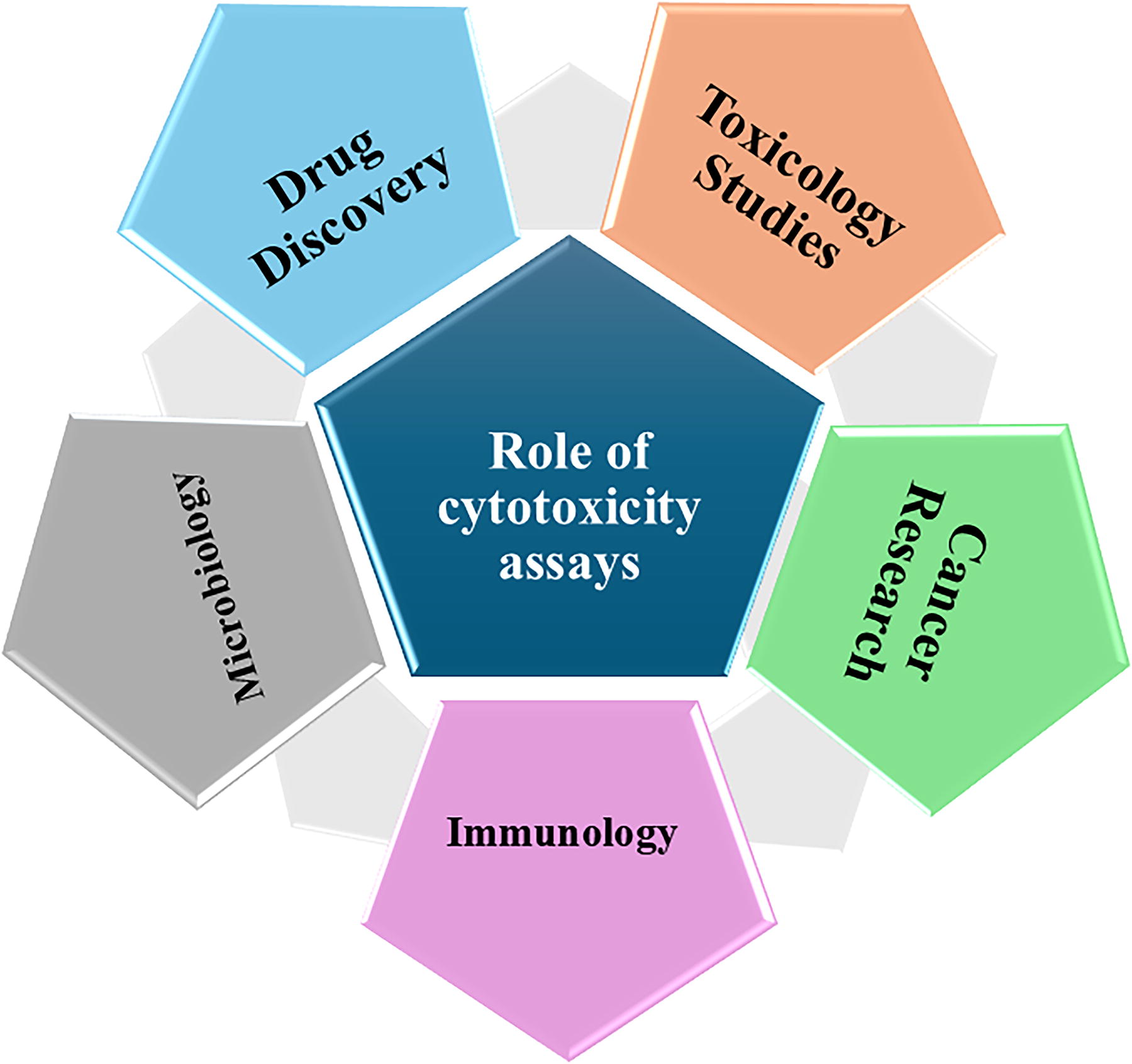
Role of cytotoxicity assays in various fields of life sciences.
Distinguishing between different types of cell death is crucial in various disease contexts, including cancer treatment evaluation, drug development, and inflammation management in neurodegenerative diseases. Cancer therapies aim to induce specific forms of cell death—such as apoptosis, necrosis, or ferroptosis—to eliminate tumor cells, with each type having a unique mechanism. The impact of these cell death pathways on disease progression and treatment responses varies accordingly. 9 In neurodegenerative diseases, neuronal death can occur via apoptosis, necrosis, or autophagy-related processes. Understanding these mechanisms not only deepens our knowledge of the disease but also informs potential therapeutic approaches, such as targeting apoptotic pathways or inhibiting necroptosis for effective treatment. 10 In regenerative medicine and wound healing, the type of cell death plays a pivotal role. Excessive necrosis can lead to inflammation and scarring, which impedes healing, while controlled apoptosis aids in clearing damaged cells and promoting tissue regeneration. In addition, environmental and chemical toxins can trigger different forms of cell death—either apoptosis or necrosis—depending on the cell type. 9 Identifying these mechanisms is essential for risk assessments, public health policies, and the development of targeted treatments. Together, understanding the diverse forms of cell death enhances our comprehension of disease mechanisms and improves therapeutic strategies. Researchers choose cytotoxicity tests influenced by several fields such as microbiology, immunology, toxicology, cancer research, and drug development, in addition to the specifics of the study issue. 8 The selected assay becomes a crucial link that guarantees the experimental results and their applicability and relevance in these ever-changing domains. Finally, cytotoxicity assays highlight the cellular reactions and possible hazards, illuminating and obscuring many aspects of drug development, toxicology, immunology, microbiology, and cancer research. These assays develop in tandem with opening of new technical horizons, offering more accuracy and productivity and advancement of scientific research. 5,7
CELL VIABILITY ASSAYS
Cell viability assays are used to assess the health and viability of cells in culture. It is commonly used to evaluate the cytotoxicity of compounds, the effects of various treatments, and the overall condition of cells. There are several types of cell viability assays, which typically measure cell metabolism, membrane integrity, or enzymatic activity (Table 1). These assays measure the ability of cells to survive, proliferate, and maintain metabolic activity in vitro under various conditions, such as exposure to drugs, toxins, or environmental stressors. Some of the commonly used techniques include MTT, MTS (3-(4,5-dimethylthiazol-2-yl)−5-(3-carboxymethoxyphenyl)−2-(4-sulfophenyl)−2H-tetrazolium), ATP assays, and fluorescent dyes that target specific cellular components or metabolic pathways. Some of these assays include parameters like mitochondrial activity, membrane integrity, or enzyme function. The choice of assay depends on various factors such as cell type, experiment objective, and required sensitivity, ultimately contributing to a comprehensive understanding of cell biology and drug discovery efforts.
Overview of Cell Viability Assays
MTT, (3-(4,5-Dimethylthiazol-2-yl)−2,5-Diphenyltetrazolium Bromide; XTT, (2,3-bis(2-methoxy-4-nitro-5-sulfophenyl)−5-carboxanilide-2H-tetrazolium); ATP, adenosine 5′-triphosphate; Calcein AM, calcein acetoxymethyl; MTS, (3-(4,5-dimethylthiazol-2-yl)−5-(3-carboxymethoxyphenyl)−2-(4-sulfophenyl)−2H-tetrazolium); G6PD, glucose-6-phosphate dehydrogenase; LDH, lactate dehydrogenase.
MTT Assay
MTT assay is a colorimetric methodology widely used in pharmaceuticals especially among cytotoxicity assays. This assay is based on the principle of cellular metabolic activity. 11 MTT is a yellow tetrazolium salt that permeates cell membrane readily. This salt travels to the mitochondria from the outer cell surface. The live cells that possess intact mitochondria contain an enzyme called succinate dehydrogenase (SDH) that converts the tetrazolium salt MTT into a distinctive purple colored formazan product 12 (Fig. 2 ). In contrast, compromised cells with impaired mitochondrial function fail to exhibit this conversion. The formazan product generated by MTT reduction is insoluble and requires the addition of a solubilizing agent like dimethyl sulfoxide. The accumulation of formazan crystals within cells serves as a direct indicator of cellular metabolic activity, which offers a tangible measure of cell viability. The MTT assay is considered a gold standard for cytotoxicity testing, and it presents several advantages, rendering it a favored choice for cell viability assessments. 13 Its simplicity and cost-effectiveness are indisputable, necessitating minimal equipment and reagents. In addition, the assay yields rapid results, facilitating timely evaluations of cellular health. Moreover, its applicability spans both in vitro and in vivo settings, augmenting its versatility across diverse experimental setups. Talking about the limitations, a noteworthy drawback is its inability to differentiate between specific cell death mechanisms, such as apoptosis and necrosis; it may not provide insights into the specific pathways leading to decreased metabolic activity. In addition, it cannot distinguish between dead cells and metabolically inactive cells. 14 MTT assay provides a single end point measurement, limiting its ability to capture dynamic changes in cell viability over time. 12 In addition, the assay’s sensitivity to interferences from factors such as cell culture media composition and incubation conditions can impact its accuracy. The formazan product generated by MTT reduction is insoluble and requires the addition of a solubilizing agent, which can have toxic effect on the cells itself. Despite these limitations, the MTT assay retains its status as a valuable tool for assessing cell viability, particularly in scenarios prioritizing simplicity, speed, and cost-effectiveness. Its widespread utilization in laboratories globally underscores its reliability and utility in the realm of cell biology research. 11,13
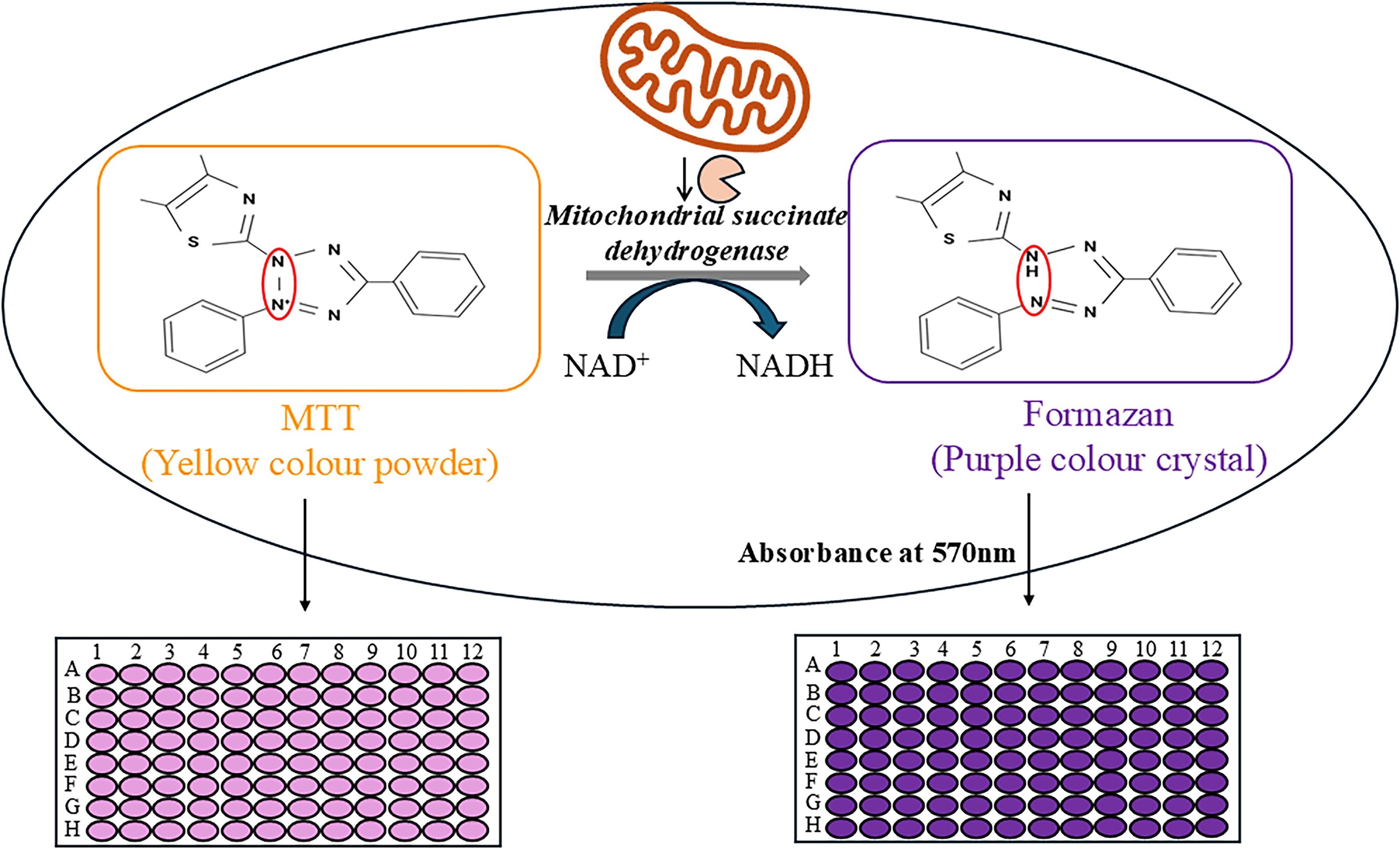
MTT assay: Yellow crystals represent MTT, whereas purple crystals represent formazan formation; the brown organelle in the cell is mitochondria. The figure explains how MTT salt is being taken up by the cells and converted inside the cell (mitochondria) using SDH into MTT formazan crystals. MTT, (3-(4,5-Dimethylthiazol-2-yl)−2,5-Diphenyltetrazolium Bromide; SDH, succinate dehydrogenase.
ATP Assay
The ATP assay is another cell viability assay based on the total ATP levels present in a cell. Since ATP is the major source of energy of all living cells, it can be directly correlated to cell metabolic activity and viability. 15 This assay harnesses the principle of cellular energy metabolism, where viable cells with intact cell membranes and mitochondria retain ATP, while compromised cells with damaged membranes release ATP into the extracellular environment and the stored ATP is quickly degraded. The sample having more ATP concentration indicates high number of living cells. Different detection methods can be used to determine the ATP levels, including colorimetric, fluorescent, and bioluminescent methods. 16 Bioluminescent detection method is generally used in the assay. In bioluminescent-based assays, ATP is converted into Adenosine diphosphate (ADP), which in turn converts luciferin into oxyluciferin with the help of luciferase enzyme. The oxyluciferin is a fluorescent compound that produces yellow light, generating a signal proportional to the ATP concentration. Colorimetric assays, on the other hand, utilize enzymatic reactions to convert ATP to a colored product, allowing for colorimetric detection. 17 The ATP assay offers several advantages, making it a popular choice for cell viability assessment. Some of the important points are its high sensitivity, high applicability, and a long-lasting signal. The assay provides real-time monitoring of cellular response, and it is compatible with all the living cells that use ATP as their major energy source. Despite several advantages, the ATP assay also bears certain limitations. One notable drawback is the sensitivity to extracellular ATP, which can be released from damaged cells or from the breakdown of dead cells. This can lead to an overestimation of cell viability if not properly controlled. 15,17
Calcein AM Assay
Calcein AM assay is a simple and widely utilized fluorometric assay to test cell viability. Calcein AM is a nonfluorescent hydrophobic compound, which is a derivative of calcein. The free carboxylic ends of calcein are fused with AM (commonly known as acetoxymethyl). This assay harnesses the principle of intracellular esterase activity. 18 Calcein acetoxymethyl ester is a cell-permeable dye that readily enters living cells due to its lipophilic nature. 19 Upon entering the cytoplasm, calcein AM is cleaved by intracellular esterase calpain, to release the highly fluorescent calcein molecule. The viable cells with intact esterase enzymes convert the nonfluorescent calcein AM dye into a highly fluorescent calcein product (Fig. 3), while compromised cells with impaired esterase function fail to do so. 19,20

Calcein AM assay: The purple rectangle represents Calcein along with AM (acetoxymethyl) marked in green circle; the AM is cleaved by esterase calpain, and the Calcein gives highly fluorescent signals.
The cell viability and esterase activity can be directly measured by the accumulation of calcein within the cells as the fluorescence intensity is directly proportional to the cell viability. The assay offers several advantages, including rapid results, high sensitivity, and a noninvasive assay. In addition, the assay works with various cell types making it a broadly applicable assay. The limitations include the inability to distinguish between specific cell death mechanisms, such as apoptosis and necrosis. 18,19 Both apoptotic and necrotic cells will exhibit reduced esterase activity, leading to decreased calcein production and potentially misrepresenting cell viability. In addition, the assay’s sensitivity to interferences from various factors, such as cell culture media composition and incubation conditions, can affect its accuracy and reproducibility. 20
MTS Assay
The MTS assay, also known as the “one-step” MTT assay, is a yellow tetrazole that permeates cell membrane readily. The assay does not require the intermittent steps that are done in the MTT assay hence giving the name one-step MTT assay. The MTS is used as a substrate by the enzyme SDH to convert it into formazan in the presence of phenazine methosulfate. 21 The assay is based on the principle of mitochondrial activity or cell metabolic activity, where viable cells with intact mitochondria enzymatically convert the tetrazolium salt MTS into a differentiable formazan product, whereas compromised/dead cells with impaired mitochondrial function exhibit a lack of such conversion. 21,22 The insoluble formazan crystals are made soluble using dimethyl sulfoxide, acidified ethanol solution, or by adding detergent solution like sodium dodecyl sulfate in diluted hydrochloric acid. 21,23 The advantage of the MTS assay is that it is more soluble and nontoxic compared with XTT (2,3-bis(2-methoxy-4-nitro-5-sulfophenyl)−5-carboxanilide-2H-tetrazolium), which allows the reuse of the cells that were taken for testing. Some of the advantages of MTS assay are its ease of use, high sensitivity, and rapid results. The limitation of the assay includes the overestimation of the cell viability, not suitable for suspension cells. The results of the assay can be altered by chemical interference as the assay depends on the mitochondrial metabolism, which can vary cell to cell. The live and dead cell counting is done manually, and it can increase the chances of human error. In addition, the absorbance method used by the MTS assay is less sensitive than fluorescent and luminescent methods. 24
Glucose-6-phosphate dehydrogenase Release Assay
The Glucose-6-phosphate dehydrogenase (G6PD) assay is a sensitive and specific fluorescence-based cell viability method for detecting Red blood cell (RBC) damage caused by G6PD deficiency. 25 This assay hinges on the principle of membrane integrity; the cells with an intact cell membrane possess the G6PD enzyme, where impaired RBC membranes fail to withstand oxidative stress, leading to a compromised and porous/leaky cell membrane, which cannot contain the enzyme and release it in the extracellular environment. The assay measures the release of G6PD, an enzyme crucial for RBC survival, into the surrounding medium following exposure to oxidative stress. In this assay, 6-phosphogluconolactone and nicotinamide adenine dinucleotide phosphate hydrogen (NADPH) are produced when G6PD oxidizes glucose-6-phosphate (G6P) in the presence of nicotinamide adenine dinucleotide phosphate (NADP) (Fig. 4). The extremely luminous resorufin molecules are produced by amplifying the generated NADPH through the diaphorase-cycling mechanism. The released G6PD is then quantified using a fluorometric method, providing a direct measure of RBC hemolysis. The advantages of the assay include high sensitivity, less interference of external factors, and broad applicability. One of the limitations of the assay is that it cannot distinguish between the specific causes of hemolysis, such as G6PD deficiency and other hemolytic disorders. 26 Another disadvantage of this assay is that it requires specialized equipment like plate reader with fluorescent detection capability for experimentation.

G6PD release assay: The Glucose-6-phosphate is converted into 6-phosphogluconolactone with the help of Glucose-6-phosphate dehydrogenase, which in turn converts NADP+ into NADPH; resazurin is given to the enzyme diaphorase, which uses the hydrogen of that NADPH to convert it into resorufin.
XTT Assay
XTT assay is a colorimetric assay that is used to access cell viability based on metabolic activity of a cell. This assay is identical to MTT assay as it covers the same principle as of MTT assay. 11 The XTT is a tetrazolium-based, yellow-colored compound, which is used in the assay. 27 This tetrazolium-based compound is given to the cells that permeates the cell wall and goes into mitochondria, where it gets enzymatically converted by NADH dehydrogenase into a distinctive orange formazan product. But the cells with compromised mitochondrial function (generally dead cells) exhibit a failure in this conversion and do not show any color change. The accumulation of formazan crystals within cells serves as a direct indicator of cellular metabolic activity (Fig. 5). The advantages of the assay include its simplicity and cost-effectiveness, which are indisputable, necessitating minimal equipment and reagents. The assay yields rapid results, enabling timely evaluations of cellular health and its applicability spans both in vitro and in vivo settings, enhancing its versatility across diverse experimental setups. 28 The limitations of the assay include its inability to differentiate between specific cell death mechanisms, such as apoptosis and necrosis. Moreover, the assay’s sensitivity to interferences from various factors, such as cell culture media composition and incubation conditions, can impact its accuracy and reproducibility. Despite these limitations, the XTT assay retains its position as a valuable tool for assessing cell viability, particularly in scenarios prioritizing simplicity, time duration, and cost-effectiveness. The widespread application in laboratories globally underscores its reliability and utility in the landscape of cell biology research. 29
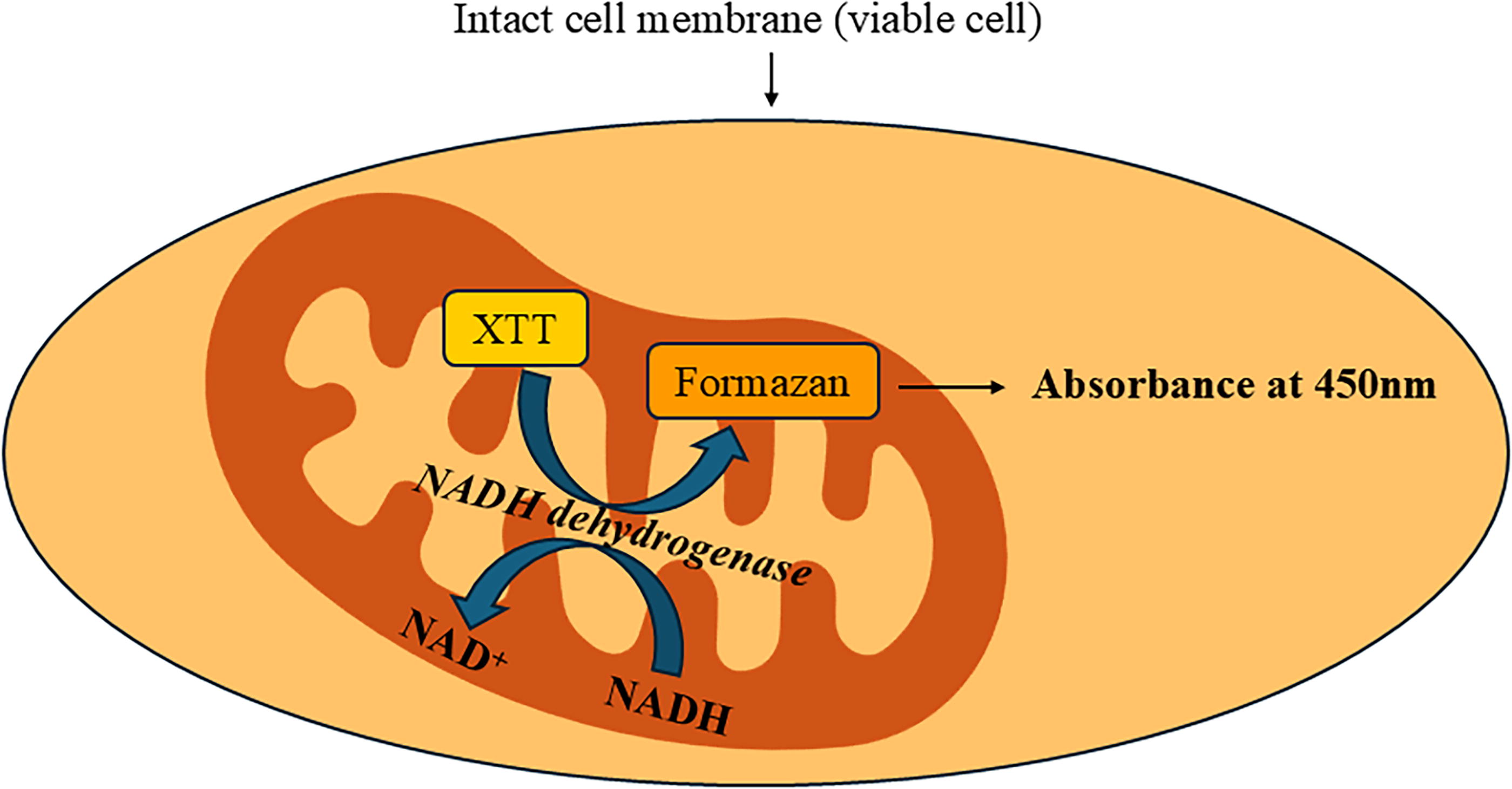
XTT assay: The yellow-colored rectangle represents XTT, which is converted into Formazan (represented as orange rectangle) by the enzyme NADH dehydrogenase. XTT, (2,3-bis(2-methoxy-4-nitro-5-sulfophenyl)−5-carboxanilide-2H-tetrazolium).
MEMBRANE INTEGRITY ASSAYS
Membrane integrity assays are an important part of cell biology as they provide insights into the structural integrity of cell membranes. These types of assays involve the use of fluorescent dyes, for example: propidium iodide (PI) or trypan blue, which selectively penetrate cells with compromised membranes, staining them in the process. Live cells with intact membranes exclude these dyes, thus remaining unstained. The researchers measure the extent of membrane damage by quantifying the proportion of stained versus unstained cells using techniques like flow cytometry or fluorescence microscopy.
Trypan Blue Assay
Within the realm of cell biology, the cell viability assessment assumes paramount importance as it serves as a foundational element in comprehending cellular health, growth dynamics, and responses to diverse stimuli. The trypan blue exclusion assay is one of the assays used to determine the number of viable cells as a straightforward, cost-effective, and widely adopted methodology. 30 This assay hinges on the fundamental principle of membrane integrity, leveraging the fact that viable cells with intact cell membrane do not allow penetration of dye, whereas the compromised and porous cell membrane of dead cells permit dye uptake, resulting in a distinct blue coloration 31 (Fig. 6). Trypan blue is a negatively charged diazo dye with a molecular weight of (approximately) 960 Daltons. The dye cannot penetrate an intact alive cell due to cell membrane integrity. However, during cellular processes such as apoptosis or necrosis, the membrane permeability increases, which facilitates the intake of trypan blue into the cytoplasm. The cell viability testing via trypan blue can also be done by flow cytometry, where the dye can be used to absorb and emit a fluorescence signal at 660 nm. The trypan blue exclusion assay boasts several merits, rendering it a preferred choice for cell viability evaluations. 32 Its simplicity and cost-effectiveness are undeniable, necessitating minimal equipment and reagents. In addition, the assay delivers quick results, enabling timely assessments of cellular health. 32,33 Along with several advantages, the Trypan Blue exclusion assay has certain limitations. One of the main constraints is its inability to differentiate between apoptosis and necrosis, distinct mechanisms of cell death. Both apoptotic and necrotic cells exhibit blue staining, offering no insight into the specific mode of cell death. 33 In addition, the assay’s sensitivity is comparatively modest when contrasted with alternative techniques such as flow cytometry, which can detect smaller populations of deceased cells. In addition, the assay includes manual cell counting, which can lead to human error in cell counting and viability testing. 31,33
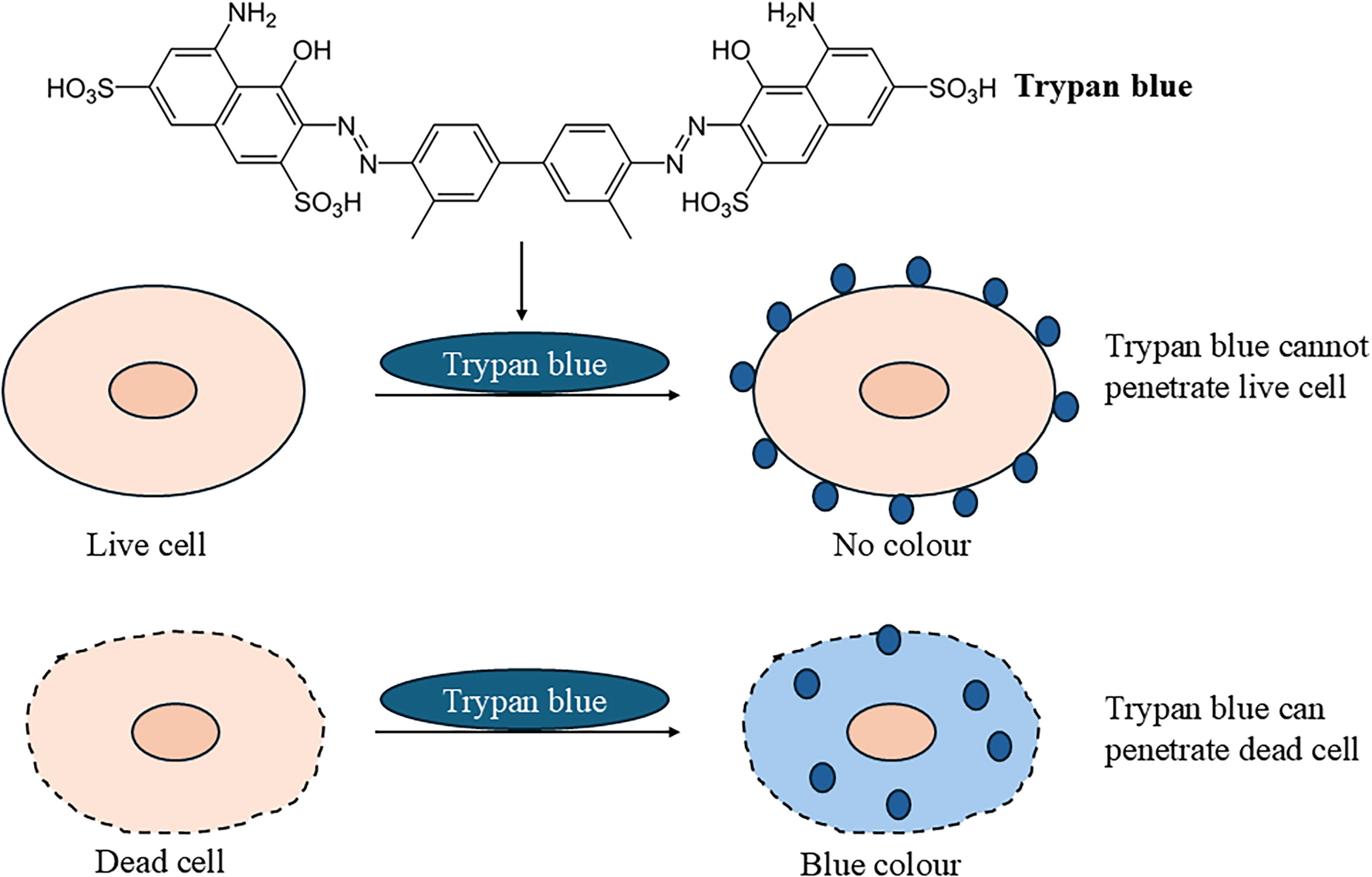
Trypan Blue assay: Blue circle represents trypan blue dye; in the figure, it is unable to penetrate live cell, but can penetrate apoptotic/necrotic cell.
LDH Release Assay
In necrosis, cell viability is compromised due to the disruption of the cellular membrane, leading to the release of intracellular contents, including enzymes such as LDH. 34 LDH assay is used to understand the dynamics of cellular health. It was initially developed for the evaluation of immune cell cytotoxicity. 35 When the cell membrane is disrupted during necrosis, the soluble cytoplasmic enzyme LDH is released into the culture media. Tumor necrosis factor (TNF) family uses the tumor necrosis factor-related apoptosis-inducing ligand receptor and Fas to initiate the necrosis process. 36 TNF activity in various cells is measured using the LDH assay. All five of LDH’s isoenzymes catalyze the same reaction, which is the oxidation of lactate to pyruvate and the reduction of NAD+ to NADH and H+. 37 This two-step procedure methodically reveals the cytotoxic effects caused by various substances or external conditions. 38 During the first stage, LDH reduces NAD+ to NADH while catalyzing the conversion of lactate to pyruvate. Then, using produced NADH, diaphorase enzymes aid in the reduction of a tetrazolium salt (INT) to a red formazan product (Fig. 7). A proportionate representation of the amount of released LDH is obtained by spectrophotometric measurement of the resulting formazan, which is measured at 490–520 nm. Light absorption is directly proportional to the number of lysed cells or cells with impaired membrane permeability. For this reason, pyroptotic cell death is indicated by an early LDH measured. Commercial LDH assay kits are used to measure the amounts of LHD in a sample of cell lysate. The LDH assay can be carried out straight in the cell culture wells and detects mild cell membrane damage without harming the population of healthy cells. 37 Although widely used, the LDH test has drawbacks. Due to its short half-life in the culture medium, LDH places time restrictions on the measurement and detection of cytotoxic effects, which affect sensitivity. Dependence on LDH activity also increases the risk of false positives from endogenous LDH activity, which calls for cautious interpretation and additional confirmatory testing. In conclusion, the LDH assay is still a suitable tool for assessing cytotoxicity and regularly used for immune cell cytotoxicity studies. The investigation of alternate approaches offers potential for a more nuanced knowledge of cellular responses to various stimuli as technology develops. 38,39
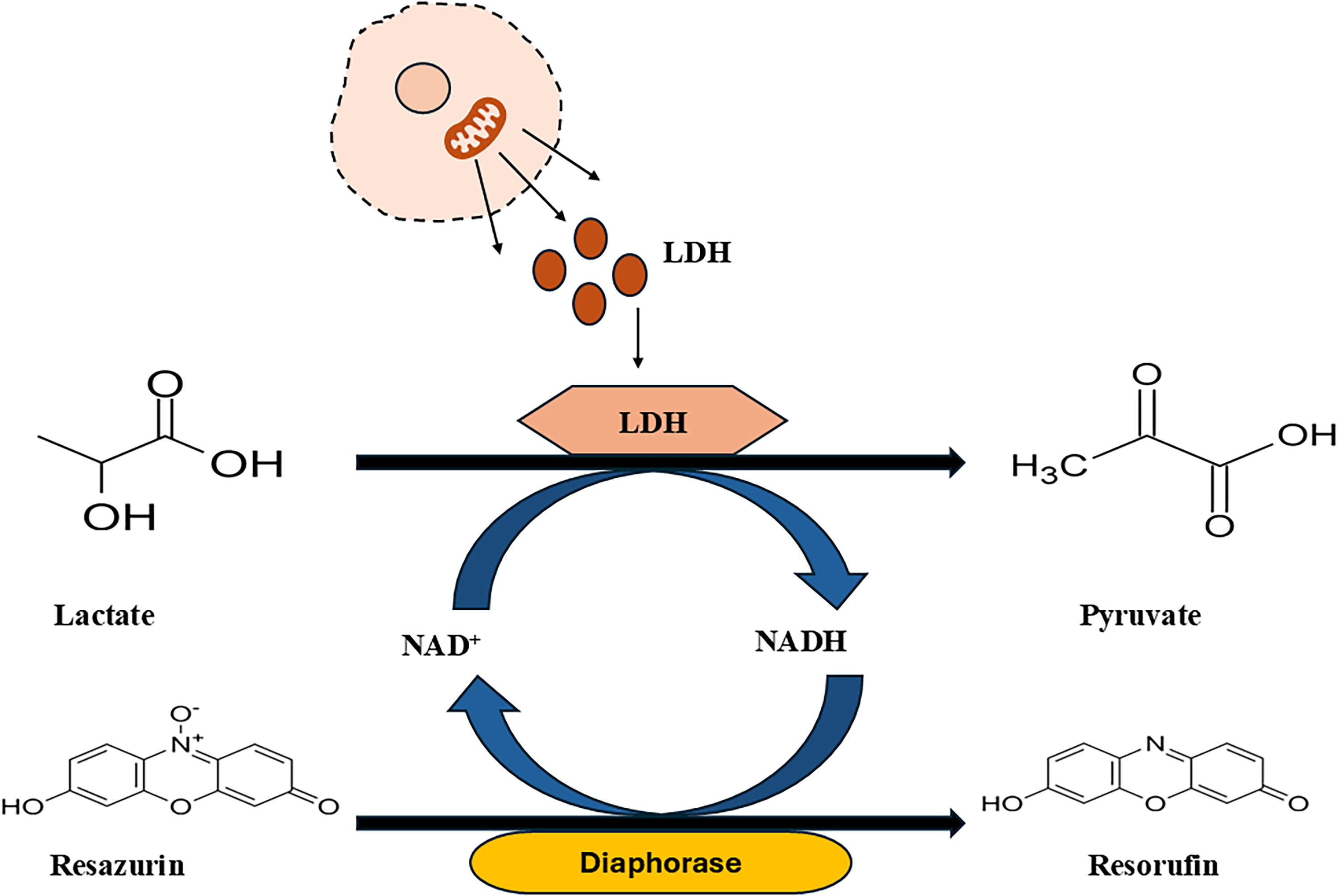
Lactate dehydrogenase release assay: The brown colored shape is the apoptotic/necrotic cell; the cell has lactate dehydrogenase that converts lactate into pyruvate giving hydrogen to NAD+ making it NADH; resazurin is given as a substrate which gets converted into resorufin by diaphorase giving florescence signals.
PI Dye Exclusion Assay
Cell viability is dynamically assessed using a methodological technique known as the PI dye exclusion assay. 40 This assay uses PI, which binds to DNA and shows unique fluorescence properties, in a Krebs–Ringer–HEPES buffer. This binding causes a change in the excitation and emission maxima, enabling a more accurate measurement of fluorescence intensity. Using a 96-well microtiter plate, a typical cytotoxicity screening setup exposes myocytes to different test agent doses. Frequent fluorescence scanning records change over time; higher fluorescence corresponds to cell death and indicates damaged cell membrane integrity. 33 The last step is to use Triton X-100 or digitonin to permeabilize the cells so that a fluorescence measurement representing 100% cell death may be made. The PI assay is not deprived of restrictions, despite its usefulness. Careful attention is necessary to curtail the possibility of test chemicals creating undesirable interference with the fluorescence marker or showing inherent fluorescence. Strict controls are necessary to guarantee test integrity and specificity, especially when working with substances that could naturally glow. In a nutshell, although the PI dye exclusion assay provides a useful real-time evaluation of cellular viability, researchers must carefully consider its limitations. Because the assay relies on fluorescence measurements, it is important to limit any interference carefully to provide accurate and trustworthy interpretation of experimental results. 41
DiBAC4 Assay
DiBAC4 (bis-(1,3-dibutylbarbituric acid) trimethine oxonol) is a voltage-sensitive fluorescent dye extensively utilized in cellular biology to monitor changes in membrane potential. 42 It operates on a very basic principle of potentially dependent accumulation or collection in cells. Due to its negative charge, DiBAC4 cannot cross an intact cell membrane with a normal negative resting potential. However, when the cell membrane depolarizes (becomes less negative inside), the dye can enter. Once inside of the cell, dye binds to the intracellular proteins resulting in a very significant increase in the fluorescence. The gradual increase in fluorescence is then used as a qualitative measure of membrane integrity, which helps in studying apoptosis. 43 However, the reliance on the integrity of the membrane potential can also be a limitation for this dye, as it may not accurately reflect changes in other cellular conditions that do not affect the membrane potential.
DNA DAMAGE ASSAYS
DNA is the primary target following exposure to any stimuli be it ultraviolet (UV) radiation, oxidative stress, or chemotherapeutic drugs. These are direct and sensitive detection of the most detrimental type of DNA damages that happen, which are DNA breaks. When combined with accurate and objective quantification, these tools are a perfect tool for monitoring the delicate balance between DNA damage and repair (Table 2).
List of Cytotoxicity Assays, Including DNA Damage Assays, Functional Assays, and Other Cytotoxicity Assays
BrdU, bromodeoxyuridine.
Comet Assay
Single-cell gel electrophoresis, also known as the comet assay, is a flexible and affordable method used in biomonitoring and genotoxicity assessment. It evaluates the unique DNA methylation, repair capabilities, and lesions of each cell. 44 Based on the principles of electrophoresis, the test induces relaxed DNA strands to move in the direction of the anode, resulting in the formation of a distinct “comet” shape. Lesion-specific enzymes improve adaptability by detecting oxidative damage, methylation, and repair effectiveness. The DNA dispersion of the comet structure quantifies cellular DNA damage. The comet assay measures the amount of damaged DNA in each sperm to provide reproductive insights and assesses genotoxicity and the efficacy of chemotherapy in cancer research. It catches temporary genetic damage without modifying DNA permanently. In the procedure, cells are embedded in agarose on a microscope slide, lysed, and formed into nucleoids with DNA loops connected to the nuclear matrix. For long-term genomic repercussions, comet assay necessitates cautious interpretation since it mostly catches temporary genetic damage. Results might be impacted by environmental sensitivity and possible electrophoresis variability. The limited adaptability stems from the application to certain DNA damage types and reliance on lesion-specific enzymes. The comet assay offers valuable information about single-cell genomic dynamics. Considering the limitations, possible variability and temporal uniqueness must be approached carefully. Comprehending these factors guarantees the efficient and wise implementation of assays in various research scenarios. 45
Micronucleus Assay
The micronucleus assay is a reliable method that can be used in both in vitro and in vivo settings to identify genetic damage caused by chemicals. This test detects micronuclei in the cytoplasm of interphase cells, which include tiny, membrane-bound DNA fragments, such as acentric chromosome fragments without a centromere, entire chromosomes broken by disruption of the mitotic machinery, and chromosomal fragments from DNA breakage. 46 It can be used to assess aneugenic and clastogenic potential of a material, ascertain compound genotoxicity, and measure DNA damage, cytostatic damage, and cell death in a variety of tissue types. Use of this assay in interpreting integrated responses to complex pollutant combinations is very beneficial. Several cell lines, such as Chinese hamster ovary or TK6 cells, primary human peripheral blood lymphocytes, and buccal exfoliated cells (BMNCyt), can be used with the micronucleus test. Several limitations need to be considered, as the robustness of the results might be impacted by the type of cell used in the experiment and the sensitivity to identify micronuclei. Proficiency is essential for correct interpretation because certain cellular formations might have characteristics like micronuclei, but from different sources. Furthermore, the sensitivity of assay to milder types of genotoxicity may be limited by its dependence on the detection of micronuclei. To get significant results, researchers must carefully plan their experiments and evaluate the results with skill. 47
TUNEL Assay
Terminal deoxynucleotidyl transferase dUTP nick-end labeling (TUNEL) is one of the most popular cytotoxicity assays that is used in molecular biology and cell biology to identify and quantify apoptotic cell death in a given population. 48 The assay is based on the principle of DNA fragmentation with exposed 3′-OH groups; as the apoptosis occurs, the DNA starts to fragment within the cell. This fragmentation of DNA is due to frequent single-stranded cuts also referred to as nicks and by the action of nucleases. This DNA fragmentation results in exposed 3′-OH group, which is then labeled with modified nucleotides with the help of terminal deoxynucleotidyl transferase. 49 It is a member of the X-family of DNA bases that is used for nucleotide addition to the free hydroxyl group in the DNA. These modified nucleotides are tagged with a fluorescent molecule or biotin. The fluorescent molecules can be visualized via fluorescent microscopy (Fig. 8). The biotin can be bound to streptavidin conjugated with a reporter to show a signal such as horseradish peroxidase. The general steps for this assay include cell fixation, permeabilization, end labeling/TUNEL reaction, and detection. Some advantages of the assay include its simplicity, sensitivity, and adaptability to various sample types. 50 But the assay has some disadvantages too; one of the major drawbacks is detection of false positive results due to nonspecific labeling. In addition, it does not distinguish between apoptosis and other forms of DNA damage and cell death. Overall, TUNEL assay is an important tool for studying apoptosis and has a lot of applications in basic research, drug discovery, and clinical diagnostics. 51
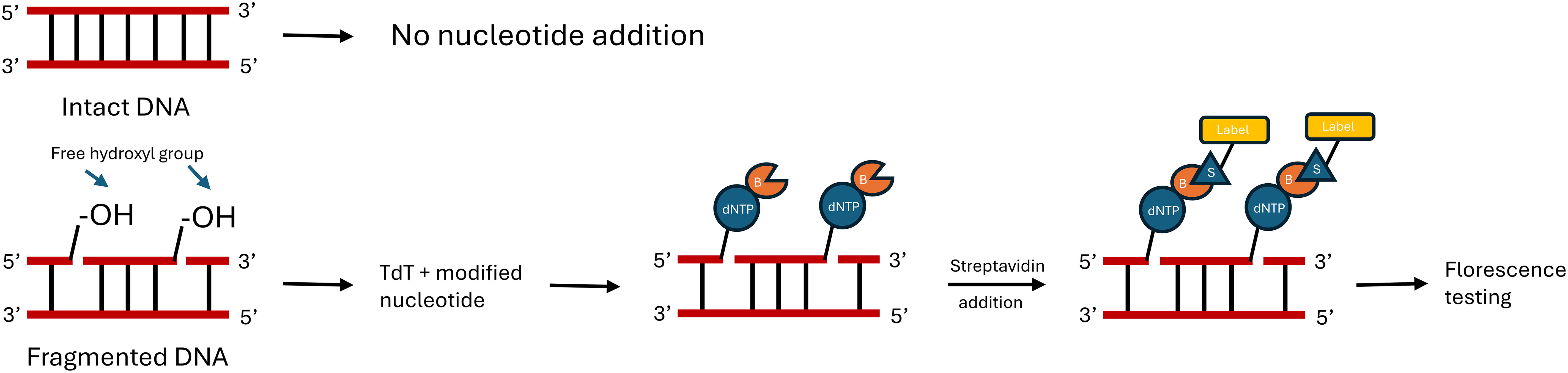
TUNEL assay: The red colored ladder represents DNA; in case of intact DNA there is no nucleotide addition, but when there are nicks in the DNA, TdT enzyme is used to add modified nucleotides that are conjugated with biotin; this biotin molecule, in turn, binds to streptavidin conjugated with fluorescent probe, which in turn is used to detect the DNA damage by detecting the fluorescence level; B, Biotin; S, Streptavidin; dNTP, Deoxyribonucleotide Triphosphate. TUNEL, terminal deoxynucleotidyl transferase dUTP nick-end labeling; TdT, terminal deoxynucleotidyl transferase.
FUNCTIONAL ASSAYS
Unlike traditional viability assays that rely solely on measuring metabolic activity or membrane integrity, functional assays provide insights into specific cellular functions or physiological processes. These assays often target key cellular functions such as proliferation, apoptosis, migration, differentiation, or specific metabolic pathways (Table 2). By evaluating these functional parameters, researchers can gain a deeper understanding of cellular responses to stimuli, screen potential therapeutics, and investigate disease mechanisms.
Alamar Blue Assay
The Alamar blue test is a flexible technique that may be used to assess the cytotoxicity and proliferation of cells in a variety of biological entities, such as yeast, mycobacteria, fungus, and bacteria. Using the luminous blue dye resazurin, this test measures cellular metabolic activity in a very detailed manner. 52 Resazurin is a blue, nonfluorescent chemical when oxidized. When the dye is introduced to cells that are actively dividing, it undergoes a chemical reduction in the growth media, resulting in the formation of the pink, highly fluorescent derivative called resorufin (Fig. 9). This change in fluorescence and color provides a numerical indicator of cellular vitality. 53 Alamar blue test is carried out by carefully controlling the incubation of cells with the resazurin dye. Spectrophotometry or fluorometry is used to measure the color change that results from metabolic processes that decrease the dye as cells multiply. The degree of color change is closely related to the metabolic activity of cells. 54 Prominent benefits of the Alamar blue test include its versatility in handling many kinds of cells, including bacteria, fungus, yeast, cultured mammalian cells, and protozoa. The technology provides researchers with flexibility according to experimental requirements since it supports both colorimetric and fluorometric detection methodologies. 55 In addition, the assay is scalable, which makes it easy to include into high-throughput screening (HTS) programs. Safer to use, the Alamar blue test shows up as a nontoxic substitute for the commonly used MTT assay. This feature not only raises its safety profile but also makes it stand out as a top option in a wide range of biological and biomedical studies where accuracy in determining cell proliferation and cytotoxicity is crucial. 56
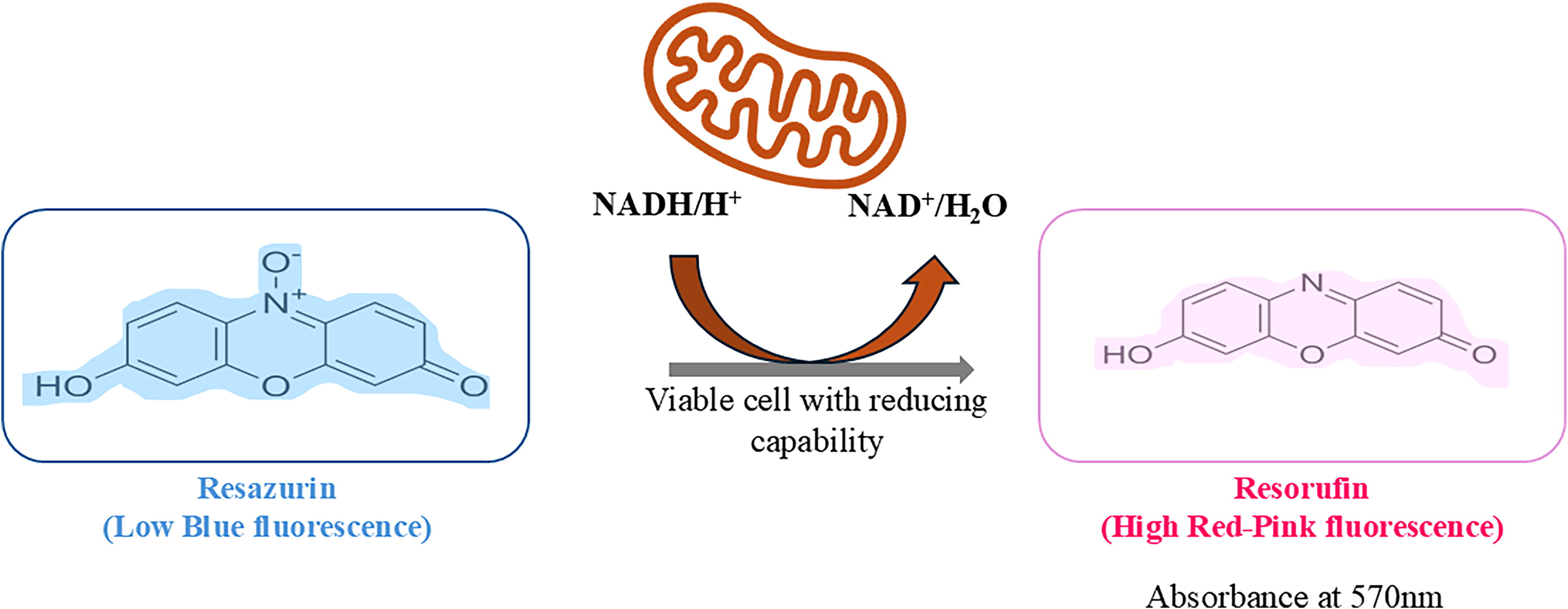
Almar Blue assay: reagent taken up by the live cell and reduces it by giving red-pink fluorescence.
Caspase Activity Assay
The caspase activity assay is a method for studying programmed cell death or apoptosis. Apoptosis is a controlled process where cells self-destruct and caspases play a role by breaking down cellular components. This assay typically uses a substance that can be detected through color changes or fluorescence when it interacts with caspases at their sites. One example of such a substance is p-nitroanilide (pNA), which produces a signal when cleaved. In the case of the caspase 3 activity assay, it uses DEVD-pNA as a substrate specific to caspase 3. When caspase 3 cleaves this substrate, the pNA part is released, resulting in a color change or fluorescent signal. 57 Moreover, the caspase activity assay has evolved to include flow cytometry techniques, which enhance the capabilities of this assay. This integration allows researchers to assess caspase activity at the cell level providing detailed insights into apoptosis within diverse cell populations. Flow cytometry-based assays often incorporate Annexin V, an occurring protein that strongly binds to phosphatidylserine (PS). In cells PS migrates to the layer of the cell membrane serving as a specific marker, for early-stage apoptosis. Annexin V binding along with measuring caspase activity allows for an examination of events related to cell death. 58 Apart from its role in understanding the aspects of cell death, the Caspase activity assay is widely used for analyses. It serves as an indicator of how cells respond to stimuli, such as drug treatments, environmental stressors, and pathological conditions. By quantifying caspase activity, we gain insights into the effectiveness of interventions that target pathways. This contributes to the development and evaluation of treatment approaches. The assays strengths lie in its sensitivity, specificity, and ability to differentiate between caspase activities. However, there are some factors to consider. It is crucial to select appropriate caspase substrates to ensure specificity for the target caspase enzyme. In addition, deciding between fluorometric detection methods depends on factors such as desired sensitivity and available equipment. In conclusion whether used in its fluorometric format or integrated into flow cytometry-based analysis systems, the caspase activity assay stands as an essential tool for unraveling the complexities of apoptosis. Its integration with Annexin V and its role as an assay further highlight its versatility, in both research and therapeutic development contexts. 59
OTHER CYTOTOXICITY ASSAYS
Bromodeoxyuridine Incorporation Assay
The Bromodeoxyuridine (BrdU) incorporation assay is a widely used technique in cell biology. It helps to visualize, track, and measure cell proliferation during the synthesis phase of the cell cycle. In this assay, BrdU, similar to thymidine, is used and replaces thymidine within the DNA molecule. On BrdU treatment, it gets incorporated into the replicating DNA during the cell cycle (Table 2). This assay has applications in studying cell proliferation, analyzing the cell cycle conducting immunochemistry experiments, and exploring neurogenesis. It offers advantages such as real-time analysis of DNA synthesis that can be applied to various types of cells. 60
Clonogenic Cell Survival Assay
The clonogenic cell survival assay is a laboratory test used to evaluate how well cells can grow and survive under stress conditions. It measures the ability of cells to form clusters after being exposed to treatments such as UV light, gamma radiation, or testing drug compounds for their toxicity. By treating the cells with varying concentrations of drugs and observing the number of clusters that form, researchers can assess how effectively the cells are able to multiply (Table 2). Plating efficiency describes the number of clusters originating from a single cell. This assay is widely used in cancer research and radiation biology with applications, including testing sensitivity to radiation studying effects on cells and investigating DNA repair mechanisms. 61 The cell surviving factor can be calculated by taking the ratio of total number of colonies observed to the number of cells seeded.
CYTOTOXICITY ASSAYS IN DRUG DEVELOPMENT
Cytotoxicity assessments play a crucial part in the complex world of drug discovery in deciphering the dynamics of safety and effectiveness related to potential therapeutic options. More than being just gatekeepers, these tests provide a thorough understanding of the substantial effect of medications on cellular viability, which is an important factor to consider when developing treatments that balance safety and efficacy. 4,8 Numerous mechanisms contribute to the complexity of cytotoxicity and each making a distinct mark on the destiny of drug-exposed cells. Necrosis, the uncontrolled rush toward cellular death, is brought on by compounds such as hydrogen peroxide and staurosporine, whereas apoptosis, the planned destruction of cells, is aided by caspases and represented by drugs such as doxorubicin and cisplatin. Intermingled with these, there is a fine balance between survival and death brought about by autophagy, a self-digestive process triggered by drugs such as rapamycin. 62 Refining drug design methodologies starts with understanding these complex systems. The goal is to target selected pathways rather than just generic cell death pathways to reduce side effects and increase the therapeutic index. The ability of cytotoxicity tests to predict outcomes is dependent on the careful selection of models. Standardized cell lines are more reproducible, but primary cells taken straight out of tissues offer the most realistic option since they mimic the intricacies of in vivo settings and allow for a thorough assessment in a variety of tissues and biological situations. 63 Difficulties arise from the translation of in vitro results to in vivo situations due to species-specific variations, pharmacokinetic complexities, and the changing tissue microenvironment. Nevertheless, despite these difficulties, cytotoxicity assays continue to be indispensable instruments, offering a preliminary screening platform and revealing the early signs of possible safety issues. Within this dynamic environment, state-of-the-art technologies surface as game-changers, redefining the parameters of cytotoxicity evaluation. Organoids bring a level of realism that was previously unattainable in traditional models by mimicking the intricacy of organ architecture. Because of their microfluidic settings and multicellular tapestry, microphysiological systems (MPS) imitate a dynamic in vitro microcosm and provide a sophisticated lens to examine the effects of drugs. Within these developments, the investigation of synergies and combination treatments takes front stage, revealing a complex web of therapeutic subtleties. The combination of medications creates a therapeutic symphony, which is meticulously choreographed by precise dose regimens, going beyond just increasing efficacy or lowering toxicity. Potential biomarkers such as staurosporine and hydrogen peroxide serve as early indications of drug-induced adversity in the search for safer therapies. Measurable molecular markers have the potential to improve safety evaluations, which lines with the personalized treatment landscapes and precision medicine principles. 64 Cytotoxicity assays, thus, transcend their role as gatekeepers; they evolve into architects shaping the future of drug development. Each assay represents a methodical exploration, painting a comprehensive portrait of safety and efficacy that transcends the superficial, unveiling the profound intricacies of cellular responses to therapeutic interventions.
CYTOTOXICITY ASSAYS IN TOXICOLOGY STUDIES
In toxicology, cytotoxicity tests play a critical role by providing a thorough assessment of cellular viability and health and by facilitating the identification of potential risks in a wide range of chemicals. These tests are essential for risk assessment, safety evaluation, and regulatory decision-making, ranging from industrial chemicals, consumer goods, and pharmaceutical prospects to environmental contaminants. 63,64 Fundamentally, cytotoxicity tests measure the ability of a substance to cause cell death or disrupt cellular activity. These tests enable researchers to determine possible toxicity and evaluate the effects of different chemicals on cellular processes by giving a quantifiable measure of cellular viability. Setting acceptable exposure limits, assessing the safety of chemicals and medications, and determining how compounds cause their harmful effects all depend on this information. Numerous cytotoxicity tests have been created, each one relevant to a particular research issue and the properties of the chemical. Desired cost-effectiveness, specificity, and sensitivity are examples of selection criteria. The trypan blue exclusion assay, 32 MTT assay, LDH release assay, 35 and flow cytometry tests are examples of common tests. An inexpensive technique for evaluating cell membrane integrity is the trypan blue exclusion assay, which shows that live cells preserve their integrity and do not absorb trypan blue. The MTT assay provides increased sensitivity in measuring mitochondrial activity. The LDH release assay measures the amount of LDH released from injured cells to quickly identify cytotoxicity. Fluorescent dyes are used in flow cytometry assays to offer a thorough assessment of cellular health. Applications for these tests may be found in the consumer goods, pharmaceutical, environmental, and workplace safety sectors. They are used in environmental toxicology to evaluate the toxicity of pollutants and to guide risk assessments and regulatory choices. 7 Cytotoxicity tests are used in occupational toxicology to assess workplace dangers and help establish safety standards and exposure limits. These assays are used in consumer product safety evaluation to evaluate different items. Cytotoxicity tests are essential in pharmaceutical toxicology for assessing the safety and effectiveness of drugs throughout the development phase and for spotting any hazardous effects at an early stage. These assays are widely used, despite the limitations of comparison of in vitro data versus in vivo results, mostly because of species differences. Furthermore, the inability to fully comprehend mechanisms makes it difficult to forecast the toxicity of novel compounds. In conclusion, cytotoxicity tests are essential techniques in toxicology that provide significant information on the toxicity of substances. 64 These tests will continue to develop as our understanding of cellular biology expands and technology progresses, becoming increasingly important in ensuring worker, product, and environmental safety.
CYTOTOXICITY ASSAYS IN BIOMEDICAL RESEARCH
Cytotoxicity tests play an important role in research as they provide valuable insights into the intricate mechanisms through which various substances, such as medications, industrial chemicals, and environmental pollutants, induce cell death or impact cell function. These tests are vital for understanding disease processes assessing the safety and efficacy of medications and identifying treatment targets. 65 By examining the effects of factors such as infections, toxins, or altered cellular processes on cell viability and function cytotoxicity, tests have proven to be immensely valuable in unraveling the underlying mechanisms behind a range of disorders. They have enhanced our understanding of illnesses like influenza, tuberculosis, and malaria by shedding light on how pathogens such as viruses, bacteria, or parasites cause tissue damage and cellular demise. In addition, these tests have contributed to the understanding of the role of stress, inflammation, and apoptosis in chronic diseases such as cancer, cardiovascular disorders, and neurological conditions. 66 In drug development processes specifically related to cancer research, cytotoxicity tests are indispensable as they enable scientists to assess the safety and efficacy of options. A crucial aspect lies in their ability to provide information about a drug's ability to selectively target cells. By identifying chemicals that can effectively kill cancer cells while minimizing side effects on cells, researchers can develop efficient cancer treatments. Cytotoxicity tests shed light on the mechanisms underlying disease states or the effects of drugs, which greatly help in identifying targets for treatment. These tests aid in discovering molecules or signaling pathways that can be modified to achieve benefits. For instance, cytotoxicity tests have played a role in identifying signaling channels that are vital for the survival and proliferation of cancer cells. Consequently, targeted medications have been developed to block these pathways and halt tumor growth. In the realm of cancer research and therapy development, cytotoxicity tests find applications such as screening drugs, evaluating drug combinations, predicting tumor sensitivity to specific medications for personalized treatments, and understanding chemotherapy resistance pathways. These tests facilitate the discovery of drug candidates, assessment of drug combinations, effectiveness prediction of tumor response to medications tailored for individual patient needs, as well as unraveling the mechanisms behind cancer cells becoming resistant to chemotherapy. 67 The impact of cytotoxicity tests on research has been transformative, as they provide insights into substance toxicity reveal disease causes and significantly advance the development of targeted and efficient treatments. The evolving role of these tests continually enhances health protection measures while expanding our knowledge about biology. 68
EMERGING TECHNOLOGIES
Microfluidic-Based Assays
In the field of cytotoxicity assays, the integration of microfluidic-based platforms has emerged as an approach. By leveraging microfluidics, these platforms enhance the precision and efficiency of studies. Microfluidic systems, which manipulate amounts of fluid, are particularly well-suited for assessing cytotoxicity (Table 3). It provides researchers with control to investigate responses in great detail. 69 One key advantage is the ability to perform assays using small sample and reagent volumes. This design feature does not align with green chemistry principles, but addresses practical challenges related to limited or expensive reagents promoting sustainability in experimental workflows. Microfluidic devices possess capabilities for transporting, mixing, and processing fluids in a manner for cytotoxicity assessments. Passive fluid control mechanisms rely on flow resistors and accelerators that mimic forces, enabling manipulation. Active microfluidics incorporate components such as micropumps and microvalves to finely regulate dynamics. These features grant researchers control over conditions ensuring reproducibility and reliability in cytotoxicity studies. Moreover, miniaturizing laboratory processes onto a chip presents a platform for HTS. It allows the evaluation of conditions. This speeds up the process of evaluating how toxic substances are to cells, which is crucial for drug development and studying toxicity. 70 Microfluidic devices create a controlled environment that allows to closely examine the cells respond to substances. By incorporating chemicals into a microfluidic device and creating precise concentration gradients over time and space, we can conduct more sophisticated cytotoxicity tests. This is especially useful for understanding the localized effects of drugs or environmental factors on cell viability. Furthermore, microfluidic systems help us explore how cells behave under confined conditions and the forces they experience. This gives us insights into their behavior in microenvironments that are relevant to our body conditions. In cancer research, this knowledge is particularly important for understanding how potential treatments selectively kill cancer cells while sparing ones. In summary, combining microfluidics with cytotoxicity tests opens up frontiers in research. The ability to control dynamics that precisely use amounts of reagents and perform HTS makes microfluidic-based cytotoxicity assays invaluable tools, for advancing drug development, toxicology studies, and our overall knowledge of how cells react to different substances. 71
Overview of Emerging Technologies
Organ-on-a-Chip Systems
Organ-on-a-chip system has become increasingly important as platforms that mimic the structure and functions of organs in laboratory settings. These scale engineered devices incorporate living cells into designed microenvironments replicating the intricate tissue architecture and physiological responses of organs. 72 By offering control over biochemical cues, organ-on-a-chip system creates a more realistic environment for studying how cells behave and respond to stimuli (Table 3). These systems have implications for drug testing, disease modeling, and personalized medicine, bridging the gap between laboratory tests and real-life studies. 73
High-Throughput Screening
HTS is a method that uses automated equipment to evaluate the biological activity of many samples, at different levels, such as model organisms, cells, pathways, or molecules. Typically, HTS involves testing 103–106 molecule compounds, including things such as chemical mixtures, natural product extracts, oligonucleotides, and antibodies. The main goal of HTS is to identify compounds called “hits” that have biological activity. 74 Key elements of HTS include designs that are compatible with automation, robotic assistance for sample handling, and automated data processing. This approach is widely used in the pharmaceutical and biotechnology industries to identify hits and lay the groundwork for optimization in drug discovery and development (Table 3). The screening process is often carried out in microtiter plates with formats such as 96-, 384-, or 1536-well plates. Quantitative HTS (qHTS) is a version of HTS where compounds are tested at concentrations to generate concentration–response curves immediately after the screening process. qHTS has gained recognition in toxicology studies since it provides a comprehensive understanding of the biological effects of chemicals. Furthermore, using qHTS aids in reducing the occurrence of negative outcomes in the screening findings, thereby improving the precision and dependability of the evaluations. 75
CHALLENGES AND FUTURE PERSPECTIVES
Challenges
Although crucial in many domains, cytotoxicity assays have a number of difficulties that affect their efficacy and consistency. A primary obstacle is the absence of uniform procedures among various laboratories and establishments. The lack of consistency in methodological approaches makes findings difficult to replicate and compare, which presents a serious problem for enterprises and researchers who depend on cytotoxicity data.
Current approaches frequently fall short when it comes to accurately forecasting in vivo results from in vitro data. The translational gap is caused by pharmacokinetic changes, species differences, and the intricate tissue microenvironment. To close this gap and provide a more precise prediction of cytotoxic effects, novel strategies that more closely resemble in vivo circumstances are needed 76 ; it is possible that the current assays fall short of capturing all the subtleties of cell responses in various tissues. The selection of cellular models has a major impact on the predictive ability of cytotoxicity tests, and there is still a need for more representative models that capture the intricacy of in vivo settings. 77 To bridge the translational gap, scientists have created cellular models and assays that more accurately mimic in vivo circumstances, considering variables such as tissue microenvironments, species variations, and pharmacokinetics. Utilizing organ-on-a-chip systems, three-dimensional (3D) cultures, and humanized models can greatly increase the predictive accuracy of cytotoxicity tests.
Future Perspectives
It is expected that advancements in cellular models would be crucial in enhancing the applicability of cytotoxicity tests. The development of organoids, MPS, and 3D cell culture methods has made it possible to simulate in vivo circumstances more accurately, improving our comprehension of how drugs react in intricate biological settings. 78 3D cell culture has improved physiological relevance making it possible to study drug responses, disease mechanisms, and cellular behaviors in a more realistic setting. Because organoids can simulate the structure and functions of particular tissues, they are useful for high-throughput drug screening and personalized medicine. 79 Their disadvantages, however, include increased expense, technical difficulty, and size and compositional variability, all of which can affect reproducibility. Larger organoids also have restricted oxygen and nutrient diffusion, leading to necrosis. 80 These drawbacks show how important it is to weigh the benefits of 3D culture systems against the real-world difficulties they present when designing experiments. In addition, recent research has started to determine the potential and constraints of zebrafish for pharmacology, toxicology, target identification, drug screening, and disease modeling. Zebrafish are unique among model systems that can be screened because of their highly conserved integrative physiology. Zebrafish have identifiable organ systems, such as livers, hearts, kidneys, pancreases, and so forth, in contrast to yeast, worms, or flies. Despite some significant variations brought about by adaptation to aquatic life, the majority of zebrafish organs function similar to those of humans and have well-preserved physiology. 81 To overcome current constraints, integrating cutting-edge technologies such as HTS and microfluidic-based assays offers great potential. A more dynamic cellular environment may be created by simulating physiological fluid flow using microfluidic systems. When combined with cutting-edge analytical instruments, HTS allows for the real-time monitoring of several cellular parameters, providing an abundance of information for thorough cytotoxicity evaluations. 82 Subsequent investigations must concentrate on creating more accurate models that consider individual differences in medication response. Approaches to personalized medicine that adjust cytotoxicity tests to patient-specific parameters might be a big step forward and guarantee a more precise evaluation of medication safety and effectiveness.
CONCLUSION
The wide range of cytotoxicity tests discussed in this article reflects the landscape of research and drug development. The various types of cell viability tests such as trypan blue and MTT assays along with advanced functional tests like caspase activity assays provide a comprehensive set of tools for assessing the health of cells and their responses to different stimuli. 11,15 By exploring assays that assess membrane viability, DNA damage, and overall cellular function, we enhance our ability to study processes and better understand how substances affect cells at a molecular and genetic level. Incorporating assays like Alamar Blue and Clonogenic cell survival assay enhances our understanding of how cells behave and respond in environments. 52,55 The significance of cytotoxicity tests in drug development, toxicology studies, and biomedical research cannot be overstated. These tests serve as gatekeepers by allowing for evaluation of drug candidates to ensure safety while also unraveling disease mechanisms. They have applications ranging from occupational toxicology to the development of safer and more effective cancer therapies. As we enter the era of emerging technologies, assays based on organ-on-a-chip systems and HTS mark a new phase in cellular research. These technologies present platforms for studying cytotoxicity in realistic environments, which offer improved predictive capabilities and expedited drug development. 62 However, despite these advancements, there are still challenges that need to be addressed requiring efforts for standardization and the creation of accurate models. Future research should focus on refining these tests addressing issues related to translatability and establishing benchmarks. In addition, exploring the synergies between tests and cutting-edge technologies could provide an understanding of cytotoxicity. 76 In essence, navigating through cytotoxicity tests involves delving into the complexities of reactions starting from assessments of cell viability to the frontiers of microfluidics and HTS.
Footnotes
ACKNOWLEDGMENT
The authors have used Microsoft PowerPoint to prepare the figures.
AUTHORS’ CONTRIBUTIONS
K.S.S.: Data curation and writing—original draft. P.B.: Data curation and writing—original draft. K.B.: Writing—original draft. S.S.: Data curation. S.C.: Conceptualization, writing—original draft, supervision, review, and editing.
DISCLOSURE STATEMENT
The authors declare no competing interests.
FUNDING INFORMATION
The authors thank the Department of Biotechnology, Govt. of India for the received Grant No. BT/PR40267/BTIS/137/67/2023.