
Abstract
Previous studies have shown that males who have AIDS are more frequently affected by infectious diseases than females. The esophagus is the organ in the digestive tube that is more commonly affected by opportunistic infections during the syndrome. The aim of this study was to assess the influence of AIDS and of gender on local immunity of the esophageal epithelium. Fragments of the esophagus from 29 autopsied women and 37 autopsied men were collected at a university hospital from 1980 to 2009 and were divided in groups with and without AIDS. The IgA-, IgG-, and IgM-positive cells and Langerhans cells (LCs) were immunostained, respectively, with anti-IgA, anti-IgG, anti-IgM, and anti-S100. The software Image J was used to measure the esophageal epithelium and to count the epithelium cellular layers. Patients with AIDS, apart from gender, showed an increase in IgA-, IgG-, and IgM-positive cells and a reduction of Langerhans cells, in thickness and in number of cellular layers in the esophageal epithelium. However, among individuals with AIDS, men presented lower secretory expression of IgA-, IgG-, and IgM-positive cells than women and more intense reduction of LCs. Women have naturally presented better local esophageal immunity than men. Although AIDS possibly causes immunological and morphological alterations in the esophageal epithelium in both genders, women have better esophageal immunity, which may explain a greater frequency of hospital admissions due to infection of men with AIDS when compared with women.
Introduction
M
Analyses of local esophageal immunity in patients with AIDS may favor an understanding of an attack in this organ during the course of the disease, since the nature of the mucous epithelium seems to be an important mechanism of infection and neutralization of HIV on these surfaces. 3
Infection by HIV induces the activation and the dysfunction of the B cells, resulting in hypergammaglobulinemia. 4 –6 Because of this, serological levels of IgA, IgG, and IgM are significantly higher in these patients. 7 The immunoglobulins in mucosas are the first line of immunological defense against infections due to their neutralizing action. 3 However, the role of humoral immunity in the control and clinical progression of HIV infection is still discussed and controversial 8 since information about the expression of B cells in mucosas of patients with HIV is scarce.
The Langerhans cells (LCs) also contribute to the local immunity of the esophagus, being present in most of the stratified squamous epithelium. 9 After contact with the antigen, the LCs are activated and become mature antigen presenter cells. 10 Studies indicate a reduction in number and possible morphological alterations of the LCs in the esophageal epithelium of patients with AIDS. 9,11
The morphology of the esophageal epithelium, especially its thickness, may be related to the onset of esophageal diseases, since the epithelial cells constitute a physical barrier, important to the defense against pathogens. 12 It has been suggested that AIDS may lead to a reduction in the thickness of the cervical 13 and esophageal 11 epithelium.
Studies carried out with hospitalized HIV patients reveal that men have longer and more frequent hospital admissions due to infectious causes than women. 14 On the other hand, women with AIDS are more commonly affected by noninfectious causes, especially by those not related to opportunist infections. 14 –17 Experimental and clinical evidence suggest that immune reactivity is more enhanced in females than in males and lymphocytes and monocytes from females show higher antigen-presenting activity and mitogenic responses. Females have higher immunoglobulin levels than males, an enhanced antibody production to both primary and secondary antigen stimulation. Because of this, males are more prone to infections. 18
The aim of this study was to assess the influence of AIDS and gender on the local immunity of the esophageal epithelium through the quantification of IgA-, IgG-, and IgM-positive cells, LCs, and the mensuration of its thickness with the determination of epithelial cellular layers as well as the evaluation of the mean cellular diameter of the epithelium.
Materials and Methods
This study was approved by the Triângulo Mineiro Federal University Research Ethics Committee on 06/08/07 under protocol number 902.
Esophageal postmortem samples of 29 women and 37 men, aged between 18 and 54 years old, autopsied by the General Pathology Division of the General Hospital of Triângulo Mineiro Federal University, in the state of Minas Gerais, Brazil, were collected from 1980 to 2009. The esophagi observed in this study were recovered from an archive in the General Pathology Division, and were transversally sectioned where the epithelium was preserved.
AIDS diagnosis was made by evidence of at least one AIDS-defining disease presented by the patient, and all patients without AIDS were HIV negative. In this study, the causes of death were grouped into cardiovascular, infectious, digestive, neoplastic, and others. 19
The samples were divided into four groups: women without AIDS (n = 16), women with AIDS (n = 13), men without AIDS (n = 15), and men with AIDS (n = 22). Cases concerning incomplete autopsy reports and patients who had Chagas' disease or cirrhosis were excluded.
Data concerning age, body mass index (BMI), cause of death, and alterations in the esophagus were collected from the autopsy reports, whereas the use of antiretroviral therapy and the values of the last viral load tests and CD4+ T lymphocyte cell counts were collected from medical records. Patients with and without AIDS were matched according to age and BMI.
The fragments were subjected to histological processing and then 4-μm-thick serial sections were cut for histochemistry and immunohistochemistry procedures.
Immunostaining of IgA-, IgG-, and IgM-positive cells and LCs was performed, respectively, with anti-IgA, anti-IgG, anti-IgM (Erviegas), and anti-S100 (DAKO) in concentrations of 1/1000, 1/1000, 1/800, and 1/400, respectively. The antigen retrieval was performed with trypsin buffer for IgA-, IgG-, and IgM-positive cells, whereas citrate buffer was used for LCs. All antibodies were incubated for 2 h (room temperature); the LSAB + System-HRP (DAKO) was used in the process of revelation and diaminobenzidine (DAB) was used as substrate-chromogen.
The quantification of IgA-, IgG-, and IgM-positive cells stained by immunohistochemistry was carried out using the KS-300 (Kontron-Zeiss) system and the results were expressed in percentages in each field.
The LCs positively stained by immunohistochemistry were counted in the whole extension of the esophageal epithelium, and were expressed as number of cells/area (mm2). The total number of LCs was determined in each field. The mature LCs presented apparent irregular shaped dendritic processes, and the immature LCs or cells with altered morphology exhibited a round shape and a reduction in the number and size of dendritic processes. 9,20 Accordingly, the percentage of immature LCs or cells with altered morphology in each field of the whole extension of the esophageal epithelium was determined.
Hematoxylin and eosin (H&E) staining was performed so as to provide morphometric analysis of the thickness and number of layers in the esophageal epithelium. For the morphometric analysis, images of the fields were captured using a video camera coupled to a light microscope. The measurements were carried out through interactive image analysis software (Image J). This software enabled the performance of five measurements in each field of the epithelium, by drawing straight lines. Below each line, the cell nuclei were counted in order to determine the number of cell layers in the epithelium. The cell average diameter was obtained by dividing the thickness by the number of nuclei in each measure. In each case, the whole extension of the esophageal epithelium was captured and quantified and the number of fields captured in each esophageal fragment ranged from 17 to 39.
Statistical analysis was carried out with SigmaStat 2.03 software. In variables with normal distribution and similar variances Student's t-test was used; otherwise, the Mann–Whitney test (T) was utilized. The differences in which the p-value was less than 5% (p < 0.05) were regarded as statistically significant.
Results
The majority of patients with AIDS, 33 (94.28%), regardless of gender, died of infection. In one (2.85%), the cause of death was cardiovascular and in one (2.85%) it was associated with other causes. With respect to patients without AIDS, irrespective of gender, the frequency of causes of death from digestive, cardiovascular, infectious, neoplastic, and other causes was, respectively, 1 (3.22%), 9 (29.03%), 11 (35.48%), 8 (25.80%), and 2 (6.45%).
In the group of patients with AIDS the T CD4+ lymphocyte count converged to 43 (11–217) (cells/mm3) in women and to 13.5 (2–109) (cells/mm3) in men, although with no significant difference (p = 0.303). The values of viral load tests converged to 74,962.00 (500.00–500,000.00) (copies/ml of blood) in women and to 198,673.50 (46,743.00–500,000.00) (copies/ml of blood) in men, once more without a significant difference between genders (p = 0.163).
Antiretroviral therapy was carried out by seven (53.84%) women with AIDS and nine (40.90%) men with AIDS. However, in most cases, the treatment was not properly conducted by the patients.
Esophageal candidiasis was reported in the autopsy records of two (15.38%) female patients with AIDS and five (22.72%) male patients with AIDS. This infection was not observed in patients without AIDS. Other patients presented esophagitis in the autopsy, however, the etiological agent was not identified in these cases.
Patients with AIDS, regardless of gender, showed a significantly higher expression of IgA-, IgG-, and IgM-positive cells in the esophageal epithelium when compared with those without AIDS. Women, with and without AIDS, presented a significantly higher expression of the analyzed immunoglobulin-positive cells when compared with men with and without AIDS, respectively (Table 1 and Fig. 1).

Immunostaining of IgA-, IgG-, and IgM-positive cells in the esophageal epithelium of a female patient without AIDS
T = 194,844.000, p < 0.001.
T = 103,203.000, p < 0.001.
T = 265,086.000, p < 0.001.
T = 138,571.500, p < 0.001.
T = 223,598.000, p < 0.001.
T = 114,921.500, p < 0.001.
T = 250,820.500, p < 0.001.
T = 115,051.500, p < 0.001.
T = 163,843.000, p < 0.001.
T = 113,612.500, p < 0.001.
T = 204,779.500, p < 0.001.
T = 144,290.000, p < 0.001.
The total number of LCs/mm2 in the esophageal epithelium was significantly lower in patients with AIDS, both male and female. Women, apart from the AIDS, showed a higher number of total LCs/mm2 in the esophageal epithelium when compared with men. However, a significant difference was observed only when men and women without AIDS were compared.
The percentage of immature or morphologically altered LCs/mm2 was increased among patients with AIDS, regardless of gender, when compared with those without AIDS. Nonetheless, this increase was significant only among women with AIDS when compared with those without AIDS (Table 2 and Fig. 2).
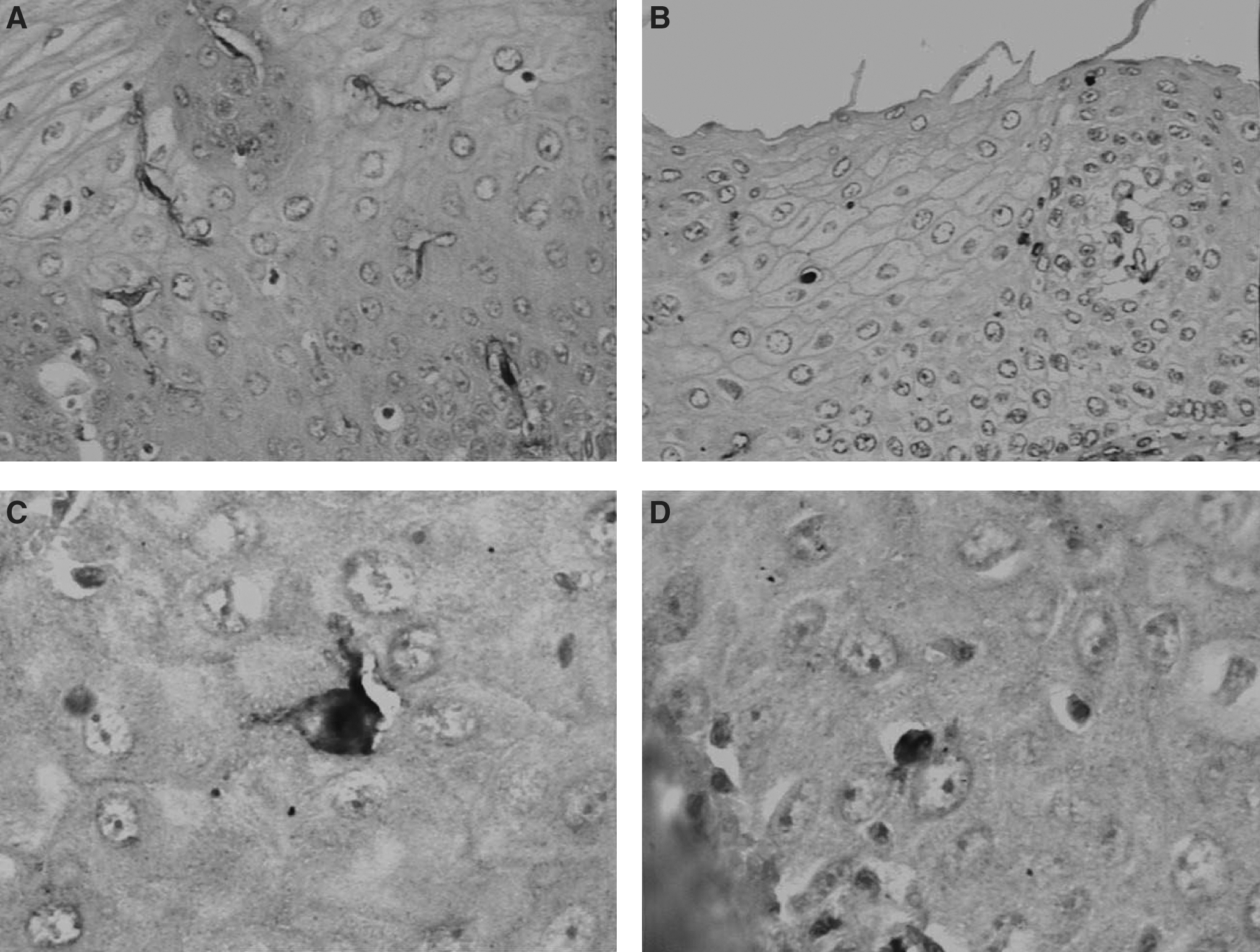
Immunostaining of Langerhans cells (LCs) in the esophageal epithelium of patients without and with AIDS.
T = 94,894.000, p < 0.001.
T = 168,052.500, p < 0.001.
T = 181,395.000, p = 0.559.
T = 349,446.500, p < 0.001.
T = 42,880.500, p = 0.014.
T = 83,323.000, p = 0.057.
T = 51,598.000, p = 0.063.
T = 113,856.500, p = 0.063.
The thickness of the esophageal epithelium was significantly reduced in patients with AIDS, regardless of gender. Women with AIDS showed a significantly thinner esophageal epithelium when compared with men with AIDS, whereas women without AIDS presented thicker esophageal epithelium than men without the syndrome, however, without significant difference (Table 3 and Fig. 3).
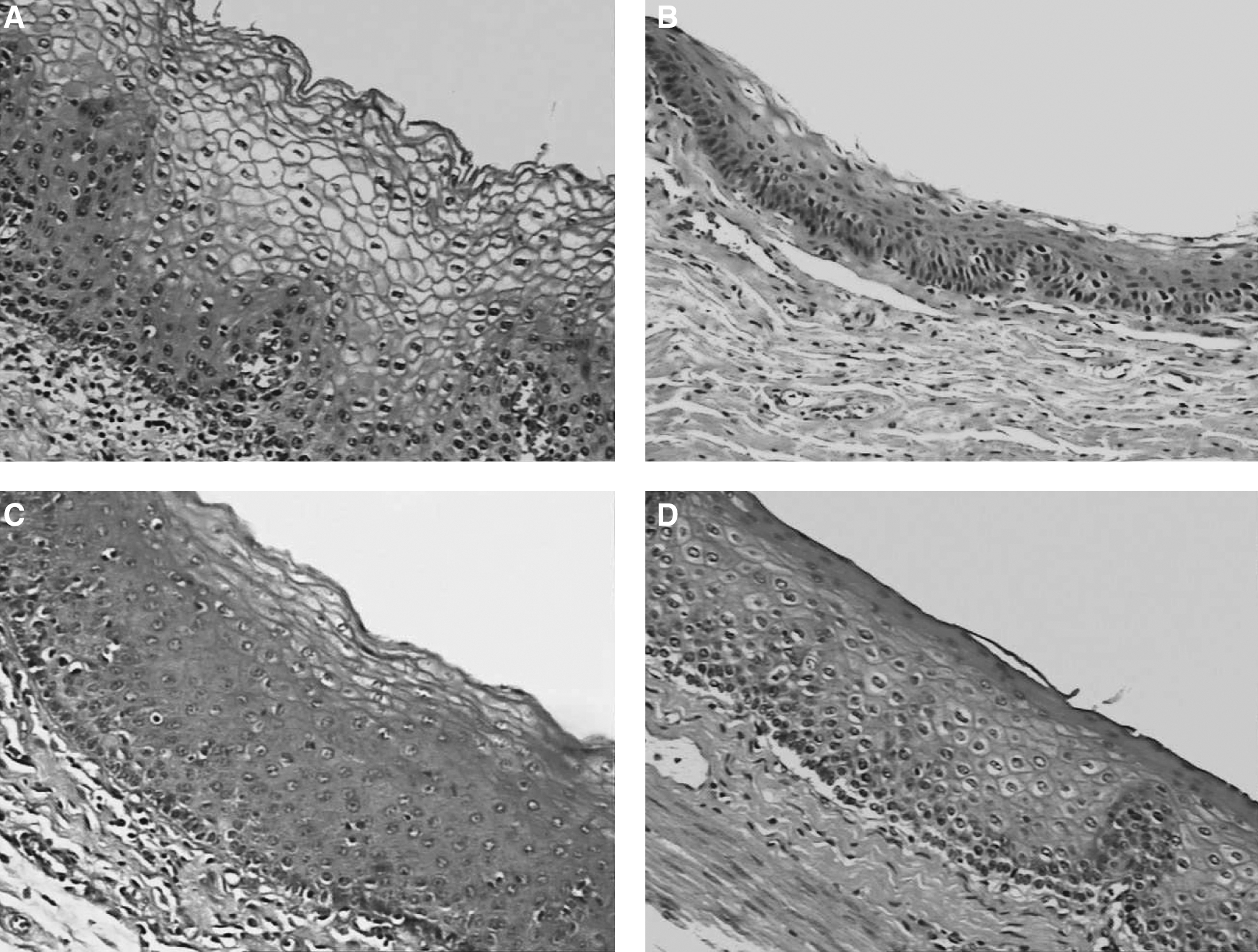
Thickness, number of cellular layers, and mean cellular diameter of the esophageal epithelium of patients without and with AIDS.
T = 2,051,560.500, p = 0.036.
T = 3,042,753.000, p = 0.384.
T = 3,080,861.000, p = 0.012.
T = 6,449,083.000, p = 0.031.
T = 1,066,706.000, p < 0.001.
T = 3,749,525.000, p < 0.001.
T = 2,343,740.000, p = 0.999.
T = 6,183,128.000, p = 0.013.
T = 1,811,043.000, p = 0.001.
T = 1,932,578.500, p = 0.001.
T = 2,139,970.000, p = 0.001.
T = 6,440,537.000, p = 0.045.
Only women with AIDS presented a significantly lower number of cellular layers in the esophageal epithelium when compared with women without AIDS. Among patients without AIDS, women presented a significantly higher number of cellular layers than men. No significant difference was observed in this parameter when women and men with AIDS were compared (Table 3 and Fig. 3).
The patients with AIDS, regardless of gender, presented a mean cellular diameter significantly larger than those who were not affected by the syndrome, and men, apart from AIDS, presented a significantly larger mean cellular diameter when compared with women (Table 3 and Fig. 3).
Discussion
A better knowledge of the silent changes in the esophageal epithelium during AIDS is valuable not only for the development of therapies, but also for a better understanding of the cure mechanisms of a large number of esophageal lesions in men and women with AIDS.
The great majority of patients with AIDS, both male and female, died of infection. Accordingly, autopsy records of immunodepressed patients indicated infectious and parasitic diseases as the main cause of death. 19
Women presented higher values of CD4+ T lymphocyte cell count and lower viral load values compared to men. Studies show lower T CD4+ lymphocyte counts in men when compared with women. These data reflect the worse immune status in men. 21
Although all patients with AIDS observed in this study have died after the worldwide introduction of highly active antiretroviral therapy (HAART), which occurred in 1997, 22 not all of them made use of the therapy, which may be explained by the fact that many of them first sought health services at an advanced stage of their disease. In comparison with men, women used antiretroviral therapy with higher frequency, in accordance with studies that show worse adherence to treatment by men. 21
Esophageal candidiasis was more frequent in patients with AIDS and in this group, women were less affected by this esophageal infection than men. Candida albicans is the most common etiological agent of esophagitis in patients with AIDS. 23 Studies with hospitalized patients with AIDS revealed that men have a longer duration of hospitalization and are more frequently admitted due to infectious causes than women. 21
A prominent pathological feature induced by the persistent HIV virus replication is the aberrant activation of the immunological system cells. Among these cells, the B cells are seriously damaged and show signs of phenotypic and functional alterations, 4,8 resulting in hypergammaglobulinemia. 24 Nevertheless, most of the excessive antibodies are not HIV antigen specific, reflecting only a generalized polyclonal activation of the B cells, caused by the HIV systemic infection. 4,25,26
In this study, a significant increase in IgA-, IgG-, and IgM-positive cells in the esophageal epithelium of patients with AIDS was observed. These data indicate that an increase in B cells happens not only in blood circulation, but also in the esophageal mucosa of patients with AIDS.
Secretory IgA, the most important effector molecule in the immune system associated with the mucosa, and secretory IgM are the first line of immunological defense against pathogenic microorganisms in mucosal surfaces, including viruses. 27 Thereby, a higher expression of IgA-, IgG-, and IgM-positive cells in the esophageal epithelium may be related to higher local immunity. However, in patients with AIDS, both cellular and humoral immunity are incapable of controlling the infection 8 and, although a proportion of the secreted antibodies has neutralizing action, 28 a great part of the increased IgA-, IgG-, and IgM-positive cells may be performing unspecific functions.
The role of humoral immunity in the control and clinical progression of the HIV infection is still highly discussed and controversial. 8 If, on the one hand, the increase of IgA-, IgG-, and IgM-positive cells in the esophageal epithelium could avoid the appearance of opportunistic infections in this organ, on the other hand, this protection seems to be inefficient, since patients with AIDS frequently show gastrointestinal manifestations deriving from opportunistic infections. 29,30
In both groups of patients, with and without AIDS, women have shown a significantly higher expression of IgA-, IgG-, and IgM-positive cells in the esophageal epithelium. These data suggest that women have stronger local esophageal immunity even during AIDS, because, despite the fact that great part of the immunoglobulins is unspecific, a proportion continues to perform neutralizing functions. 28
In the sample observed, women with AIDS compared with women without AIDS showed a decrease in the number of LCs and a higher incidence of immature or morphologically altered cells in the esophageal epithelium. On the other hand, men with AIDS compared with men without AIDS only presented a reduction in the number of these cells.
Studies suggest that the reduction in number as well as the attenuation of the dendritic process of the LCs in patients with AIDS may be related to a loss in the function of these cells, mainly the presentation of antigens. This may contribute to the decrease of local esophageal immunity in these individuals. 9,11 Women, apart from AIDS, presented a higher number of LCs in the esophageal epithelium when compared with men, which could be evidence of better esophageal immunity in women.
In the cases of patients with AIDS there was a reduction in the thickness of the esophageal epithelium; therefore, the epithelial cells that form a physical barrier, important for a defense against pathogens, 12 may have this function reduced in patients with AIDS. Women without AIDS have shown greater epithelial thickness than men without AIDS; nevertheless, during the infection by HIV, the reduction of the esophageal epithelium thickness appears to be more intense in women.
The epithelial cells in the esophagus of patients with AIDS may have been reduced in number due to apoptosis, since the cytopathic effect of the infection by HIV is associated with this process of cellular death. 31 However, in the present study, cellular apoptosis in the esophageal epithelium might be occurring more intensively in women affected by the syndrome, as the reduction of epithelial cellular layers was observed only among women with AIDS. There was no difference between genders when this parameter was analyzed in patients with AIDS.
The mean epithelial cellular diameter was significantly larger in patients with AIDS when compared with those without the disease. Cytopathic mechanisms of HIV correlates with the flow of the osmotically active monovalent cations K+ and Na+ into infected cells because the virus is able to induce disturbances in membrane ionic permeability. 32,33 Increased intracellular ion content is expected to be associated with increased water influx into the cell to balance osmolarity, thereby expanding the total volume of cells. 33 The most common type of cytopathic effects observed in vitro is a process termed balloon degeneration whereby cells swell up beyond the limits of their membrane integrity and lyse. 34 –37 Cell swelling in this case appears to be irreversible in most cells, though it has been hypothesized that those cells that can overcome these alterations in cell volume may survive to become a population of chronically infected cells. 35 Men presented a significantly larger mean cellular diameter than women, which indicates that the alteration in volume as a consequence of HIV cytopathic effect seems to be more severe in men.
In accordance with the analyses carried out, women naturally presented better local esophageal immunity than men. AIDS may possibly cause immunological and morphological alterations in the esophageal epithelium in both genders; nonetheless, women preserve better local esophageal immunity, according to the majority of the parameters observed. Therefore, patients with AIDS, especially men, need special attention from health professionals, since they develop a more severe reduction of local esophageal immunity, which may lead to an increase in susceptibility of these individuals to opportunistic infections in the esophagus.
Footnotes
Acknowledgments
This study was conducted at General Pathology Division of Triangulo Mineiro Federal University, Uberaba, Minas Gerais, Brazil, with grants from Conselho Nacional de Desenvolvimento Científico e Tecnológico (CNPq), Coordenação de Aperfeiçoamento de Pessoal de Nível Superior (CAPES), Fundação de Amparo à Pesquisa do Estado de Minas Gerais (FAPEMIG), and Fundação de Ensino e Pesquisa de Uberaba (FUNEPU).
Author Disclosure Statement
No competing financial interests exist.