
Abstract
We previously demonstrated that a commercially available natural product preparation with high content (>90%) of theaflavin derivatives (TFmix) exhibited potent anti-HIV activities. Here we developed a TFmix gel formulation as a topical microbicide candidate. The effect of TFmix on the amyloid fibril formation of semen-derived enhancer of virus infection (SEVI) peptide was detected by transmission electron microscopy. The toxicity of the TFmix gel was evaluated using human vaginal and cervical epithelial cell lines and rabbit vaginal irritation models, respectively. Levels of proinflammatory cytokines (IL-1β, IL-6, IL-8, and TNF-α) and immunoregulatory cytokines (IL-10 and GM-CSF) in cervicovaginal lavages (CVLs) were measured by ELISA kits. Proliferating cell nuclear antigen (PCNA) immunostaining was performed to evaluate inflammation in the vaginal tissues. TFmix gel could degrade SEVI-specific amyloid fibrils and showed low cytotoxicity to epithelial cells of the female reproductive tract. No apparent cervicovaginal toxicity was observed at any time point evaluated following the intravaginal administration of TFmix gel to rabbits, whereas application of N-9 gel resulted in damage to the vaginal epithelium. Neither proinflammatory nor immunoregulatory cytokine production was triggered by TFmix gel. Only low expression of PCNA was observed in vaginal tissues of TFmix gel-treated rabbits. The concentration of TFmix in plasma was very low (below the lower limit of quantitation) 1 h after a single vaginal administration of TFmix gel. However, TFmix was still detected in the cervicovaginal lavages (CVLs) 6 h after treatment, indicating that it could be retained in the vaginal cavity for a long period of time. With its potent anti-HIV-1 activity, marked stability at acidic condition, low mucosal toxicity, and lack of systemic absorption, TFmix gel can be considered as an inexpensive and safe microbicide candidate for the prevention of HIV sexual transmission.
Introduction
A
Microbicides are products designed for vaginal or rectal insertion prior to intercourse to prevent HIV acquisition without causing unacceptable toxic effects. 6 A recent clinical trial report showed that antiretroviral tenofovir gel effectively decreased HIV incidence by 39% (54% with high adherence) when used 12 h before or after sexual intercourse. 7 Clinical studies suggest that genital inflammation and loss of integrity of the cervicovaginal epithelium are critical risk factors associated with the incidence of HIV-1 infection. Several microbicides have failed in clinical trials because they did not fully protect against HIV acquisition or they increased the likelihood of HIV infection as a result of toxicity to the vaginal mucosal surface. For example, the detergent nonoxynol-9 (N-9), which is popular as a contraceptive, showed inflammatory lesions in the genital tract mucosa after repeated and frequent use. 8,9 A cellulose sulfate-based microbicide (6% gel) also failed to prevent vaginal HIV-1 transmission and may even have increased the risk of HIV acquisition in a Phase III clinical trial, 10 possibly because cellulose sulfate selectively reduced epithelium-associated colony-forming units of lactobacilli and disturbed cytokine production in the presence of bacterial colonization. 11 Therefore, as a safety measure in formulating microbicide candidates, it is essential to evaluate parameters that may increase the risk of HIV-1 sexual transmission. Additionally, it is estimated that health systems could save up to $2.7 billion by the widespread use of microbicides in low-income countries, indicating the urgent need for the production of inexpensive microbicides. 12
Historically, tea has been one of the most widely consumed beverages in the world. Much of its biological activity has been primarily ascribed to polyphenols. Tea polyphenols, which are mainly catechins and theaflavins, have recently become a focus of attention because of their antibacterial, 13,14 antitumor, 15,16 antiinflammatory, 17 –20 antioxidative, 21,22 antiproliferative, 23 and proapoptotic 24,25 effects. In addition to these effects, the antiviral activity of tea polyphenols has been studied and reviewed in many publications. For example, (–)-epigallocatechin gallate (EGCG) induced inhibition of HIV-1 infectivity by preventing the attachment of HIV-1-glycoprotein 120 (gp120) to the CD4 molecule. 26 –28 However, most of these studies focused on EGCG, the major polyphenol in green tea, while only limited investigation has been carried out on theaflavins.
Theaflavins are the most abundant polyphenols, accounting for 2–6% of the dry weight of solids in brewed black tea. This polyphenol is thought to give black tea its medicinal value, and it can be fractionated into four main components: theaflavin (TF1), theaflavin-3-gallate (TF2A), theaflavin-3'-gallate (TF2B), and theaflavin-3,3'- digallate (TF3). All of these possess a benzotropolone skeleton that is formed from cooxidation of selected pairs of catechins, one with a vic-trihydroxyphenyl moiety, and the other with an ortho-dihydroxyphenyl structure. Theaflavins showed more potent anti-HIV-1 activity than catechins in our earlier report. 29 We found that theaflavin derivatives could effectively inhibit HIV-1 entry into target cells by blocking the formation of the gp41 six-helix bundle required for HIV membrane fusion, effectively inhibiting viral entry. However, it is difficult to purify the individual theaflavins and the purified compounds are unstable. Fortunately, we found a commercially available natural product preparation with a high content (>90%) of theaflavin derivatives, designated TFmix. Besides its low production cost, TFmix is highly stable under a low pH environment. This property is particularly advantageous for development as a microbicide, as it is well known that healthy women maintain an acidic pH in the range of 4.0 to 5.0 in the vaginal microenvironment, which enhances the natural defense mechanism. We also demonstrated previously that TFmix exhibited potent antiviral activity against laboratory-adapted and primary HIV-1 strains as well as T-20-resistant HIV-1 strains. Mechanism studies suggest that TFmix mainly inhibits HIV-1 entry by targeting gp41, since it is effective in inhibiting gp41 six-helix bundle (6-HB) formation and HIV-1 envelope protein-mediated cell–cell fusion. 30 Therefore, based on the stability and efficacy of the anti-HIV profiles, TFmix, which contains a mixture of all four theaflavin derivatives, represents a promising microbicide candidate for the prevention of sexually transmitted HIV-1.
In the present study, we aimed to develop a vaginal gel consisting of 2% TFmix as an anti-HIV-1 microbicide. The structural integrity of the vaginal epithelium, the expression of proliferating cell nuclear antigen (PCNA), and the local production of cytokines in the cervicovaginal lavages (CVLs) of TFmix gel-treated rabbits were studied.
Materials and Methods
Reagents and cell lines
TFmix, a mixture of theaflavin derivatives (TF1, TF2A, TF2B, and TF3) with 90% of the theaflavin contents, was provided by the Yingshili Natural Plant Co. (Zhejiang, China). The individual theaflavins, named TF1, TF2A, TF2B, and TF3, were obtained from ChromaDex (Irvine, CA). EGCG was purchased from the Xunjian Natural Plant Co. (Fujian, China). N-9 was purchased from China Medicine University Pharmaceutical Co. (Nanjing, China). Carbopol 974P was of pharmaceutical grade and was kindly provided by the Lubrizol Co. (Wickliffe, OH). XTT and phenazine monosulfate (PMS) were purchased from Sigma (St. Louis, MO). HPLC grade acetonitrile was purchased from Merck Co. (Germany). Ultrapure water was produced in the laboratory using a Milli-Q Water Purification System (Millipore, USA). All other chemicals were analytic grade. A stock solution of TFmix was prepared at a concentration of 13.95 mM of 0.1% DMSO and stored at −20°C until use. VK2/E6E7, Ect1/E6E7, and End1/E6E7 cells were originally from the American Type Culture Collection (ATCC) (Manassas, VA).
HPLC analysis of TFmix contents
TFmix contents were determined by reversed phase-high performance liquid chromatography (RP-HPLC) (Waters Alliance 2690 Separations Module, 2487 Dual Wavelength Absorbance Detector), and data acquisition and analysis were preformed with the Empower Software Systems (Nihon Waters, Tokyo, Japan). Chromatographic separation was performed on a Shim-pack VP-ODS column (150 mm×4.6 mm) at room temperature. The mobile phase consisted of eluent A (2% acetic acid in ultrapure water) and eluent B (acetonitrile:ethyl acetate of 21:3, v/v) at a flow rate of 0.8 ml/min. The elution was programmed as follows: initial, 18% B; gradient to 26% B in 40 min. 31 Twenty microliters of sample was injected into the column and monitored at 280 nm by a UV-detector.
Preparation of TFmix vaginal gel formulation
TFmix gel was prepared by mixing TFmix with several pharmaceutical excipients, such as Carbopol 974P, glycerin, and propylene glycol. First, Carbopol 974P was dispersed in an appropriate amount of water with gentle stirring. The pH of the Carbopol was adjusted with triethanolamine. After complete hydration of Carbopol 974P by the vehicle, glycerin was added and mixed completely. Then 2 g of TFmix was added to a solution containing 15% propylene glycol and methyl hydroxybenzoate as preservative (0.05% in total gel, w/w). Ethanol (20%) was added as solubilizers/solvents to improve TFmix solubility in making the gel (0.1% in total gel, w/w). The mixture was slowly incorporated into the Carbopol 974P gel base by stirring continuously until homogeneity was reached. Finally, purified water was added to attain the total weight of 100 g with continuous stirring. The pH of the final gel formulation was 4.1 (range 4.0∼4.7). The placebo gel was prepared in the same way and using the same chemicals as the TFmix gel, except that it contained no TFmix. The N-9 gel and the placebo gel were considered as positive and negative control groups, respectively. The formulation of the N-9 gel contained nonoxyinol, polyvinyl alcohol, sodium carboxymethyl cellulose, glycerol, and ethyl hydroxybenzoate.
In vitro release study
Release characteristics of the TFmix gel formulation were evaluated by using a dialysis membrane (10 kDa MWCO; Pierce, USA) and Franz-type diffusion cells with a receptor compartment volume of 17.5 ml and an effective diffusion area of 1.77 cm2. Simulated vaginal fluid 32 (pH 4.2) was used as the receptor medium, and 1 g of TFmix gel was placed on the donor side. The receptor medium cell was shaken on a magnetic stirrer at 300 rpm throughout the assay to ensure sink conditions. The temperature in both compartments was maintained at 37.0±0.5°C with a thermostatic water bath circulating through the double glass walls of the jacketed diffusion cell. At predetermined time intervals (0.5, 1, 2, 3, 4, 5, 6, 7, 8, and 9 h), samples (1 ml) were withdrawn from the receiver compartment and replaced with an equal volume of fresh buffer. All samples were analyzed by HPLC. The cumulative release amounts of the TFmix of the formulation were plotted against the square root of time.
Effect on amyloid fibril formation of SEVI peptide
To observe whether TFmix could disrupt amyloid fibril formation, we used the transmission electron microscopy method described before. 33 Briefly, a synthetic peptide named semen-derived enhancer of virus infection (SEVI), which was derived from prostatic acidic phosphatase (PAP) sequence 248–286, was dissolved in phosphate-buffered saline (PBS) at 10 mg/ml, incubated at 37°C on a rotating mixer to induce fibril formation, and monitored by thioflavin T staining. To observe whether TFmix could disrupt PAP-derived amyloid fibril formation, SEVI was incubated in the presence (final concentration of 8 mg/ml and 2 mg/ml, respectively) or absence of TFmix at 37°C for 36 h. Subsequently, 5 μl of mixture was adsorbed for 2 min onto 200-mesh carbon-coated copper grids (Electron Microscopy Sciences, Hatfield, PA). Grids were subsequently stained with 2% aqueous uranyl acetate (Electron Microscopy Sciences) for 60 s. Fibrils were visualized with a Tecnai 12 transmission electron microscope (Philips/FEI, USA).
Cytotoxicity of human vaginal and cervical epithelial cells
The cytotoxicity of TFmix to immortalized human vaginal cells (VK2/E6E7), ectocervical cells (Ect1/E6E7), and endocervical cells (End1/E6E7), which were kindly provided by Dr. Fichorova at Harvard Medical School, was measured by the XTT assay as described elsewhere. 34 These three cell lines are from normal human vaginal, ectocervical, and endocervical epithelia immortalized by expression of human papillomavirus 16/E6E7. The cell lines maintain the differentiation characteristics of their tissues of origin. 35,36 Briefly, 100 μl of TFmix at graded concentrations was added to equal volumes of cells (5×10⁵/ml) in wells of 96-well plates. After incubation at 37°C for 4 days, 50 μl of XTT solution (1 mg/ml) containing 0.02 μM PMS was added to each well and incubated for 4 h. Absorbance was recorded on an ELISA reader (Tecan) at a wavelength of at 540 nm and a reference wavelength of 620 nm. The effect of TF on growth inhibition was assessed as the percent of cell viability, where cells with no treatment were considered as control. The 50% cytotoxicity concentrations (CC50) were calculated by using CalcuSyn software, kindly provided by Dr. T.C. Chou at Sloan-Kettering Cancer Center (New York, NY). 37
Animal studies
Thirty adult nonpregnant New Zealand white (NZW) female rabbits (mean age: 7±1 months; mean body weight: 4.0±0.5 kg) were provided by the Animal Breeding Center of the Southern Medical University (Guangzhou, China). All animals were acclimated for 1 week under a 12 h light and 12 h dark cycle at 20±2°C with a relative humidity of 50±10%. Animals were housed individually in stainless steel cages. Tap water and rabbit food pellets were available ad libitum. The study was approved by the Institutional Animal Care and Use Committee of the Southern Medical University. These rabbits were used for the following system absorption study, skin irritation test, and vaginal irritation test.
System absorption of TFmix
The systemic absorption of TFmix after vaginal application of TFmix gel was assessed by measuring the TFmix concentration in the blood as previously described. 31 Blood samples were collected in tubes containing heparin from TFmix-treated animals at 0, 15, 30, 60, 90, and 120 min after a single dose. Heparinized blood samples were immediately centrifuged to separate the plasma fraction from the whole blood. Fifty microliters of EGCG (purity>98%) was added to each plasma sample (200 μl) as an internal standard. Then, each plasma sample was homogenized with 80 μl of 6% perchloric acid and vortexed for at least 1 min. The resulting supernatant (20 μl) was injected into the HPLC column after centrifugation. Analytical methods were validated for accuracy, recovery, precision, and lower limit of quantitation (LLOQ) determinations. The LLOQs of theaflavin (TF3), theaflavin-3-gallate (TF2A), theaflavin-3′-gallate (TF2B), and theaflavin-3-3′-digallate (TF1) are 2.50, 4.32, 3.88, and 6.46 μg/liter, respectively. An individual value below the LLOQ was assigned as zero for calculation of the means. The HPLC chromatograms were compared with control plasma extracts spiked with known amounts of TFmix.
TFmix levels in cervicovaginal lavages (CVLs)
The gel formulations were administered into the vagina using a 12-cm flexible catheter, inserted up to its 8 cm mark. The body weights of rabbits and clinical observations were recorded daily, including swollen vulva areas, blood-stained urine, and soft stools. Sterile saline (500 μl) was used to repeatedly bathe the cervix and vagina before collection of the CVLs, which were then centrifuged at 1,000 rpm for 15 min at 4°C to separate the soluble supernatants from cell debris and frozen at −80°C until further use. The supernatants were used to determine cytokine levels by ELISA. 38 CVLs were collected from the vagina of TFmix-treated animals at 0, 15, 30, 60, 90, and 120 min after a single dose. The concentration of TFmix in the CVLs was measured as described above.
Skin irritation testing
Skin irritation testing of gel formula containing TFmix (2%, w/w) was also investigated. Six rabbits were used for this test, and each animal served as its own control. On day 0 of the test period, hair was clipped from the back (about 10% of the total body surface area) of each rabbit. The left side (about 3×3 cm) served as a test site for TFmix gel, while the right side served as a control site for N9 gel or placebo gel. On day 1 of the test period, TFmix gel (1 g) was applied to the rabbits' back skin, and the site was covered. After 24 h of exposure, the coverings were removed, and the application sites were rinsed with distilled water. The animals were examined for the presence of erythema and edema according to the Draize dermal irritation scoring system. 39
Rabbit vaginal irritation test
The rabbit vaginal irritation model 40 was used to evaluate the toxicity and inflammation associated with application of TFmix gel. Fifteen rabbits were divided into three different groups for testing: (1) TFmix gel, (2) placebo gel without TFmix, and (3) a gel formulation containing 4% N-9. The placebo gel and the N-9 gel were considered as positive and negative control groups, respectively. Once daily the rabbits were intravaginally administered 1 ml of N-9 gel, TFmix gel, or placebo gel using a 12-cm flexible catheter introduced up to its 8-cm mark for 14 consecutive days. The body weight of rabbits and clinical observations were recorded daily. Rabbits were sacrificed using CO2 asphyxiation on day 15. Their vaginas were surgically excised and medially opened. After recording the microscopic observation (swelling, redness, and bleeding), cervicovaginal tissues were cut at the upper, middle, and lower portions of the abdominal vagina and processed for histopathology. Anatomical observation and histological evaluation of edema, congestion, epithelial erosion, and leukocyte cell infiltration were performed. The irritation scores were assigned based on the semiquantitative scoring system of Eckstein et al. 41
Immunohistochemical staining of proliferating cell nuclear antigen (PCNA)
PCNA immunostaining was performed to evaluate the inflammation status of vaginal tissue sections (5 mm) using the PCNA kit (Boster Inc., Wuhan, China). Briefly, vaginal tissue sections were deparaffinized in xylene and rehydrated through descending grades of alcohol. The endogenous peroxidase activity was removed by treating the tissues with 3% H2O2 in methanol for 10 min and rinsing in PBS for 5 min. The nonspecific binding was blocked with 5% bovine serum albumen (BSA) for 20 min at room temperature, and tissues were then incubated overnight at 4°C with a 1:100 dilution of mouse antihuman PCNA monoclonal antibody. Slides were washed twice in PBS and incubated for 1 h at room temperature with 1:100 dilutions of goat antimouse secondary antibodies conjugated to horseradish peroxidase (HRP). The bound HRP complexes were developed using DAB staining. The sections were counterstained with hematoxylin and then mounted for visualization. Positive controls consisted of sections of mouse intestine known to express PCNA. The sections stained with secondary antibody alone served as negative controls. Immunohistochemical staining was visualized using a Nikon Eclipse 55i light microscope (Nikon Inc., Japan) with a high resolution camera.
Cytokine determination
The rabbits were dosed on three consecutive days, and CVLs were collected with 500 μl saline at 24 h intervals before each treatment and on 14 consecutive days after the last application. CVLs were measured as described above. 38 The levels of proinflammatory cytokines (IL-1β, IL-6, IL-8, and TNF-α) and immunoregulatory cytokines (IL-10 and GM-CSF) in CVLs were measured by using commercial ELISA kits (Rapidbio Lab System, CA). The catalog numbers of ELISA kits are as follows: rabbit Interleukin-1 (Cat. A1010A0501), rabbit Interleukin-6 (Cat. A1010A0504), rabbit Interleukin-8 (Cat. A1010A0505), rabbit Interleukin-10 (Cat. A1010A0506), rabbit TNF-α (Cat. A1010A0510), and rabbit G-CSF (Cat. A1010A0563). The TNF-α assay was processed immediately after sample collection, given the intrinsic instability of this cytokine. All procedures were performed as recommended by the manufacturer. Cytokine concentrations were calculated by quadratic regression analysis based on logarithmically transformed optical densities. Interference of compounds with cytokine ELISA was ruled out by spiking various compound concentrations with cytokine standards provided by the manufacturer.
Statistics
SPSS 13.0 software was used for statistical analysis. Data were analyzed by one-way ANOVA followed by LSD multiple comparison or Dunnett's multiple comparison tests. p<0.05 was regarded as statistically significant. For cytokines, concentrations in the lavage samples were converted to log10 concentrations and compared with those for the placebo gel and the N-9 gel group.
Results
TFmix gel formulation
The total content for the four theaflavin derivatives (TF1, TF2A, TF2B, and TF3) in the TFmix preparation by weight was 89.9%. The average molecular weight (716.6) was used to calculate the molar concentration of the TFmix. The process of optimizing the levels of Carbopol 974P, glycerin, ethanol, and propylene glycol was performed by assessing the in vitro release profiles and viscosity for TFmix gels. Based on the preformulation observations, the optimal formulation of TFmix gel contained 2% TFmix, 1% Carbopol 974P, 0.75 % glycerin, 15% propylene glycol, and 0.05% methyl hydroxybenzoate (w/w).
The in vitro release properties of TFmix from 2% TFmix gel were evaluated by Franz-type diffusion cells. As shown in Fig. 1, more than 50% of the drug was released at 3 h. The release profiles of TFmix were best described by the Higuchi model through fitting the data into different kinetic equations, including zero order, first order, and Higuchi models. 42 A linear relationship (r=0.9882) was obtained when the cumulative amounts of TFmix released from the formulations were plotted against the square root of time.

The cumulative percent release profiles of 2% TFmix gel. Simulated vaginal fluid (pH 4.2) was used as the receptor medium that was maintained at 37°C with stirring at 300 rpm. Each sample was tested in triplicate under two independent experiments. Data are depicted as mean±SD.
TFmix gel interfered with the fibril formation of SEVI
Peptides in human semen, like PAP248-286, are capable of forming amyloid fibrils. This kind of peptide, termed SEVI, can potently enhance HIV infection. Therefore, SEVI is regarded as one of the factors promoting HIV-1 sexual transmission. 43 As shown in Fig. 2A, PAP248-286 at a concentration of 8 mg/ml could form larger, typical fibrillary structures and was highly stable over a time period of 36 h. TFmix at a concentration of 2 mg/ml formed small aggregates (Fig. 2B). When PAP248-286 (8 mg/ml) was incubated with TFmix (2 mg/ml), no fibrils were seen, even after 36 h, by transmission electron microscopy (Fig. 2C). This result showed that TFmix could interfere with the amyloid formation of SEVI, which may abrogate semen-mediated enhancement of HIV-1 infection.
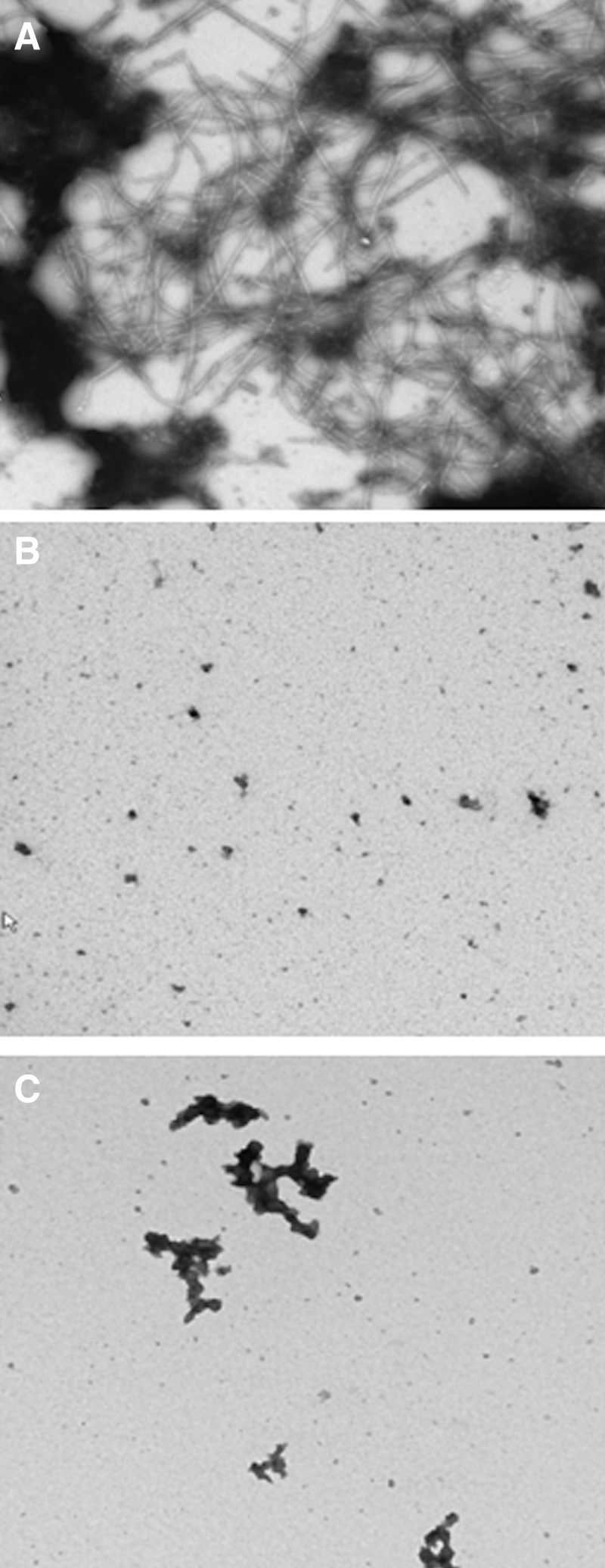
Effect of TFmix on SEVI-specific amyloid fibrils was analyzed by transmission electron microscopy.
TFmix had low in vitro cytotoxicity on human vaginal and cervical epithelial cells
The efficacy of a microbicide depends on striking a balance between safety and the biological mechanism of action. Therefore, we assessed the cytotoxicity of TFmix on human vaginal and cervical epithelial cells by the XTT assay. As shown in Table 1, TFmix had low in vitro cytotoxicity with a selection index (SI) more than 100 when compared with the anti-HIV activity reported previously. 30
SI, selection index. The IC50 of TFmix for inhibiting HIV-1IIIB infection is 0.15 μg/ml. 30 Each sample was tested in triplicate under two independent experiments. Data are depicted as mean±SD.
Application of TFmix gel did not cause skin irritation
In the 24 h closed patch testing, dermal application of TFmix did not reveal any positive reaction or signs of toxicity. There was almost no erythema persisting 72 h after opening the patch. Erythema faded away quickly, and the skin was back to normal within a short period. Neither erythema nor edema was observed on the control site of any rabbit.
TFmix gel did not cause significant cervicovaginal inflammation
The histopathological examination of cervicovaginal tissues was performed after repeated intravaginal application of TFmix gel. The results were compared with N-9 gel-treated and placebo groups. TFmix gel was administered intravaginally once daily to three female rabbits (1 ml/day) for 14 days. Light microscopic examination demonstrated that the TFmix gel-treated group retained the structural integrity of the vaginal epithelium, in which the lamina propria and squamous epithelium (SE) appeared to be intact. Additionally, the results showed the absence of leukocyte infiltration in the representative upper and lower regions of the rabbits' vaginal tissues (Fig. 3C). However, N-9 was clearly identified as the most irritating compound with multifocal damage of the vaginal mucosa showing congestion, edema, inflammatory infiltration, and epithelial disruption (Fig. 3B). Table 2 summarizes the cumulative RVI scores of histological changes in three different regions of rabbit vaginal tissue after 14 days of intravaginal application.

Photomicrographs showing section of cervicovaginal tissue in groups treated with placebo gel
Mean±SD values representing the cervicovagina, mid-vagina, and urovagina of the three rabbits in each group. N-9 (4%) was used as a positive control. Semiquantitative scoring criteria were adapted from Eckstein et al. 41 Individual score: 0=none, 1=minimal, 2=mild, 3=moderate, and 4=intense. Correlation to human irritation potential: total score <8 acceptable, 9 to 10 marginal, and ≥11 unacceptable. Data were analyzed by Dunnett's test for multiple comparisons (*** p<0.001).
TFmix gel did not cause abnormal cytokine production in CVLs
Cytokine levels in CVL supernatants of the above samples were measured by ELISA. Following the intravaginal administration of TFmix gel to rabbits (1 ml/day), the levels of various proinflammatory cytokines (IL-1β, IL-6, IL-8, and TNF-α) and immunoregulatory cytokines (IL-10 and GM-CSF) were not statistically different compared with placebo controls (Fig. 4B). In contrast, the N-9 gel induced an obvious inflammatory reaction. Only three consecutive applications of N-9 were sufficient to trigger the release of proinflammatory and immunoregulatory cytokines, as shown in Fig. 4A. In particular, the levels of IL-6 were found to be higher than other cytokines. CVL cytokine levels agreed with cumulative mucosal irritation scores after multiple applications (p<0.01).
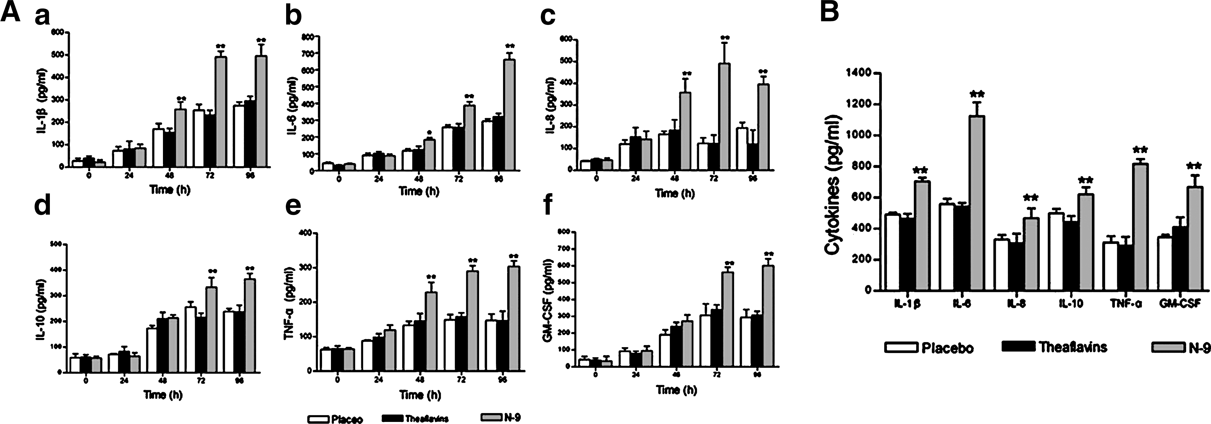
Cytokine expression in cervicovaginal lavages (CVLs) before and after vaginal administration of TFmix gel.
Intravaginal TFmix gel did not cause cellular hyperplasia
PCNA expression was observed by the immunohistochemical method. Figure 5 shows the PCNA staining pattern of representative vaginal sections of rabbits administered N-9 gel, TFmix gel, or pacebo gel aginally for 14 consecutive days. No significant differences in PCNA positivity were noted in the epithelium or stromal cells of TFmix-treated tissues (Fig. 5C).
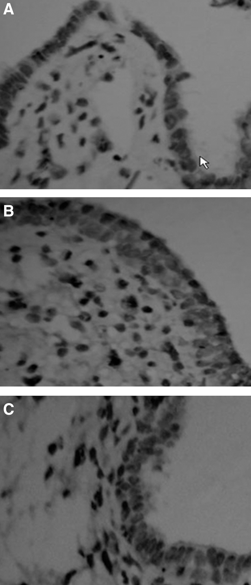
Immunohistochemical (IHC) localization of proliferating cell nuclear antigen (PCNA). Representative PCNA-positive and hematoxylin-counterstained, paraffin-embedded sections of rabbit vaginal tissues treated intravaginally with negative gel
No systemic absorption of TFmix in the gel
The systemic absorption of TFmix in the gel applied intravaginally was studied in vivo in NZW rabbits. Blood was collected at intervals of time after a single administration of 2% TFmix gel. As demonstrated in Fig. 6A, EGCG and four components of TFmix could be clearly separated using established HPLC conditions. The retention time of EGCG was 6.58±0.03 min. The four theaflavin derivatives, including TF1, TF2A, TF2B, and TF3, were identified with retention times of 23.42±0.26, 29.38±0.17, 32.18±0.24, and 34.2 5±0.28 min, respectively. The blood plasma samples were prepared 1 h after intravaginal administration of 1 ml TFmix gel. Individual values below the LLOQ of theaflavin derivatives were assigned a value of 0 for the calculation of the means. As shown in Fig. 6B, the TFmix levels in the plasma 1 h after a single vaginal administration of TFmix gel were often below the LLQQ, suggesting that there is no significant systemic absorption of TFmix. HPLC profiles of CVL samples were prepared 1 h and 6 h after treatment (Fig. 6C and D). In the CVLs at 6 h, a detectable concentration of TFmix components can still be observed. This suggested that the anti-HIV activity of TFmix can be maintained up to 6 h.
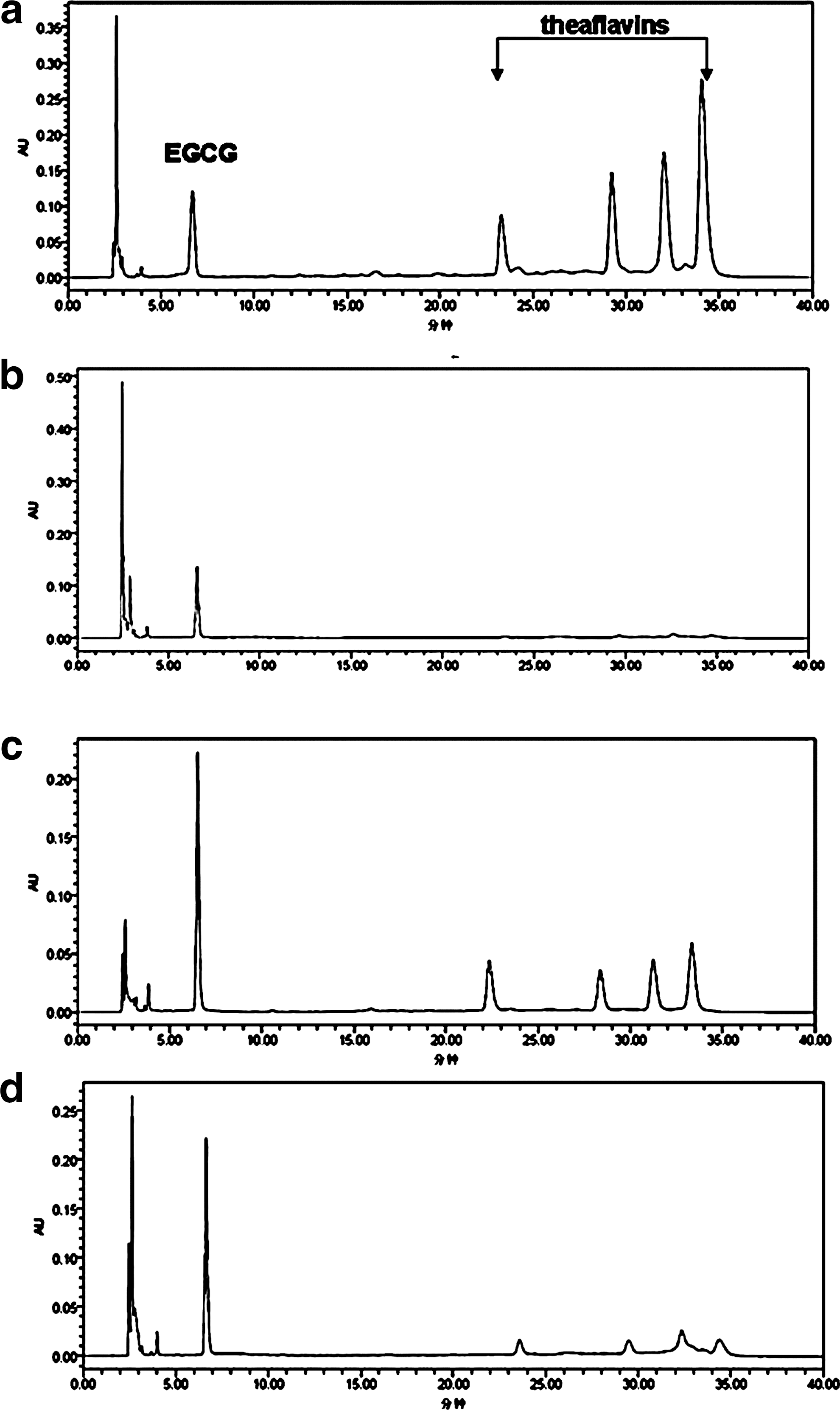
Representative HPLC chromatogram of TFmix in plasma and CVL samples.
Discussion
The use of natural compounds to prevent and treat acute and chronic diseases, such as HIV/AIDS, is attracting increased attention. 44 Tea has been widely used by people all over the world and is generally recognized as being safe. 45 Theaflavins are the major components of tea polyphenols in brewed black tea. Therefore, the theaflavin derivatives mixture (TFmix) as an anti-HIV microbicide candidate is expected to be safe and inexpensive. The U.S. FDA has approved the marketing of sinecatechins (Veregen), a botanical drug product, for the treatment of external genital and perianal warts. Sinecatechins combine catechins and other green tea components and are derived from the green tea leaves of Camellia sinensis. 46,47 Cellular infection and the use of cellular machinery are necessary for the production of new viruses. Therefore, blocking the entry of HIV into target cells should be an effective way of neutralizing HIV infectivity. Accordingly, HIV entry inhibitors show promise as topical strategies or "virustats" to prevent mucosal transmission of HIV infection. 48,49 We previously reported that theaflavin derivatives in black tea could inhibit HIV-1 entry by targeting gp41. 29 We further assessed the inhibitory activity of TFmix on infection by laboratory-adapted HIV-1 strains and drug-resistant strains. The results suggest that TFmix has a broad spectrum of antiviral activity. 30 Interestingly, TFmix could efficiently inhibit the fibril formation of SEVI peptide and abrogate its enhanced effect on HIV-1 infection, similar to the reported effect of EGCG. 33,43 Therefore, TFmix is an attractive candidate for the development of a vaginal topical microbicide to prevent the sexual transmission of HIV-1.
In preformulation studies, we adjusted the concentration of the active components in the formulation, including Carbopol 974P, glycerin, and propylene glycol. These pharmaceutical excipients were selected because of their safety, physical characteristics, bioadhesive properties, versatile consistencies, and solubility. They have all been used in the preparation of a variety of topical medications. For example, propylene glycol is widely used to permeate skin in many transdermal therapeutic systems. Twenty percent EtOH was used as solubilizers/solvents to improve the solubility of TFmix in water. Carbopol, a commercially available polyacrylic acid polymer, has been recognized as a standard vaginal mucoadhesive polymer that presents several advantageous properties contributing to its broad use in investigational and commercially available vaginal drug formulations. 50 Here, Carbopol 974P was selected as a thickening agent to obtain a gel with desirable viscosity and biocompatibility with vaginal mucosa. Based on these observations, the optimal formulation of TFmix gel contained 2% TFmix, 1% Carbopol 974P, 0.75% glycerin, and 0.05% methyl hydroxybenzoate (w/w).
The dissolution data of TFmix fit the Higuchi's model well (r=0.9882), presenting a biphasic pattern. As shown in Fig. 1, more than 50% of the drug was released at 3 h. Additionally, 2% TFmix gel shows good physical stability with no sedimentation, no layer separation, and no color change at the end of 6 months of storage at accelerated conditions (40°C and 75% room humidity), as well as at room temperature (data not shown). This demonstrates that TFmix vaginal gel can be prepared simply and that the quality is controllable. Additionally, skin testing performed on New Zealand white rabbits demonstrated that TFmix gel resulted in only slight irritation, suggesting its suitability for further studies.
The failure of N-9 and cellulose sulfate in Phase III clinical trials has given investigators ample warning that a safety evaluation of a microbicide candidate should be carried out as early as possible. Here we first assessed the effect of TFmix on the viability of VK2/E6E7, Ect1/E6E7, and End1/E6E7 cells (Table 1) by XTT assay. Results revealed low toxicity to the human vaginal and cervical epithelial cells. We next assessed the safety of TFmix gel in vivo using a rabbit model. TFmix was compared with placebo and N-9 gel using the following parameters and measures: cell morphology, the structural integrity of the vaginal epithelium, and titers of proinflammatory and immunoregulatory cytokines in CVLs.
The cumulative RVI scores reflected potential changes across the regions from the stratified squamous to columnar epithelium. The TFmix gel-treated group achieved a score of 6.2 after once daily intravaginal administration for 14 days, indicating the presence of minimal irritants (Table 2). Since homology exists between rabbits and humans with respect to the irritation potential of vaginal products, the score of TFmix-treated animals is well below the acceptable range for human usage (total score 8), suggesting that the TFmix gel is not toxic to human mucosal tissues. It should be further noted that TFmix gel did not induce apparent irritation and/or inflammation in rabbit vaginal epithelium, as determined by histological examination.
Proinflammatory cytokines in the CVLs, which may amplify viral replication in infected cells, serve as sensitive markers for vaginal mucosal epithelial damage in identifying and/or screening out suitable candidate microbicides before they enter Phase I clinical trials. Furthermore, they can reflect important biological changes that occur in the vagina as a result of microbicide administration. 51,52 N-9 was found to induce a number of cytokines and chemokines measured in human CVLs, including IL-1α, IL-1β, and IL-8. 9 It was also found to induce the proinflammatory cytokines and immunoregulatory cytokines in the rabbit model. 51 Here, several biomarkers and inflammatory mediators in the CVLs of rabbits, such as proinflammatory cytokines (IL-1β, IL-6, IL-8, and TNF-α) and immunoregulatory cytokines (IL-10 and GM-CSF), were monitored in response to vaginal application of TFmix gel. No significant difference in cytokine levels was observed between the placebo and TFmix-treated groups, indicating that the TFmix gel did not induce irritation and/or inflammation in the vaginal epithelium. Cytokine levels in CVLs agreed with cumulative mucosal irritation scores after multiple applications (p<0.01).
PCNA, a DNA polymerase, was used as a marker for cellular proliferation. Here, we used immunohistochemistry for PCNA to visualize proliferative activity of inflammatory cells or vaginal epithelial cells in paraffin-embedded vaginal tissue sections of control and TFmix-treated rabbits. PCNA-positive cells identified by their darkly stained nuclei were detected in both vaginal epithelial and stromal cell nuclei of all vaginal tissues examined. No significant PCNA signal was observed in the cell nucleus of the TFmix-treated group (Fig. 5C), indicating that repeated intravaginal use of TFmix did not cause cellular inflammation or hyperplasia in the vaginal epithelium. The immunohistochemical localization of PCNA corresponded to the histopathology results.
Taken together, these results indicate that repeated intravaginal administration of TFmix gel, even at concentrations higher than 105-fold over its in vitro anti-HIV IC50 value, did not cause significant vaginal irritation in the rabbit model. Furthermore, when the formulation was applied intravaginally to female rabbits once daily (with a dose of 1 ml/day) for 14 days, no effect was observed on the hematology, coagulation, serum chemistry, body weight of animals, and relative organ weights (data not shown).
The effectiveness of a vaginal microbicide is dependent on the bioadhesion of the formulation and the bioavailability of the drug. In particular, bioadhesive gels should be retained in the vaginal cavity for prolonged intervals while maintaining biological activity. When we investigated the systemic absorption of TFmix from our TFmix gel formulation applied intravaginally, the results showed the persistence of biologically significant concentrations of TFmix in the vagina for >6 h, which supports the development of TFmix as a microbicide for once daily application or 12 h before/after sexual intercourse.
In conclusion, the present study has shown that TFmix is a potential candidate for topical microbicide development owing to its proven in vitro and in vivo efficacy and safety profiles, as well as its physical and chemical properties.
Footnotes
Acknowledgments
This study was supported by the Natural Science Foundation of China (U0832001), the National Key Science and Technology Special Project (2012ZX10001-007-009), and the Foundation for Key Laboratory of Prevention and Control of Emerging Infectious Diseases of Guangdong Higher Education Institutes (KLB09007).
Author Disclosure Statement
No competing financial interests exist.