
Abstract
Human immunodeficiency virus type 1 (HIV-1) viral genes nef and tat play an important role in disease progression. In this study we characterized the Nef and Tat proteins from a group of HLA-B57 typed pediatric perinatally infected long-term survivors (LTS) with ≥10 years of infection. We identified 19 therapy-naive LTS after screening 250 children from an Indian pediatric cohort. Nef and tat amplified from plasma virus showed that all the LTS harbored HIV-1 subtype C. The two B57+ children showed mutations, deletions, and insertions in experimentally defined B57 epitopes in the virus that are likely to be escape mutants. Only GW12 (GPGVRYPLTFGW) and YY9 (YTPGPGIRY) were conserved, while the remaining 90% (18/20) of the epitopes showed some degree of mutations. The most variable epitopes were RR15, SE15, QP15, KF9, HW9, YT9, and GF15. To our knowledge this is the first study from India in which characterization of Nef and Tat from LTS has led to information on genetic alterations in these genes that are associated with slow disease progression, and can provide an important lead in future studies.
T
Several studies have indicated the role of viral factors nef and tat gene products in nonprogression of disease. 3,6,7 The function of the nef gene is down-regulation of the host antigen presentation to HLA class I receptors, down-modulation of CD4 receptors, and formation of a complex with p21-activated protein kinase 2 (PAK2), thus stimulating viral replication and enhancing the infectivity of the HIV-1 virion. 8,9 Large deletions and premature stop codons pertaining to nonfunctional Nef are significantly associated with disease progression. 10 On the other hand, Tat is involved in various intracellular and extracellular functions during viral infection contributing to viral pathogenesis. 11 Studies have established a correlation between the presence of anti-Tat antibodies and progression to severe immunodeficiency. 7
To understand the genetic architecture of these two viral accessory genes, we analyzed viral proteins Nef and Tat from previously determined (based on HIV-1 pol and env genes) HIV-1 subtype C obtained from HLA-B57 typed perinatally infected long-term survivors (LTS) from a south Indian pediatric cohort. To our knowledge this is the first study from India in which mapping of Nef and Tat is done to investigate the association between genetic variability and slow disease progression.
Long-term surviving children were identified after screening 250 children from an Indian pediatric cohort followed at the St. John's hospital pediatric infectious diseases clinic. A single peripheral blood sample was collected between December 2007 and October 2011 from 19 LTS who were therapy-naive and had a duration of infection of ≥10 years. LTS included 10 long-term nonprogressors (infection ≥10 years and CD4 count >500 cells/mm3) and 9 slow progressors (infection ≥10 years and CD4 count <500 cells/mm3). Additionally, we collected blood samples from 11 therapy-naive normal progressors with an average duration of infection of 6 years and median CD4+ T cell count of 254 cells/mm3 (IQR: 228–380). The viral genes were amplified from plasma virus using reverse transcriptase polymerase chain reactions (RT-PCR) followed by conventional nested PCR. Nef gene PCR was carried out using the external forward primer UN_NEF1F 5′-CAARGAATTTGTAGAGCTATCYG-3′ (HXB2 position 8724–8747) and the reverse primer UN_NEFCR 5′-GCTTATATGCAGCATCTGAGG-3′ (HXB2 position 9497–9518).
The cycling conditions include one cycle at 95°C for 3 s, followed by 39 cycle at 95°C for 20 s, 55°C for 30 s, and 72°C for 30 s, and finally one cycle at 72°C for 30 s. Nested PCR was carried out with internal forward primer: UNNEF2F 5′-AAGAATAAGACAGGGCTTTGAAGC-3′ (HXB2 position 8759–8780) and a common reverse primer UNNEFCR. The cycling conditions included initial denaturation at 95°C for 3 s, followed by 30 cycles at 95°C for 20 s, 55°C for 30 s, and 72°C for 30 s, followed by one cycle of final extension at 72°C for 5 min. The Tat gene was amplified as described by us previously.
12
The purified PCR products were subjected to bidirectional sequencing using internal primers. Phylogenetic analysis, intrapopulation divergence (nucleotide distance from an ancestral subtype C sequence downloaded from the Los Alamos database,
Patient characteristics were as follows: among the LTS, 57% (11/19) were female, with a median CD4+ T cell count of 540 cells/mm3 (IQR: 262–769) and mean (SD) viral load of 5.2 (0.7) log10 copies/ml. HLA-B57 was positive in two individuals.
We successfully amplified and sequenced 19 nef and 17 tat from the Indian LTS. The nef and tat genotyping confirmed the identity of all samples to be HIV-1 subtype C. All the study sequences demonstrated well-separated branches indicating the absence of PCR cross-contamination (Fig. 1). Multiple sequence alignment of B57-positive individuals, for 20 experimentally predefined B57-CTL epitopes (source Los Alamos Immunology Database,
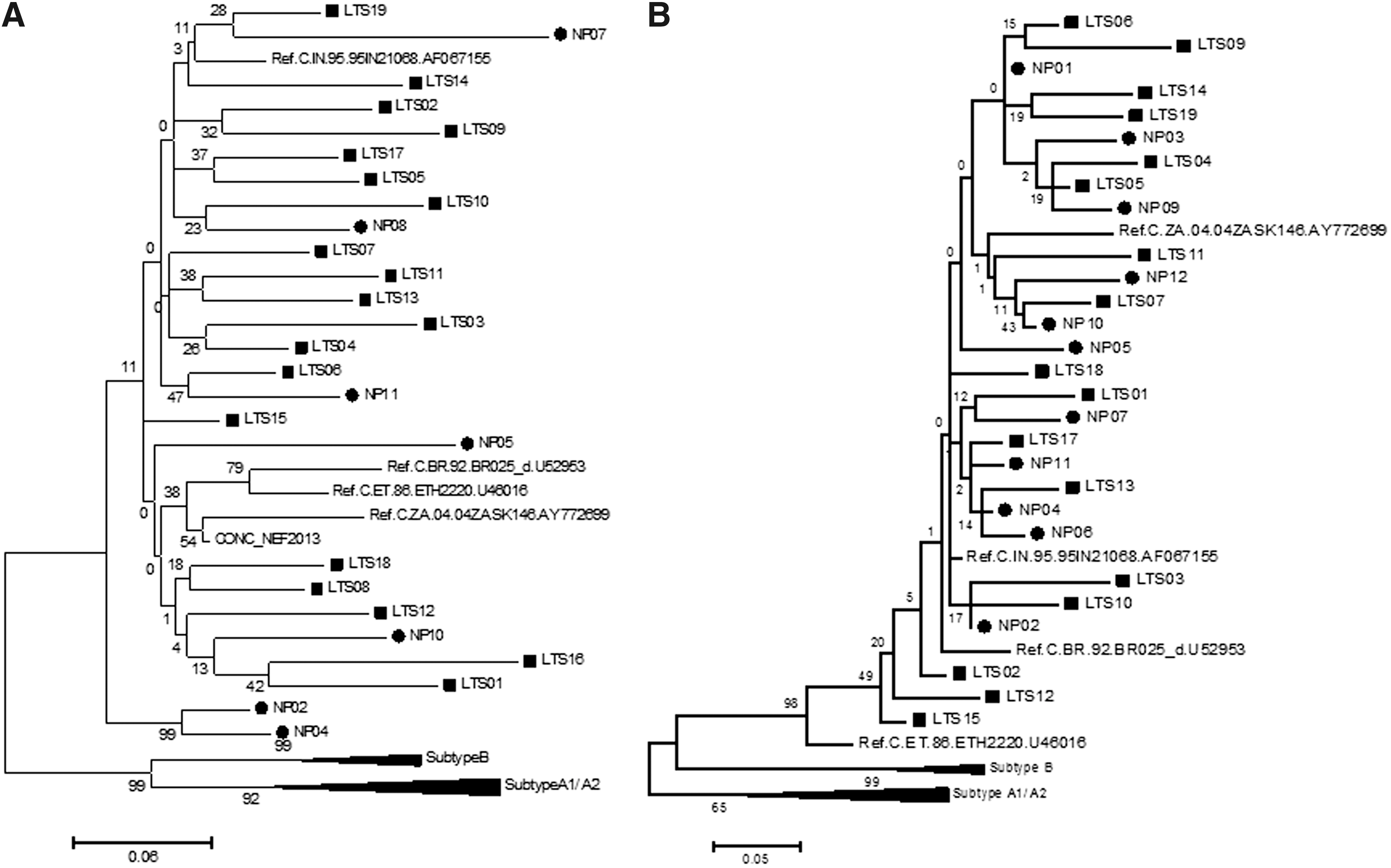
Phylogenetic analysis. Neighbor joining phylogenetic tree with 500 bootstrap dataset for nef
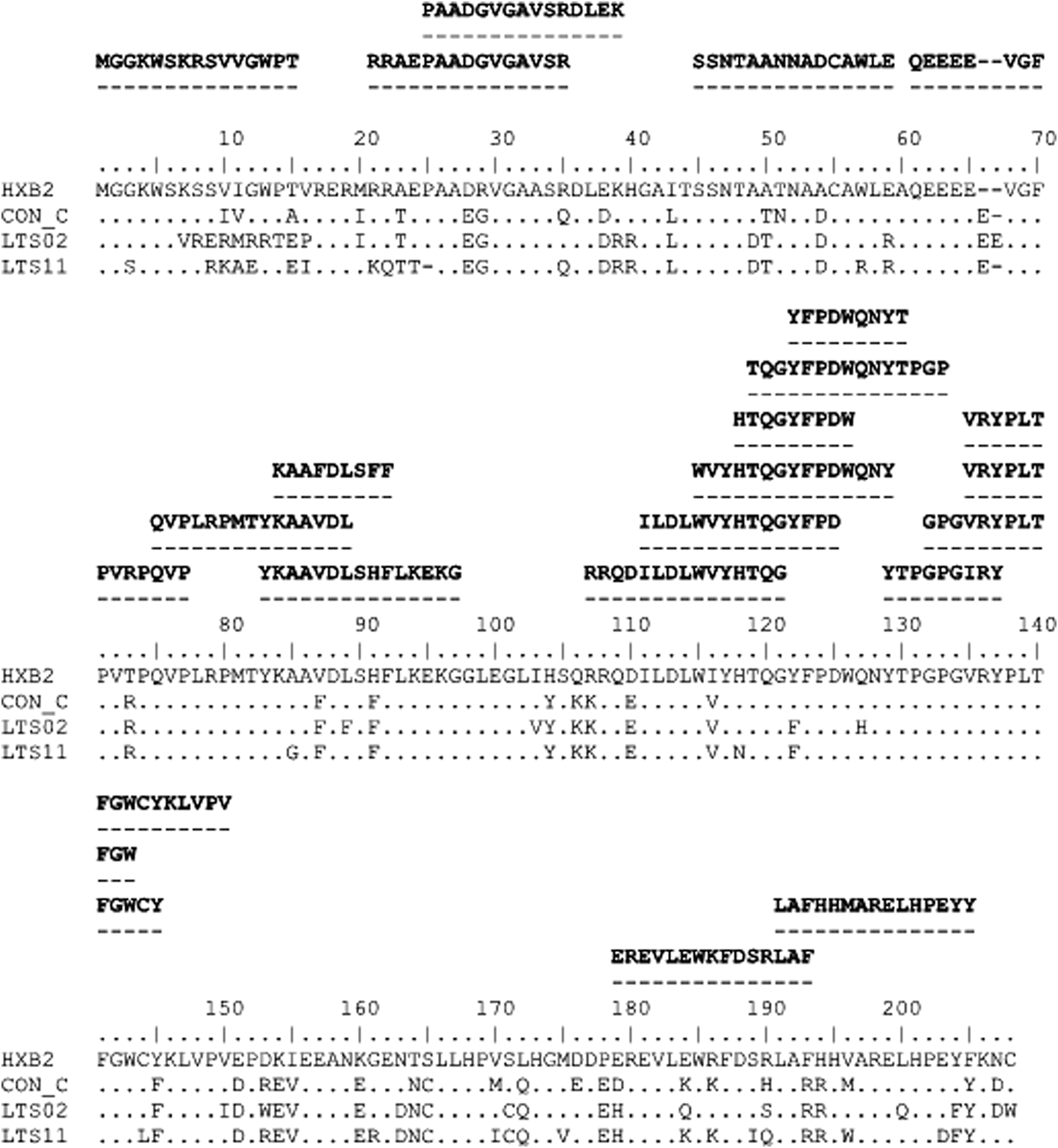
Multiple sequence alignment of B57-positive long-term survivors (LTS). The Nef sequences from Indian LTS with B57+ were aligned with HXB2 (subtype B reference) and consensus C (downloaded from the Los Alamos database). Experimentally predefined B57-CTL epitopes (source Los Alamos Immunology database,
Wild type (denoted as WT) is the subtype B sequences. Consensus C (denoted as Con_C).
Studies have shown that CD8+ T cell escape mutations in HIV-1 are rarely present in resting CD4+ T cells within the proviral compartment but commonly occur in plasma viruses due to the presence of novel escape mutants. 15 Our results provide a glimpse of the concept that mutations seen in the B57 epitopes in subtype C sequences from the plasma virus of Indian LTS may possess escape mutants. Although this study is limited by its small sample size, the findings present valuable pilot data to explore this concept further in larger studies.
In conclusion, for the first time we have described the genetic architecture of Nef and Tat variations in Indian subtype C-infected LTS. The variability of the B57 epitopes seen in two B57+ LTS suggests that this may be the novel viral escape mutations that are present in plasma virus and that may be the reason for viremia inside the body. Further studies on functional analysis to understand the role of these mutants and viral escape are ongoing.
Footnotes
Acknowledgments
The authors would like to thank the staff at the Infectious Disease Clinic for their generous help with field work and sample and data collections. We would also like to thank the study participants. The study was partially funded by Child Health Foundation and Indian Council of Medical Research.
Author Disclosure Statement
No competing financial interests exist.