
Abstract

R
We have previously demonstrated that although unstimulated resting CD4+ T cells are relatively resistant to HIV infection, incubation of resting CD4+ T cells with chemokines that bind the chemokine receptors, CCR7, CXCR3, and CCR6, which are all highly expressed on resting CD4+T cells, facilitates latent infection in vitro. 2,3 Pretreatment with the chemokine CCL19, the ligand for CCR7, facilitated rapid nuclear localization and efficient integration. This was in part mediated via CCL19-induced changes in the actin cytoskeleton. 3 Our aim here was to visualize the early events following HIV entry into a resting CD4+ T cell.
We used fluorescent HIV to infect CCL19-treated resting CD4+ T cells to track the migration of virus to the nucleus. Resting CD4+ T cells were incubated with 30 nM CCL19 and infected at an MOI of 0.1 with HIVNL4.3 that was dual labeled with green fluorescent protein (GFP) fused to HIV Vpr and S15tomato fused to the viral membrane. GFPVpr associates with HIV cores and tracks HIV complexes in the cytoplasm of target cells. S15tomato is a fluorescent variant of S15mCherry that is no longer visible when virus particles fuse to target cells. 4 Therefore, HIV that has fused and entered a cell can be identified as GFPVpr+ S15tomato−. 4
Two hours after the addition of virus, cells were fixed and filamentous actin (F-actin) was stained using Alexa Fluor 350.phalloidin (Molecular Probes). The cells were placed in a Labtek II chamber slide and images were captured in a z series using a CoolSNAP camera (Photometrics) on a DeltaVision microscope (100×1.4 numerical aperture oil immersion lens, Applied Precision). Images were deconvolved using softWoRx deconvolution software (Applied Precision). A single z section of a deconvolved image is shown. Cells were also cultured for 4 days and quantification of integrated DNA was performed using Alu-LTR real time PCR. 3 Reverse transcriptase (RT) was quantified in supernatant. The frequency of integrated HIV was 92,000 copies/106 CD4+ T cells and there was minimal RT production consistent with latent infection.
In the image shown in Fig. 1, fused HIV complexes (GFPVpr+ S15tomato−) that have penetrated cortical F-actin at the cell periphery and are located in the resting CD4+ T cell cytoplasm can be seen. We are now examining how HIV interacts with other components of the cell cytoskeleton and defining key cellular proteins required for rapid transit of HIV to the nucleus in CCL19-treated resting CD4+ T cells.
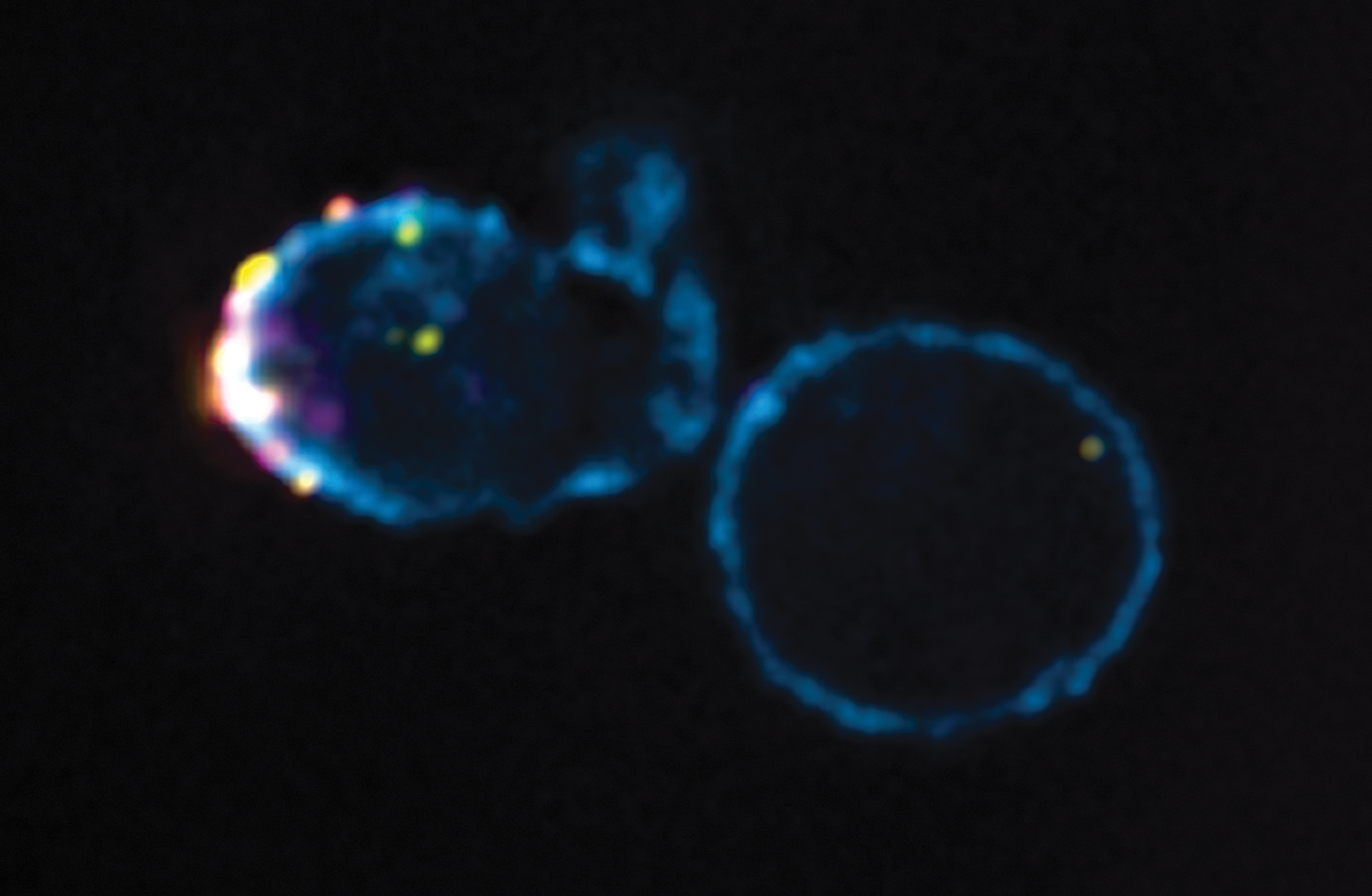
Fused HIV in the cytoplasm of CCL19-treated resting CD4+ T cells. Resting CD4+ T cells pretreated with 30 nM CCL19 were infected for 2 h with HIVNL4.3 dual labeled with GFPVpr (yellow) and S15tomato (majenta). Fixed cells were stained for F-actin using Alexa Fluor 350.phalloidin (cyan) and imaged on a DeltaVision microscope (100×). Fused HIV complexes (GFPVpr+ S15tomato−) that have penetrated past cortical F-actin at the cell periphery into the cytoplasm are shown in a deconvolved z section of CCL19 cells.
Footnotes
Acknowledgments
The authors acknowledge the facilities and scientific and technical assistance of Monash Micro Imaging, Monash University, Victoria, Australia. This research was supported by NIH Bridging Grant 1R56AI95073-1A1 and a U19 Grant to the DARE Collaboratory AI096109.