
Abstract
T
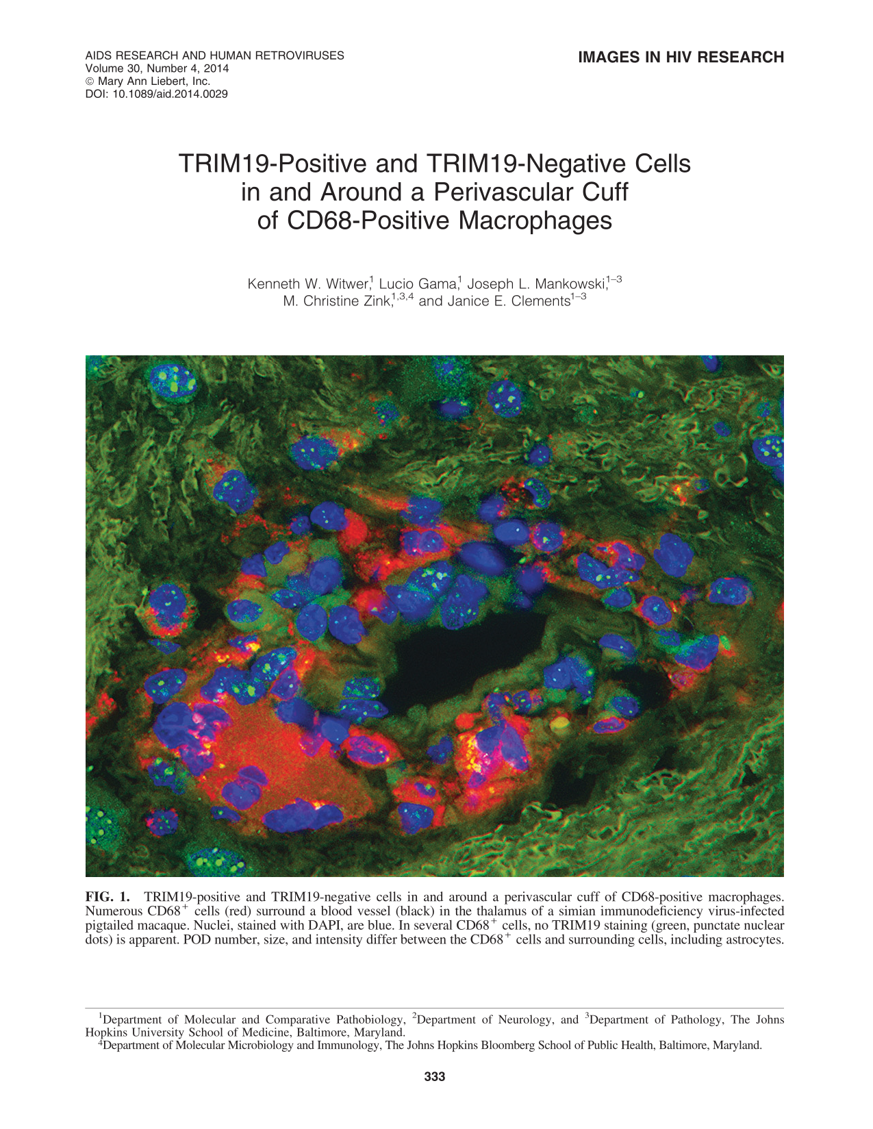
Using immunohistochemical techniques to stain thalamic thin sections, we observed a striking and coordinated modulation of TRIM19 bodies in response to SIV infection of the central nervous system (CNS). Following infection, significantly more PODs per nucleus were found in astrocytes, the most abundant cell of the CNS (unpublished data). Astrocytes are susceptible to infection but are usually not productively infected. The upregulation of TRIM19 was observed during acute infection and development of encephalitis, when virus is most abundant, tracing a course similar to what we have described previously for the cytokine response. 4 In Fig. 1, astrocytes include several of the cells near or at the cuff with high, blown-out levels of TRIM19 (green, as labeled with an Alexa 488-conjugated anti-PML antibody kindly provided by Gary Hayward).
TRIM19-positive and TRIM19-negative cells in and around a perivascular cuff of CD68-positive macrophages. Numerous CD68+ cells (red) surround a blood vessel (black) in the thalamus of a simian immunodeficiency virus-infected pigtailed macaque. Nuclei, stained with DAPI, are blue. In several CD68+ cells, no TRIM19 staining (green, punctate nuclear dots) is apparent. POD number, size, and intensity differ between the CD68+ cells and surrounding cells, including astrocytes.
Interestingly, similar fluctuations were not observed in macrophages in the brain. Myeloid lineage cells that enter the brain from the periphery are an important source of virus in the CNS and are responsible for retroviral entry within days of infection. Macrophages contribute to pathogenesis both by hosting viral replication and by shaping a damaging inflammatory milieu. Perivascular cuffs of infiltrating macrophages are a hallmark of progression to CNS disease. In our assays, TRIM19 was not detected in some infiltrating CD68+ macrophages (labeled red). Although astrocytes displayed TRIM19 responses consistent with interferon (IFN)-mediated upregulation, macrophages did not. Some infected macrophages contained few or no visible PODs.
Based upon these results, it is tempting to speculate that low levels or the absence of TRIM19 nuclear bodies may contribute to a permissive environment for lentiviral replication in perivascular macrophages, while high levels of TRIM19 help to restrict replication in astrocytes. However, a causal relationship is not demonstrated by these observations, and our unpublished work with in vitro models—with all the caveats these must bear—has admittedly not supported a major role for TRIM19 in retroviral restriction. Indeed, perhaps the most striking message of the illustration is that cells only tens of microns distant from each other display such different responses to similar inflammatory signals.
Footnotes
Author Disclosure Statement
No competing financial interests exist.