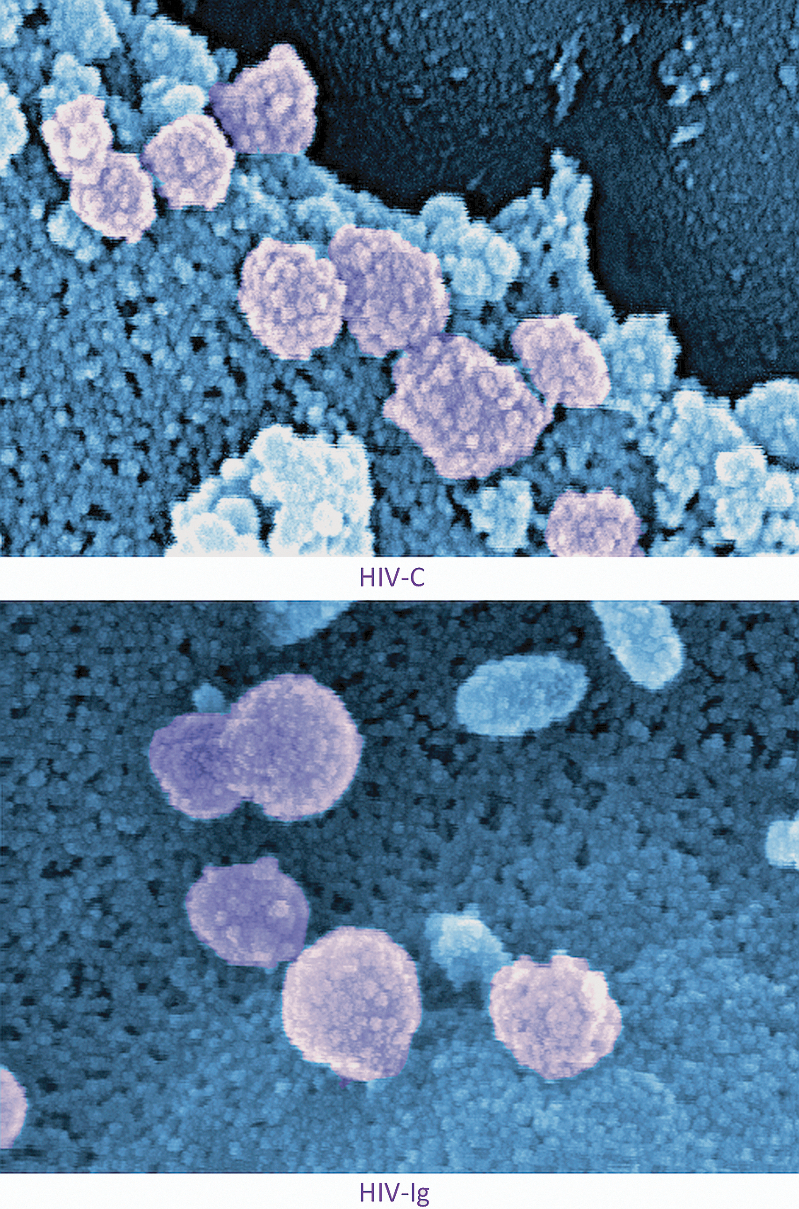
We previously demonstratedin vitro and ex vivo that dendritic cells (DCs) exposed to IgG-opsonized HIV (HIV-Ig), as found after seroconversion and in HIV-infected individuals,1 showed an impaired productive infection or were not infected at all and exerted a significantly attenuated CD8+ T cell-stimulatory capacity.1,2 In contrast, complement-opsonized HIV (HIV-C) acted as an endogenous adjuvant for DC-mediated induction of retrovirus-specific cytotoxic T lymphocytes (CTLs) and efficiently bypassed the restriction mechanisms in DCs, thereby causing a high productive infection of DCs.2 Complement opsonization immediately occurs following entry of HIV-1 via mucosal surfaces since the virus expresses a complement-activating domain in its envelope glycoprotein and is spontaneously surrounded by covalently bound complement fragments.3 These structures will ligate the abundant complement receptors 3 and 4 [CR3 (CD11b/CD18), CR4 (CD11c/CD18)] expressed on DCs more likely than other pattern recognition receptors on DCs, i.e., C-type lectins or Toll-like receptors as illustrated earlier by our group.4 Recently, the role of the complement system in protection against viral infections has been gaining importance and as shown by us, dendritic cell-mediated cellular immunity also depends on complement opsonization of the pathogen. Figure 1 shows human myeloid dendritic cells (blue) exposed to complement-opsonized HIV (Fig. 1: upper image: HIV-C purple) or IgG-opsonized HIV (Fig. 1: lower image: HIV-Ig purple). Covalently bound complement fragments surround the virus like a cloak (Fig. 1: upper image) thus masking the envelope glycoproteins—the structure seems rougher compared to the smooth viral surface of IgG-opsonized HIV (Fig. 1: lower image) and the size of the particle bigger. In addition to causing an enhanced DC infection and DC-mediated CTL induction, complement coating of HIV hampered interactions with C-type lectins expressed on DCs.4 Nevertheless, CD4 and a chemokine coreceptor were essential for productive DC infection.4 Antibody-opsonized HIV (Fig. 1: lower image) will more likely bind to FcγRII expressed by DCs, which might explain the differential handling of C- and IgG-coated virus by DCs. Scanning electron microscopy of DCs (blue) interacting with differentially opsonized HIV (purple) identifies the differences in structure and dimension of the virus depending on the opsonization with complement (Fig. 1: upper image) or specific IgGs (Fig. 1: lower image), respectively. A magnification of 200,000×was used for image acquisition.
Human myeloid dendritic cells.
Footnotes
Author Disclosure Statement
No competing financial interests exist.
References
1.
PoschW, CardinaudS, HamimiC, FletcherA, MuhlbacherA, LoackerK, et al.: Antibodies attenuate the capacity of dendritic cells to stimulate HIV-specific cytotoxic T lymphocytes. J Allergy Clin Immunol, 2012; 130:1368–1374 e1362.
2.
WilflingsederD, BankiZ, GarciaE, PruensterM, PfisterG, MuellauerB, et al.: IgG opsonization of HIV impedes provirus formation in and infection of dendritic cells and subsequent long-term transfer to T cells. J Immunol, 2007; 178:7840–7848.
3.
StoiberH, BankiZ, WilflingsederD, and DierichMP: Complement-HIV interactions during all steps of viral pathogenesis. Vaccine, 2008; 26:3046–3054.
4.
PruensterM, WilflingsederD, BankiZ, AmmannCG, MuellauerB, MeyerM, et al.: C-type lectin-independent interaction of complement opsonized HIV with monocyte-derived dendritic cells. Eur J Immunol, 2005; 35:2691–2698.