
Abstract

S
However, questions remain concerning the initial recognition of HIV-1 by pDCs. Human pDCs are characterized by the expression of CXCR4, CCR5, and CD4 in contrast to mouse pDCs that do not express CD4. Thus, we question whether CD4 and/or its coreceptors are involved in HIV recognition by human pDCs. Previous studies showed that blocking of the CD4/gp120 interaction totally abrogated pDC activation by HIV-1, 4 demonstrating that CD4 is a major actor in this process. However, CXCR4 or CCR5 may also play a role in the initial recognition of the virus.
We show here (Fig. 1) that human purified pDCs cultured with HIV-1MN (X4 tropic) and CXCR4 blocker (AMD-3100) become activated by expressing membrane TRAIL similar to pDCs cultured with HIV-1 alone. In contrast, pDCs cultured with HIV in the presence of soluble CD4 do not express membrane TRAIL. Using a three-dimensional (3D) viewer from ImageJ we compiled more than 40 stacks per cell, allowing a 3D view of the cells and proteins. We observe that unstimulated pDCs (left cells) express high levels of membrane CD4 (purple), and we can distinguish TRAIL (green) under this CD4 crown. Thus, pDCs were exposed to HIV-1 and AMD-3100 for 2 h, and supernatants were removed. pDCs were then cultured overnight and stained with CD4 and TRAIL (center). We observe that CD4 molecules are endocytosed into HIV-exposed pDCs and that TRAIL is expressed on the membrane despite the presence of the CXCR4 inhibitor. Thus, AMD-3100 does not block TRAIL expression and CD4 endocytosis, demonstrating that the initial recognition of HIV by pDCs is due to CD4/virus binding and not to a coreceptor/virus interaction. In contrast, we found that pDCs cultured with the mix of HIV and soluble CD4 did not express membrane TRAIL, and CD4 was expressed on the cell membrane (right). These latter results highlight a major difference between endocytosis and infection.
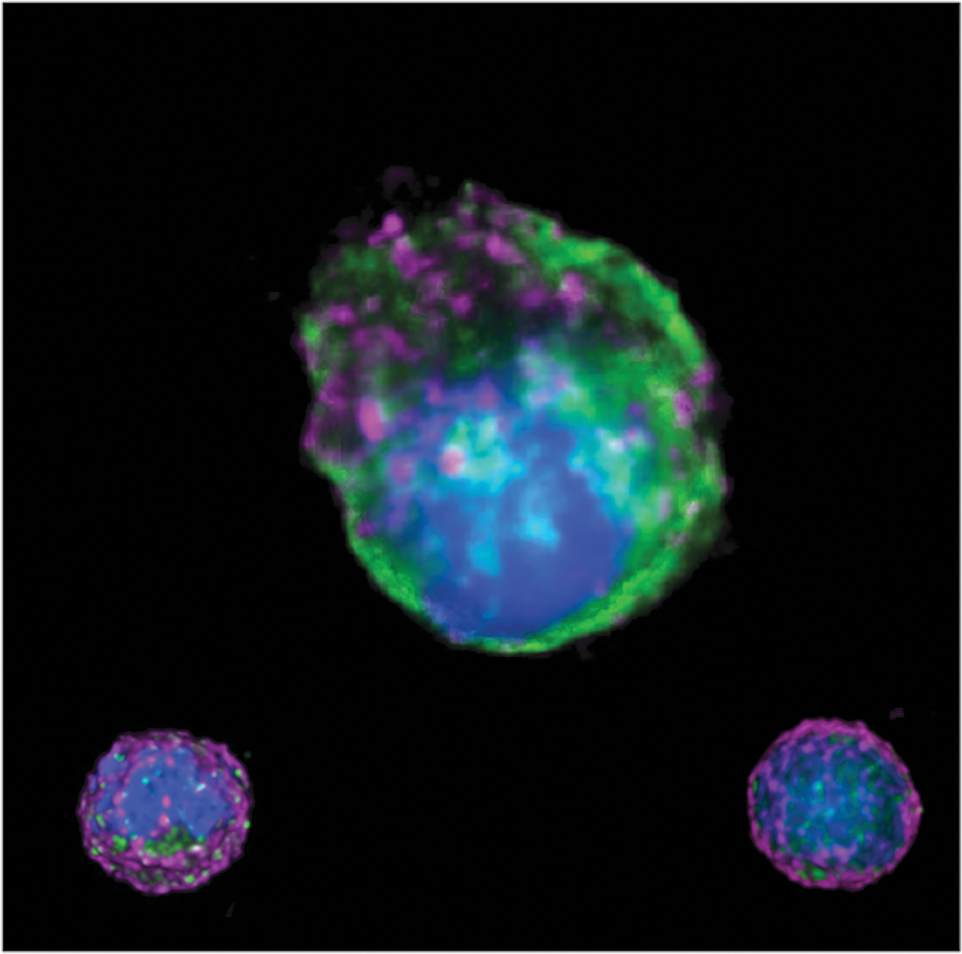
CD4 and Tumor Necrosis Factor-Related Apoptosis Ligand (TRAIL) localization in human plasmacytoid dendritic cells (pDCs) by three-dimensional (3D) microscopy. Human pDCs purified from healthy donors were cultured in the absence or presence of HIV and AMD-3100 or soluble CD4 for 2 h. Supernatants were removed and cells were cultured overnight. pDCs were stained with antibodies against TRAIL (green) and CD4 (purple); the nucleus was stained with DAPI (blue). Cells were acquired using a 3D microscope. Analysis was performed using a 3D viewer from ImageJ. Forty stacks were compiled to make a 3D view of the cells. Unstimulated pDCs (small cell, left) were covered by a CD4 crown (purple) and intracellular TRAIL (green). Cells cultured with HIV-1 in the presence of AMD-3100 (center) expressed membrane TRAIL (green) and CD4 (pink) was detected in the intracellular compartment. In contrast, pDC cultured with HIV and soluble CD4 did not express membrane TRAIL (right) and CD4 was expressed on the cell membrane.
Our findings may help to explain why the majority of pDCs become activated and not infected by HIV. Indeed, the binding of gp120 to CD4 is a fast process involving only two proteins. In contrast, CXCR4 binding to gp120 is a complex and slow process, thus it is statistically less frequent than CD4-gp120 binding. This is confirmed by in vitro experiments, in which more that 99% of pDCs become activated and less that 1% become infected by HIV.
Footnotes
Author Disclosure Statement
No competing financial interests exist.