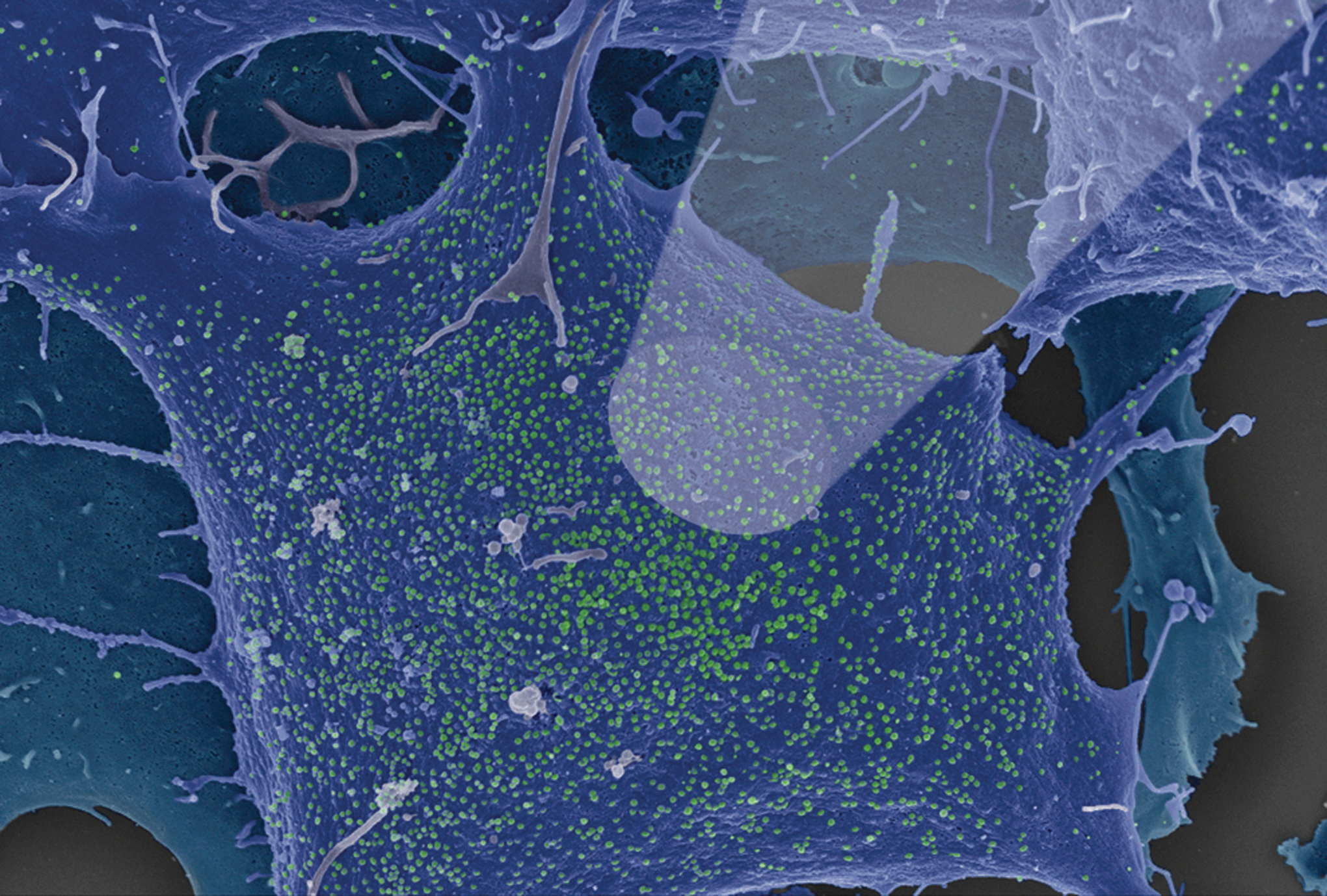
Transfection of human epithelial kidney cells (HEK293T) with a vector encoding the human immunodeficiency virus (HIV) is an easy way to produce infectious virus particles. These cells can be easily transfected with high transfection efficiencies. The main advantage is that these cells do not express the viral receptor CD4 and one of the coreceptors such as CCR5 or CXCR4. The produced virus is therefore not able to infect cells in this culture, ensuring that mutations in the viral genome will not occur. The basis of most HIV-1 molecular clones is the vector pNL4-3.1 To trace the infection of cells by immunofluorescence techniques, the vector pHIV-1 JR-FL Gag-iGFP expressing the Gag-internal green fluorescent protein (iGFP) was created.2 There the GFP sequence is flanked by viral protease cleavage sites.2 In the vector pNL43-V3-92TH014.12-IRES-eGFP GFP is expressed separately but from the same RNA transcript by an introduced IRES sequence.3 In this case GFP is found in the whole transfected cell, not only in the virus particles. Using scanning electron microscopy we observed budding structures at the plasma membrane on cells transfected with pNL43-V3-92TH014.12-IRES-eGFP (Fig. 1). To confirm that GFP expression occurred in the same cells, we performed correlative microscopy. Using the inverted cLSM Zeiss 780, GFP-positive cells were identified and subsequently investigated by scanning electron microscopy. Budding viruses were detected only in GFP expressing cells. These results demonstrate that the correlative technique provides more reliable data than using microscopy modes independently.4
The scanning electron microscopy image shows human embryonic kidney (HEK293T) cells producing human immunodeficiency virus (HIV-1). The cells were transfected with the plasmid pNL43-V3-92TH014.12-IRES-eGFP, fixed in glutaraldehyde, dehydrated in ethanol, critical point-dried, and treated with gold/palladium. The image was taken with a Leo 1530 Gemini and was manually colorized. The spot light illustrates the laser beam from the confocal laser scanning microscope (cLSM).
Footnotes
Acknowledgments
The picture was colored by Andrea Schnartendorff and Hans Günther Bredow, Robert Koch Institute. The work was supported by the Peter and Traudl Engelhorn Foundation, Germany with a grant to Daniel Ivanusic and Jung-Stiftung für Wissenschaft und Forschung, Hamburg/Germany. We thank Prof. Frank Kirchhoff for the vector pNL43-V3-92TH014.12-IRES-eGFP.
Author Disclosure Statement
No competing financial interests exist.
References
1.
AdachiA, GendelmanHE, KoenigS, et al.: Production of acquired immunodeficiency syndrome-associated retrovirus in human and nonhuman cells transfected with an infectious molecular clone. J Virol, 1986; 59(2):284–291.
2.
ChenP, HubnerW, SpinelliM, and ChenB: Predominant mode of human immunodeficiency virus transfer between T cells is mediated by sustained Env-dependent neutralization-resistant virological synapses. J Virol, 2007; 81:12582–12595.
3.
MunchJ, RajanD, SchindlerM, et al.: Nef-mediated enhancement of virion infectivity and stimulation of viral replication are fundamental properties of primate lentiviruses. J Virol, 2007; 81(24):13852–13864.
4.
MadelaK, BanhartS, ZimmermannA, et al.: A simple procedure to analyze positions of interest in infectious cell cultures by correlative light and electron microscopy. Methods Cell Biol, 124 (in press).